

 PDF
PDF Citation
 Print
Print


INTRODUCTION
Vancomycin is the first-line treatment for methicillin-resistant Staphylococcus aureus (MRSA) infections, which cause diseases, such as bacteremia, endocarditis, pneumonia, and osteomyelitis [1]. However, vancomycin dosing can be challenging because of large inter- and intra-individual variability in pharmacokinetic (PK) parameters and efficacy, resistance, and nephrotoxicity issues, which justify therapeutic drug monitoring (TDM) [2, 3]. Historically, the trough vancomycin concentration has been recommended as the target index; however, more recently, increasing evidence supporting area under the concentration–-time curve (AUC)-guided TDM has led the American Society of Health-System Pharmacists, Infectious Diseases Society of America, and Pediatric Infectious Diseases Society to recommend using the AUC as the target PK index in their recently revised U.S. consensus guidelines [4].
As a methodology for AUC-guided TDM, the revised U.S. guidelines recommend a Bayesian approach using “Bayesian software programs embedded with a PK model based on richly sampled vancomycin data as the Bayesian prior” [4]. To accurately estimate and predict the AUC using the Bayesian method, along with the proper selection of a priori PK parameters of the population model, it is essential to collect observational data on drug concentrations [5, 6]. As the blood sampling strategy for vancomycin TDM, two PK samples are recommended by the revised U.S. guidelines (i.e., 1–2 hours post-infusion and at the end of the dosing interval) for estimating the AUC using the Bayesian approach [4]. The recent Japanese vancomycin TDM guidelines also recommend two-point measurements [7]. These recommendations are mainly based on evidence from simulation studies that compared estimated AUC values from limited concentration data with reference AUC data from rich concentration data mainly acquired from published vancomycin PK studies [8-10]. However, the methodology for estimating AUC in the clinical setting remains controversial and is being actively researched [11].
Since January 2020, Ewha Womans University Seoul Hospital (Seoul, Korea) has implemented an internal TDM policy recommending that a post-distributional peak concentration be measured in addition to the trough concentration when requesting clinical PK consultation service (CPCS) for vancomycin to better predict concentration and AUC values by the Bayesian method. However, it remained unknown whether peak concentration data would be helpful in an actual clinical setting with more error factors than in PK study settings. We evaluated the performance of concentration and AUC prediction using Bayesian software based on two types of input concentration data (i.e., trough concentration alone vs. both trough and peak concentrations).
MATERIALS AND METHODS
Patients and TDM data
This was a retrospective study of patients for whom CPCS for vancomycin was requested between January 2020 and March 2021 in Ewha Womans University Seoul Hospital, an academic hospital in Seoul, Korea. Patient clinical data, including age, sex, weight, height, vancomycin administration history, renal replacement therapy history, vancomycin TDM data (concentration and sampling time), serum creatinine concentrations, and timing of creatinine measurement, were collected from hospital electronic health records and CPCS request forms. The following inclusion criteria for patient and TDM data were applied to evaluate the predictive performance in adult patients without renal impairment: (1) adult patients (≥18 years); (2) no renal replacement therapy during vancomycin dosing; (3) estimated glomerular filtration rate (eGFR) ≥60 mL/min/1.73 m2 as calculated using the Chronic Kidney Disease Epidemiology Collaboration (CKD-EPI) equation [12] at each TDM occasion; (4) TDM data within the acceptable time interval (0–1 hour before the start of infusion for the trough concentration; 1–3 hours after the end of infusion for the peak concentration); (5) patients with TDM data on two occasions in a ≤1-week interval. If a patient had TDM data on multiple occasions, data from the first two occasions were used. This study was approved by the Institutional Review Board of Ewha Womans University Seoul Hospital (approval No.: SEUMC 2020-06-018). The requirement for patient consent was waived due to because of the retrospective design of the study design.
Hospital setting and TDM practice during the period for study data
The hospital has 653 beds. When requesting CPCS for vancomycin, the internal TDM recommendation was using both trough (0–30 minutes before infusion) and peak (1–2 hours after the end of infusion) samples to better estimate the PK parameters. The request form included vancomycin dosing history, blood draw times (year, month, day, hour, and minute) for trough and peak concentrations, and dialysis information. The clinical laboratory provided information on the recommended blood collection times for trough and peak concentrations in the CPCS request form. If the blood draw time was suspected to be recorded incorrectly, the laboratory staff or pharmacist contacted the ward nurses by phone to obtain accurate sampling information. The recommended dosing regimen targeted a minimum concentration (Cmin) of 15.0–20.0 mg/L for severe MRSA infection cases (10.0–15.0 mg/L for other cases) and 24-hour AUC (AUC24)>400 mg∙hr/L for all cases. Vancomycin concentrations were measured using the Architect iVancomycin assay (Abbott, Wiesbaden, Germany) on an Architect i1000SR analyzer (Abbott). Serum creatinine concentrations were measured using CREA reagent (Beckman Coulter, Brea, CA, USA) on an AU5822 Automated Clinical Chemistry Analyzer (Beckman Coulter) based on the Jaffe method. Cmin and AUC24 were predicted based on simulations using the commercial Bayesian software, MwPharm++ v.1.9.0.338 (Mediware, Prague, Czech Republic).
AUC estimation by first-order PK analytic equations
For the collected data, first-order PK analytic equations were used to estimate the AUC when it was considered that the corresponding concentrations were drawn at the steady state (after at least three regular vancomycin doses). Three cases on the 1st occasion were excluded from the analysis. We used the two equations reported by Pai, et al. [13]:
where Ke is the elimination rate constant calculated using peak and trough concentrations, Cetrough is the forward-extrapolated concentration at the end of the dosing interval, Ceoi is the back-extrapolated concentration at the end of infusion, and Csoi’ is the back-extrapolated concentration at the start of infusion assuming an infusion duration of zero. Although the peak and trough concentrations were obtained around a single dose (rather than in the same dose cycle), the measured trough concentration was used as a substitute for the trough concentration in the next cycle (the same cycle as the peak concentration). The equations were named Eq. (4) and Eq. (5), as in the referenced paper.
AUC and concentration prediction using the Bayesian software
Bayesian individual PK parameter estimation and time–-concentration curve simulations were conducted using MwPharm++, which is used in routine practice. We used the built-in two-compartment model with the population PK parameters for adults based on the Dutch Association of Hospital Pharmacists monograph [14]. Briefly, the population PK parameters (mean±SD) as Bayesian priors were V1=0.21±0.04 L/kg, kelr=0.00327± 0.00109 hr–1/(mL/min/1.73 m2), k12=1.12±0.28 hr–1, and k21=0.48±0.12 hr–1, where V1 is the volume of distribution of the central compartment, kelr is the rate constant for renal elimination from the central compartment, k12 is the rate constant for elimination from the 1st (central) to the 2nd (peripheral) compartment, and k21 is rate constant for elimination from the 2nd to the 1st compartment. The metabolic elimination rate constant from the central compartment (kelm) was fixed at 0.0143 hr–1. The relationship between kelr and renal clearance (CLr) is CLr (L/h/kg)=kelr [h–1/(mL/min/1.73 m2)]×CLcr (mL/min/1.73 m2)×V1 (L/kg), where CLcr is creatinine clearance calculated using the Cockcroft–Gault equation and is normalized to the body surface area [15]. The individual PK parameters were estimated using two types of input concentration data (i.e., trough concentration alone vs. both trough and peak concentrations) for each TDM occasion.
Concentration-time curves were obtained using estimated individual PK parameters, and the AUC value over the dose cycle corresponding to each TDM occasion was calculated. The AUC value estimated using the TDM data at the corresponding dose cycle is represented as AUCestimated. AUCestimated was normalized to 24 hours for agreement evaluation by dividing the AUCestimated for the dose cycle by the dosing interval and multiplying by 24 and is represented as AUC24estimated. The AUC value predicted for the 2nd occasion using the TDM data from the 1st occasion is represented as AUCpredicted. The input concentration data type for individual PK parameter estimation is indicated, for example, AUCestimated[T&P] (AUC value estimated by fitting the trough and peak concentrations on the corresponding occasions) and AUCpredicted[T] (AUC value on the 2nd occasion predicted by fitting the trough concentration on the 1st occasion). Predicted concentration values on the 2nd TDM occasion using the data from the 1st TDM occasion are represented as Cpredicted. The measured concentration is expressed as Cmeasured.
Evaluation of predictive performance and statistical analysis
The median difference, median percentage difference, and Pearson’s correlation coefficient (r) were calculated to investigate the relationships among AUC24estimated[T], AUC24estimated[T&P], AUC24Eq.(4), and AUC24Eq.(5). The AUC24estimated results were divided into three groups (subtherapeutic, <400 mg∙hr/L; therapeutic, 400–600 mg∙hr/L; and toxic, >600 mg∙hr/L) based on the target range (assuming a minimum inhibitory concentration of 1 mg/L) suggested by the U.S. consensus guidelines [4]. Agreements were compared using the weighted Cohen’s kappa.
The ability of Bayesian modeling for predicting concentration or AUC values on the 2nd TDM occasion using the data from the 1st TDM occasion was evaluated in terms of bias and imprecision. The prediction error (PE) was defined as where ValuePredicted is the concentration or AUC value of the vancomycin dose cycle on the 2nd TDM occasion predicted by estimated individual PK parameters using the previous concentration data and ValueReference is the value of Cmeasured, AUC estimated using the first-order PK analytic equations, or Bayesian AUCestimated using TDM data from the 2nd TDM occasion. The median PE (MDPE) for bias and median absolute PE (MDAPE) for imprecision were calculated as predictive performance parameters: MDPE=median (of PE) and MDAPE=median (of |PE|). To evaluate the relative bias or imprecision of predictors from the two input concentration data types, the median of paired differences was calculated: ΔPEi=PEi (trough) - PEi (trough and peak) and ΔAPEi=|PEi (trough)| - |PEi (trough and peak)|. The 95% confidence interval (CI) for the performance parameters was calculated using the bootstrap percentile method (5,000 bootstraps). The 95% CIs for the medians of ΔPE and ΔAPE were also calculated, and an interval not including zero was considered a significant difference.
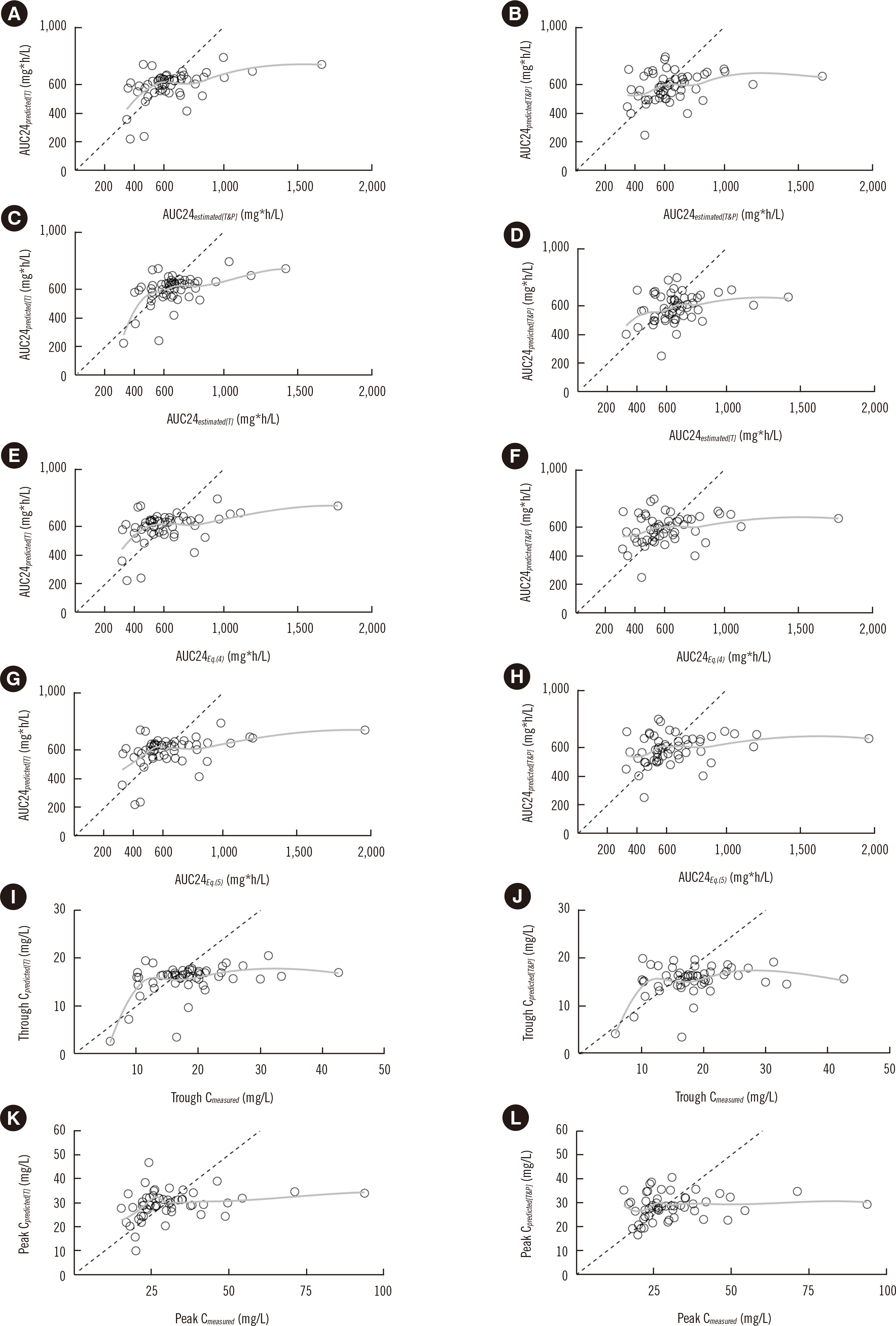
Multiple variable graphs with dots and lines were drawn to show trends in estimated AUC24 according to the TDM occasion and dosage. Scatterplots with locally estimated scatterplot smoothing curves were used to show the relationships between predicted and reference values. Bland–Altman plots were used to visually explore the differences between estimated AUC24 values. R version 4.0.4. (R Foundation for Statistical Computing, Vienna, Austria) was used for statistical analysis and to calculate the performance metrics.
RESULTS
Characteristics of patients and TDM data
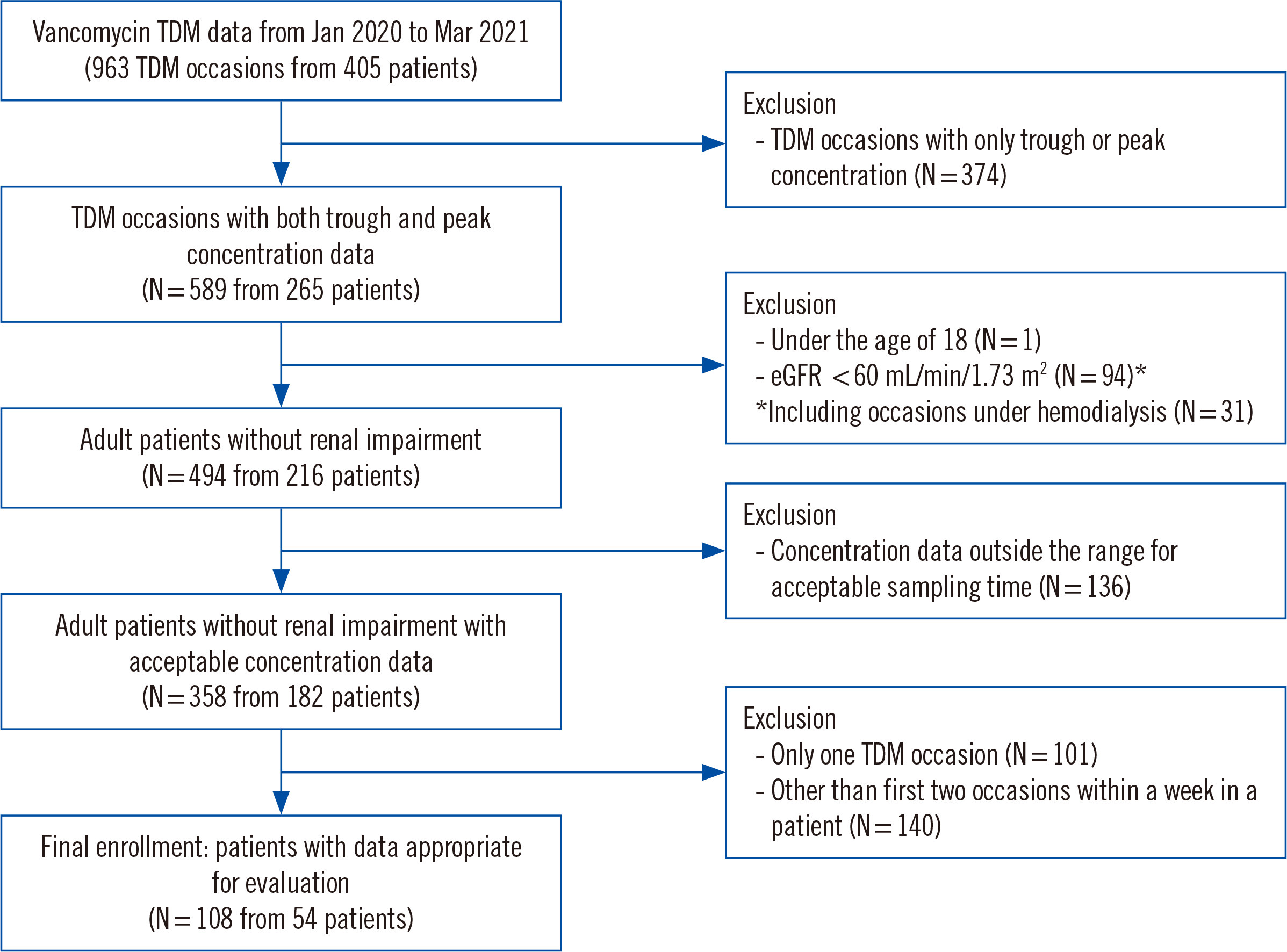
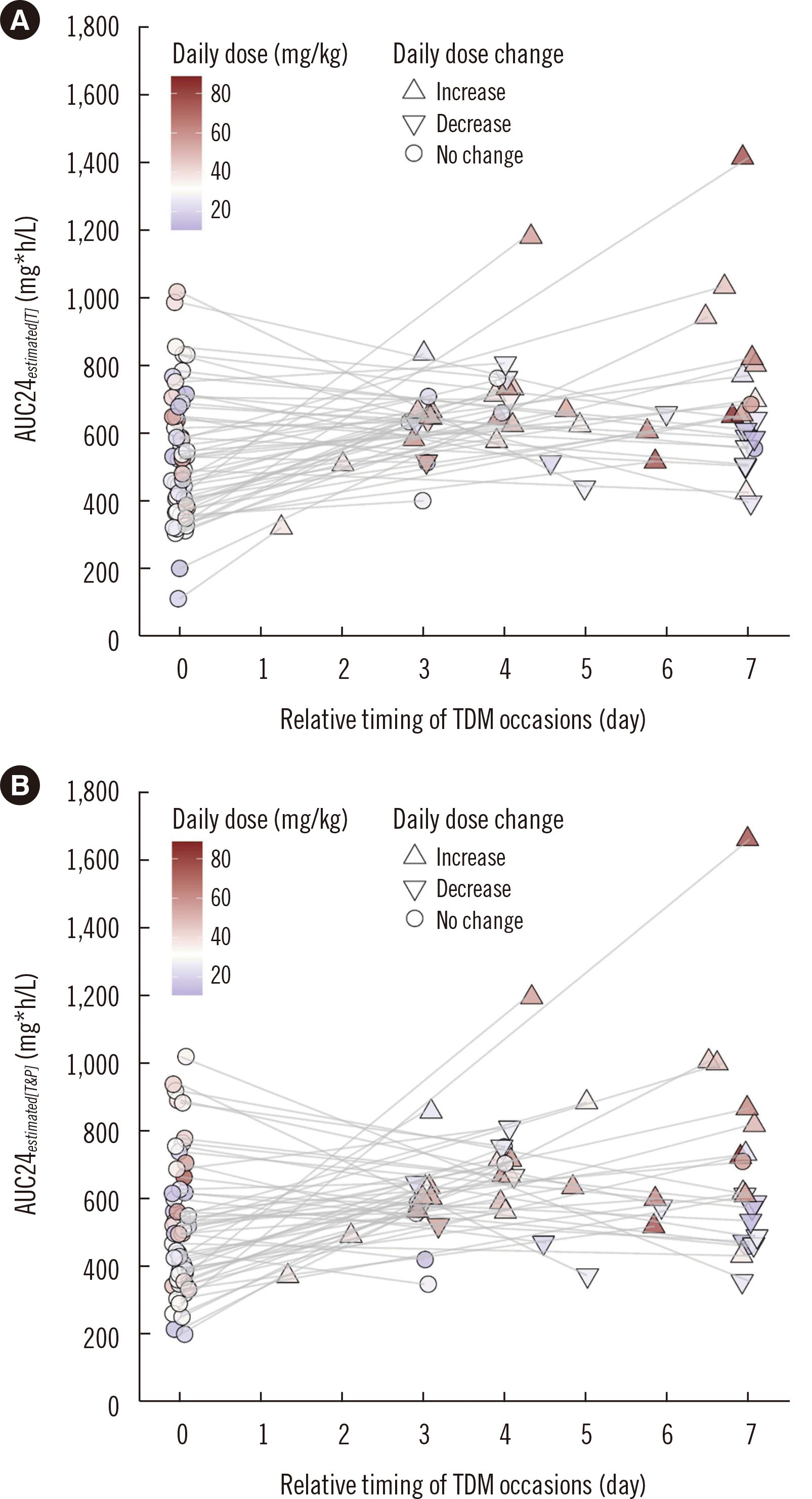
Among 963 vancomycin TDM occasions for 405 patients, two-point concentration data were available for 589 occasions; 108 TDM occasions for 54 patients met the inclusion criteria. All patients were Korean and received intravenous vancomycin via intermittent infusion. A detailed flow diagram of study patient and TDM data inclusion is shown in Fig. 1. The median (Q1 to Q3) age was 71.5 (range, 59–81) years, and 26 (48.1%) patients were male. Thirty-eight patients had complicated infections: 24 bacteremias and eight CNS infections, including two meningitis, two osteomyelitis, and four pneumonia cases. Others had intra-abdominal infections (N=10), skin and soft-tissue infections (N =4), or urinary tract infections (N=2). The median (Q1 to Q3) difference in serum creatinine concentrations between 1st and 2nd occasions was –0.04 (–0.10–0.06) mg/dL. The median (Q1 to Q3) difference in eGFR between 1st and 2nd occasions was 2 (–4–7) mL/min/1.73 m2. No case had a change in creatinine concentration at the acute kidney injury level (a ≥50% or ≥0.3 mg/dL increase in the serum creatinine concentration) [16]. The median (Q1 to Q3) time between trough samplings on the 1st and 2nd TDM occasions (TDM interval) was 105.3 (73.5–168.0) hours. Detailed characteristics of the patients are shown in Table 1. Estimated AUCs at all TDM occasions over time along with information on the vancomycin dosage are shown in Fig. 2. Sampling and infusion time data were recorded at limited time points (Supplemental Data Fig. S1). The modes of trough sampling, infusion end, and peak sampling time relative to the infusion start time were 0, 60, and 120 minutes, respectively.
Agreement between AUC24estimated[T] and AUC24estimated[T&P]
AUC24estimated[T] strongly correlated with AUC24estimated[T&P], with r=0.956 (P<0.001). The median (Q1 to Q3) difference between AUC24estimated[T] and AUC24estimated[T&P] was 27.7 (–10.9–50.5) mg∙hr/L in total, 27.7 (–12.6–48.6) mg∙hr/L on the 1st occasion, and 24.6 (–5.7–50.4) mg∙hr/L on the 2nd occasion. The median (Q1 to Q3) percentage difference between AUC24estimated[T] and AUC24estimated[T&P] was 2.4% (–1.0%–4.7%) in total, 5.2% (–2.8%–10.0%) on the 1st occasion, and 3.6% (–1.1%–9.2%) on the 2nd occasion. The differences are depicted in Bland–Altman plots in Supplemental Data Fig. S2. In analysis of the agreement based on clinical categories, AUC24estimated[T] and AUC24estimated[T&P] showed 84.3% agreement (85.2% on the 1st occasion and 83.3% on the 2nd occasion), with a weighted kappa value of 0.80 (Table 2).
Agreement between AUC24 estimated using Bayesian modeling and using first-order PK analytic equations
AUC24estimated[T] strongly correlated with AUC24Eq.(4) and AUC24Eq.(5), with r=0.902 (P<0.001) and 0.879 (P<0.001), respectively. The median (Q1 to Q3) difference between AUC24estimated[T] and AUC24Eq.(4) and AUC24Eq.(5) was 59.8 (14.8–104.3) mg∙hr/L and 38.1 (–15.0–83.6) mg∙hr/L, respectively. AUC24estimated[T&P] strongly correlated with AUC24Eq.(4) and AUC24Eq.(5), with r=0.985 (P<0.001) and 0.977 (P<0.001), respectively. The median (Q1 to Q3) difference between AUC24estimated[T&P] and AUC24Eq.(4) and AUC24Eq.(5) was 36.7 (14.1–48.1) mg∙hr/L and 12.1 (–8.5–21.1) mg∙hr/L, respectively. The differences are shown in Bland–Altman plots in Supplemental Data Fig. S3. In analysis of the agreement based on clinical categories, AUC24estimated[T] showed 71.4% agreement (weighted kappa=0.65) with both AUC24Eq.(4) and AUC24Eq.(5), whereas AUC24estimated[T&P] showed 86.7% agreement (weighted kappa=0.83) with both AUC24Eq.(4) and AUC24Eq.(5).
Predictive performance of Bayesian modeling for AUC or concentration values on the next occasion
The performance of Bayesian modeling using TDM data from the 1st occasion in predicting AUCs or measured concentrations on the 2nd occasion is summarized in Table 3. When evaluated based on AUCestimated[T&P], AUCestimated[T], AUCEq.(5), and trough Cmeasured, the predicted values using the trough concentration alone were less biased and less imprecise. When evaluated based on peak Cmeasured, the predicted values using both trough and peak concentrations were less biased, but slightly more imprecise. The PEs using the trough concentration alone were significantly positively biased compared with those using both trough and peak concentrations. The absolute PE using the trough concentration alone was significantly smaller when evaluated based on AUCestimated[T]. The relationships between the predicted and reference values are depicted in scatter plots in Fig. 3.
DISCUSSION
We evaluated the concentration- and AUC-predictive performance of Bayesian modeling using retrospective TDM data from routine hospital practice. We found that using both trough and peak concentrations did not improve the predictive performance and consider three main reasons for this result. First, the increase in predictability when using peak concentration data may have been so small that it was difficult to observe in the small study cohort. Second, the population PK model used may not be appropriate for the study population and TDM data. The difference in AUC estimation accuracy according to the combination of input data may be affected by the population PK model [9]. This issue will be further discussed in the limitations section. Third, there may have been errors in the clinical data. Clinical data, including the drug concentration and sampling and dose administration times, are prone to errors that may significantly affect the Bayesian estimation of individual PK parameters [6, 17, 18]. Inaccuracies in time recordings were suspected to be a significant cause of invalidity of peak concentration data because peak concentrations may be more dependent on the actual infusion end time and may show more significant changes over time than trough concentrations.
Our results contradict those of previous studies using richly sampled PK study data supporting two-point measurements [8-10]. These previous studies calculated reference AUC values using five to eight vancomycin concentration data points in a dose cycle. They compared estimated AUCs with the reference AUC, and all showed a more considerable variability in errors from AUCs estimated using the trough concentration alone than in those estimated using trough and peak concentrations. However, these studies evaluated a small number of subjects, including healthy individuals, and may not represent the clinical patient population. In addition, they used time–-concentration data from a strictly monitored PK study with an accuracy that may be difficult to achieve in a real clinical setting. Along with the evaluation of richly sampled PK data, Oda, et al. [10] evaluated 28 adult clinical patients with peak and trough concentrations, showing that the AUC estimated using the trough concentration alone was unbiased against estimates using both trough and peak concentrations. However, they concluded that AUC estimation using the trough concentration alone should be avoided when treating invasive MRSA infections in critically ill patients with large variability in PK due to decreased accuracy.
Olney, et al. [19] compared AUCs estimated by the Bayesian method using the trough concentration alone with those estimated using a first-order equation and the Bayesian method using trough and peak concentrations. While there was a strong correlation and agreement between the AUCs estimated by the two-concentration Bayesian method and first-order equation (r=0.963), with 87.4% agreement across clinical categories, the trough concentration Bayesian method demonstrated a weaker correlation (r=0.823) and 76.8% agreement. The correlation between the trough and two-concentration Bayesian method was strong (r=0.931), with 88.5% agreement. Although our and their results on these metrics cannot be directly compared because they are largely affected by the AUC range and distribution, our results are very similar to theirs, with the degree of correlation and agreement being in the order of two-concentration Bayesian method and first-order equations (r=0.985 and 0.977; 86.7% agreement) >trough and two-concentration Bayesian methods (r=0.956; 84.3% agreement) >trough concentration Bayesian method and first-order equations (r=0.902 and 0.879; 71.4% agreement).
Along with a comparison between estimated AUCs, we evaluated the prediction performance for AUCs and concentrations on the next TDM occasion. Prediction performance metrics for subsequent concentrations after incorporating the patient’s vancomycin concentrations have been used to evaluate Bayesian models and/or the usefulness of incorporated concentration data [20-22]. To our knowledge, this was the first study to evaluate the performance of Bayesian modeling in vancomycin AUC prediction for the next occasion. The predicted AUC using the trough concentration alone showed a higher value than that using both trough and peak concentrations, showing a bias (MDPE) that is measured differently according to the reference AUC. Their ΔAPEs were not significant, except in AUCestimated[T], and even MDAPEs using trough concentrations alone were better than those for reference values such as AUCestimated[T&P], AUCestimated[T], and AUCEq.(5). Therefore, we tentatively concluded that using both trough and peak concentrations did not improve the AUC-predictive performance. We suspect that inaccuracies in the time records in the clinic are a major explanation for this result.
Although the importance of time recording has been steadily highlighted in routine practice, time data recorded by general medical staff still need to be more accurate. The time data in our study were limited to specific times and were unrealistic. Given that planned rather than actual sampling times are often recorded in a clinical setting, discrepancies between actual and documented antimicrobial infusion administration times would not be surprising [23]. Compliance with the recommended blood sampling time was also insufficient. Approximately 27.5% (136 out of 494) of TDM data were excluded for being outside the defined acceptable time range. Previous studies have also reported such non-compliance [24, 25]. Although AUC estimation by the Bayesian approach could be less susceptible to concentration errors than PK analytic equations [26], for proper application of AUC-guided TDM, TDM data accuracy should be ensured first [5]. However, without practical, well-resourced support, including tools, education, and staffing, it may not be easy to improve or audit the accuracy of TDM data. The role of the laboratory is also crucial in increasing the clinical usefulness of TDM [27, 28].
Our study had a significant limitation in that the population PK model used may not be ideal for the Korean population. We utilized used the built-in population PK model of MwPharm++, which was developed based on a population of a different race. Moreover, our study population had a high proportion of elderly patients, who may have different PK properties [29]. In addition, the information on how the default PK parameters were generated is unclear, making it difficult to evaluate their appropriateness for our study population. However, using the built-in population PK model of MwPharm++ is not exceptional in Korea [30], and our study reflects the real-world situation where TDM analysts in the clinic often have to rely on readily available models.
Several Korean-specific population PK models for vancomycin, which may have been more appropriate for our study population, have been published [31-33]. Differences between the built-in MwPharm++ model and the Korean-specific models include variability in the structural model, covariates, and PK parameter values. For example, when compared with the PK parameters from a recent study ausing a two-compartment model assuming a typical patient with a weight of 60 kg and a CLcr of 72 mL/min, the built-in MwPharm++ model has a larger clearance (2.82 vs. 3.15 L/h), a smaller central compartment volume (31.8 vs. 12.6 L), larger intercompartmental clearance (11.7 vs. 14.1 L/h), and smaller peripheral compartment volume (75.4 vs. 29.4 L) [33]. These differences in population PK parameters can result in a significant difference in the Bayesian AUC estimate [22]. However, these Korean-specific population models have not been validated as a priori information for Bayesian AUC estimation. Additionally, the use of customized population PK models in a Bayesian software program may not be feasible in most clinics. Considering this situation, further development of a relevant population model or validation of AUC estimation using published models is required to implement proper AUC-guided dosing using the Bayesian approach in Korea, and the dissemination of a software tool with a validated population PK model is required to facilitate the use of AUC-guided dosing in clinical practice. Similar work has been carried out by a Japanese expert group [10].
This study had several other limitations. First, the reference AUC value was not measured based on rich sample data. However, it is nearly impossible to acquire rich and accurate sample data in the clinical setting. The AUC estimated by the Bayesian approach using the two concentration data points from the next TDM occasion may be the best reference value that is practically feasible in a clinical setting. We also evaluated the predictive performance with other reference values to provide a balanced perspective. Second, we did not assess performance in comparison with other population PK models; therefore, generalization may be limited. Third, inaccuracies in time recordings were presumed to have caused the non-superiority of using peak and trough data; however, we did not measure this directly. Fourth, as this was a retrospective study, the patients were selected according to data availability, and sampling bias from the intended population (general hospitalized patients treated with vancomycin) may have occurred. Finally, we should have evaluated cost implications, which may be critical in determining the TDM strategy. A recent study in the U.S. showed significant institutional cost benefits of the Bayesian method over trough-guided dosing [34]. Similar research is needed in Korea.
In conclusion, while there was a significant difference between AUC24 values estimated using trough alone and both trough and peak concentration data, the usefulness of peak concentration data for predicting the AUC on the next occasion by the Bayesian program was not demonstrated. These results bring into question the practicality of peak sampling for AUC-guided dosing. However, as the study was conducted in a specific setting, different results may be obtained depending on the patient population, population PK model suitability, and data accuracy; therefore, caution should be exercised in result interpretation. Detailed operation protocols should be researched and suggested to allow proper implementation of AUC-guided TDM.
 XML Download