Journal List > Cancer Res Treat > v.55(3) > 1516083302

Yoon, Han, Lee, Choi, Choi, Kim, and Kim: Intraindividual Comparison of MRIs with Extracellular and Hepatobiliary Contrast Agents for the Noninvasive Diagnosis of Hepatocellular Carcinoma Using the Korean Liver Cancer Association–National Cancer Center 2022 Criteria

Abstract

Purpose

The aim of the present study was to evaluate the per-lesion sensitivity and specificity of the Korean Liver Cancer Association–National Cancer Center (KLCA-NCC) 2022 criteria for the noninvasive diagnosis of hepatocellular carcinoma (HCC), with intraindividual comparison of the diagnostic performance of magnetic resonance imaging with extracellular agents (ECA-MRI) and hepatobiliary agents (HBA-MRI).

Materials and Methods

Patients at high risk for HCC who were referred to a tertiary academic institution for hepatic lesions with size ≥ 10 mm between July 2019 and June 2022 were enrolled. A total of 91 patients (mean age, 58.1 years; 76 men and 15 women) with 118 lesions who underwent both ECA-MRI and HBA-MRI were eligible for final analysis. The per-lesion sensitivities and specificities of the KLCA-NCC 2022 criteria using ECA-MRI and HBA-MRI were compared using McNemar’s test.

Results

The 118 lesions were 93 HCCs, 4 non-HCC malignancies, and 21 benign lesions. On HBA-MRI, the “definite” HCC category showed significantly higher sensitivity than ECA-MRI (78.5% vs. 58.1%, p < 0.001), with identical specificity (92.0% vs. 92.0%, p > 0.999). For “probable” or “definite” HCC categories, there were no differences in the sensitivity (84.9% vs. 84.9%, p > 0.999) and specificity (84.0% vs. 84.0%, p > 0.999) between ECA-MRI and HBA-MRI.

Conclusion

The “definite” HCC category of the KLCA-NCC 2022 criteria showed higher sensitivity in diagnosing HCC on HBA-MRI compared with ECA-MRI, without compromising specificity. There were no significant differences in the sensitivity and specificity of “probable” or “definite” HCC categories according to ECA-MRI and HBA-MRI.

Introduction

Hepatocellular carcinoma (HCC) is the most common primary malignant liver tumor, and is unique in that imaging is widely utilized to make diagnoses in clinical practice [1,2]. Several guidelines have been proposed worldwide for the noninvasive diagnosis of HCC, with criteria varying based on geographic area and population [1,36]. Magnetic resonance imaging (MRI) using either an extracellular agent (ECA) or hepatobiliary agent (HBA), along with contrast-enhanced computed tomography, is the primary imaging modality utilized for the diagnosis of HCC [4,5,7,8]. Major international guidelines consider non-rim arterial phase hyperenhancement (APHE) and washout appearance on the portal venous phase (PVP) or delayed phase (DP) of ECA-MRI, or on the PVP of HBA-MRI as radiologic hallmarks for the diagnosis of HCC [1,4,7,8]. The Korean Liver Cancer Association–National Cancer Center (KLCA-NCC) 2018 criteria expanded the definition of washout appearance to include washout on the DP or hepatobiliary phase (HBP) of HBA-MRI, with the intention of achieving a high sensitivity in the diagnosis of HCC [6].
The recently updated KLCA-NCC 2022 criteria maintained the expanded definition of washout appearance on HBA-MRI, with additional changes to the “probable” HCC category, which corresponds to the category LR-4 of the Liver Imaging Reporting and Data System [5]. In the earlier KLCA-NCC 2018 criteria, “probable” HCC was diagnosed based on the presence of at least one item from each of the two categories of ancillary imaging features, regardless of the presence or absence of APHE [6]. In the updated KLCA-NCC 2022 criteria, “probable” HCC can be more easily diagnosed when a nodule with APHE presents with at least one item from any of the two groups of ancillary imaging features [8]. This change was based on reports that nodules with APHE show an increased probability of being HCC or having a higher risk of progressing into HCC than those without APHE [810].
There have not yet been reports on the diagnostic performance of the updated KLCA-NCC 2022 criteria, owing to its recent implementation. Moreover, no intraindividual analyses directly comparing the diagnostic performance of ECA-MRI and HBA-MRI based on the KLCA-NCC 2022 criteria have been reported. Therefore, the objective of the present study was to evaluate the per-lesion sensitivity and specificity of the recently published KLCA-NCC 2022 criteria for the noninvasive diagnosis of HCC, with an intraindividual comparison of the diagnostic performance of ECA-MRI and HBA-MRI.

Materials and Methods

1. Study population

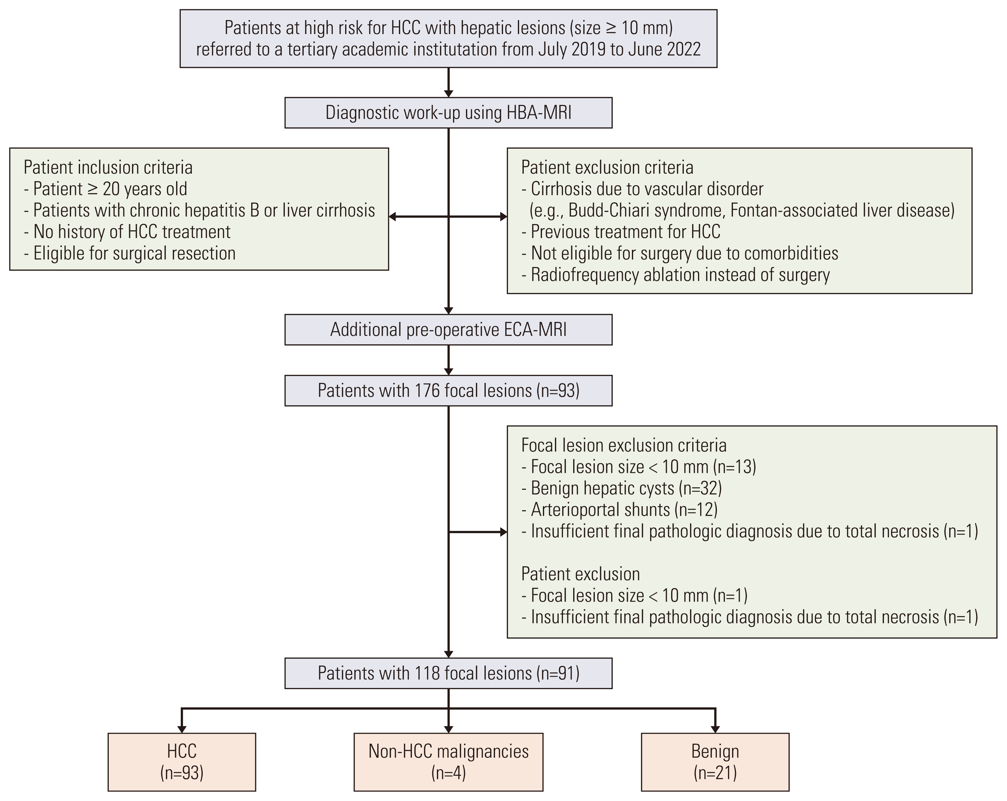
Between July 2019 to June 2022, we consecutively enrolled patients at high risk for HCC with hepatic lesions with a diameter ≥ 10 mm who had been referred to our institution (Fig. 1). The inclusion criteria were as follows: (1) patients aged ≥ 20 years, (2) patients at high risk for HCC with chronic hepatitis B or liver cirrhosis, (3) patients with no history of HCC treatment, and (4) patients eligible for surgical resection. The exclusion criteria were as follows: (1) patients with cirrhosis due to vascular disorders, such as Budd-Chiari syndrome or Fontan-associated liver disease, (2) patients not eligible for surgery due to comorbidities, and (3) patients who received radiofrequency ablation instead of surgery. HBA-MRI was performed first, as it is currently preferred over ECA-MRI at our institution for patients suspected of having hepatic malignancy. Two of the authors (D.H.H. and G.H.C.), who did not participate in the imaging review session, determined the surgical indication according to the size, number, and location of tumor. After identifying eligibility for inclusion in the present study based on the HBA-MRI findings, ECA-MRI was performed on those patients who consented. A researcher (M-J.K. with 29 years of experience in abdominal radiology), who did not participate in the main image analysis, reviewed both MRI scans and compiled a list of eligible patients according to the focal lesion inclusion and exclusion criteria.

2. MRI exams

MRI exams were performed using 3.0-Tesla (T) systems (Skyra, Verio, Prisma Fit, Magnetom Lumina, or Magnetom Vida, Siemens Healthineers, Erlangen, Germany; Achieva, Ingenia, Ingenia CX, or Ingenia Elition, Philips Healthcare, Best, Netherlands; and Signa HDxt, Signa Architect, or Discovery MR750w, GE Healthcare, Waukesha, WI). The scan protocol included the acquisition of dual-echo T1-weighted gradient-echo images (in- and opposed-phase), T1-weighted three-dimensional gradient-echo images with dynamic contrast enhancement, navigator-triggered single- or multi-shot T2-weighted images, and diffusion-weighted images (DWI) at various b-values. Dynamic T1-weighted imaging was performed before and after the administration of either ECA (gadobutrol, Gadovist, Bayer Pharma AG, Berlin, Germany) or HBA (gadoxetate disodium, Primovist, Bayer Pharma AG). Arterial phase scanning was initiated using the test bolus or bolus tracking technique, after which PVP and DP images were obtained using ECA-MRI. For HBA-MRI, PVP, DP, and 20-minute delayed HBP images were obtained using HBA-MRI. Detailed parameters of the MRI sequences are listed in S1 Table.

3. Image analysis

All MRI exams were evaluated by two abdominal radiologists—J.K.Y. and S.L. with 3 and 10 years of experience in reading liver imaging, respectively. The radiologists were blinded to the final diagnosis of each lesion; however, they were informed that the study population consisted of patients at high risk for HCC. Readers were not blinded to the contrast agent used for the liver MRI due to the obvious differences in images. Readers independently reviewed the ECA-MRI and HBA-MRI simultaneously without washout period. Any discrepancies regarding imaging findings were resolved by consensus of the two readers. The consensus data were then used for the primary image analysis and overall characterization for each observation. All MRI scans were retrieved from a picture archiving and communication system (Centricity Radiology RA 1000, GE Healthcare, Chicago, IL).
The radiologists assessed the images for the presence or absence of the radiological hallmarks of HCC (APHE and washout appearance), ancillary imaging features, marked T2 hyperintensity, and targetoid appearances, based on the KLCA-NCC 2022 criteria [8]. Per the KLCA-NCC 2022 guidelines, a lesion showing marked T2 hyperintensity was classified as benign. Targetoid appearance was evaluated on dynamic contrast-enhanced studies or DWI sequences. Excluding the benign category and targetoid appearance lesions, each lesion was categorized as “definite” HCC, “probable” HCC, or an “indeterminate” nodule, in a stepwise manner [8]. “Definite” HCC was defined as a lesion with both APHE and washout appearance on the PVP, DP, or HBP phases [8]. Lesions that did not meet the noninvasive diagnostic criteria for HCC were diagnosed as “probable” HCC if the following criteria were met: (1) APHE with at least one ancillary imaging feature suggesting malignancy in general (group A: mild-to-moderate T2 hyperintensity, high signal intensity on DWI, or threshold growth) or favoring HCC in particular (group B: enhancing or non-enhancing capsule, mosaic architecture, nodule-in-nodule appearance, or fat or blood products in mass), or (2) no APHE with at least one ancillary imaging feature from each group.
Threshold growth was not evaluated in the present study due to the lack of prior computed tomography or MRI examinations for comparison. When imaging features did not fulfill the aforementioned criteria, the lesion was categorized as “indeterminate” [8]. After independent categorization by each radiologist, the inter-reader agreement was evaluated.

4. Reference standards

The diagnoses of HCC and non-HCC malignancies were confirmed by pathology (n=97). Benign diagnoses were determined based on the presence of typical imaging features (n=9) or stability on diagnostic imaging for at least 2 years (n=12).

5. Statistical analysis

All analyses were performed using commercial software (R ver. 4.2.1, R Foundation for Statistical Computing, Vienna, Austria). The per-lesion estimates of the diagnostic performance (sensitivity, specificity, positive predictive value, and negative predictive value) were calculated, and the sensitivities and specificities of the KLCA-NCC 2022 criteria using ECA-MRI and HBA-MRI were compared using McNemar’s test. Inter-reader agreement was evaluated using Cohen’s unweighted κ coefficient. The κ values were interpreted as follows: none-to-slight, 0.00–0.20; fair, 0.21–0.40; moderate, 0.41–0.60; substantial, 0.61–0.80; and almost perfect agreement, 0.81–1.00 [11]. A two-sided p-value < 0.05 was considered statistically significant.

Results

1. Characteristics of patients and lesions

The baseline characteristics of the 91 patients included (118 lesions; mean age, 58.1 years; 76 men and 15 women) are shown in Table 1. Hepatitis B (81.3%) was the predominant etiology for the underlying liver disease. There were 40 (44.0%) liver cirrhosis patients. The median modified end-stage liver disease score was 7 (interquartile range, 6 to 8). Hepatic lesions were categorized based on ECA-MRI and HBA-MRI findings according to the KLCA-NCC 2022 criteria (Table 2). A total of 56 lesions (47.4%) were diagnosed as “definite” HCC based on ECA-MRI, while 75 lesions (63.6%) were diagnosed as “definite” HCC based on HBA-MRI. In contrast to the 27 lesions (22.9%) diagnosed as “probable” HCC based on ECA-MRI, seven lesions (5.9%) were diagnosed as “probable” HCC based on HBA-MRI. There were 14 (11.9%) and 13 (11.0%) focal lesions with targetoid appearances on ECA-MRI and HBA-MRI, respectively. The pathologic results of these lesions are shown on S2 Table. The median interval between HBA-MRI and operation was 24 days (interquartile range, 16 to 32 days).

2. “Definite” HCC category based on ECA-MRI and HBA-MRI

The per-lesion diagnostic performances of the “definite” HCC category based on the KLCA-NCC 2022 criteria are shown in Table 3. Based on HBA-MRI, the “definite” HCC category showed a significantly higher sensitivity for the diagnosis of HCC than ECA-MRI (78.5% vs. 58.1%, p < 0.001), although with identical specificity (92.0% vs. 92.0%, p > 0.999) (Fig. 2). In the subgroup analysis based on lesion size, HBA-MRI showed significantly higher sensitivity than ECA-MRI (10–19 mm, 72.7% vs. 36.4%, p=0.005 and ≥ 20 mm, 81.7% vs. 70.0%, p=0.020, respectively) without differences in specificity (10–19 mm, 95.0% vs. 95.0%, p > 0.999 and ≥ 20 mm, 80.0% vs. 80.0%, p > 0.999, respectively).

3. “Probable” or “definite” HCC categories on ECA-MRI and HBA-MRI

The per-lesion diagnostic performances for the “probable” or “definite” HCC categories of KLCA-NCC 2022 criteria are shown in Table 4. There were no differences in the sensitivity (84.9% vs. 84.9%, p > 0.999) and specificity (84.0% vs. 84.0%, p > 0.999) between ECA-MRI and HBA-MRI. Subgroup analyses based on lesion size showed no difference between ECA-MRI and HBA-MRI in sensitivity (10–19 mm, 84.8% vs. 84.8%, p > 0.999 and ≥ 20 mm, 85.0% vs. 85.0%, p > 0.999, respectively) or specificity (10–19 mm, 85.0% vs. 85.0%, p > 0.999 and ≥ 20 mm, 80.0% vs. 80.0%, p > 0.999, respectively).

4. Radiologic hallmarks of HCC on ECA-MRI and HBA-MRI

Radiologic hallmarks of the 93 HCC lesions observed on ECA-MRI and HBA-MRI are shown in Table 5. APHE was more frequently observed on ECA-MRI than HBA-MRI in all HCCs (83.9% vs. 79.6%, p=0.219) and in the subgroup of lesions sized 10–19 mm (84.8% vs. 69.7%, p=0.063), but without statistically significant difference. On the other hand, the HBA-MRI showed more frequent washout appearance than ECA-MRI in all lesions (98.9% vs. 72.0%, p < 0.001) and in subgroup analyses based on lesion size (10–19 mm, 100.0% vs. 48.5%, p < 0.001; and ≥ 20 mm, 98.3% vs. 85.0%, p < 0.001, respectively) with statistical significance.

5. False positive diagnoses for HCC

There were two false-positive cases which were categorized as “definite” HCC on both ECA-MRI and HBA-MRI. These cases were confirmed as combined hepatocellular-cholangiocarcinomas (cHCC-CCAs). For the “probable” HCC category, there were two false-positive cases which were confirmed as dysplastic nodules.

6. Inter-reader agreement

The inter-reader agreement for the final categorization and imaging feature determination based on the KLCA-NCC 2022 criteria is shown in S3 Table. Lesion categorization based on the KLCA-NCC 2022 criteria showed almost perfect agreement for both ECA-MRI and HBA-MRI (κ=0.82 and κ=0.83, respectively). The radiologic hallmarks of HCC, ancillary imaging features, marked T2 hyperintensity, and targetoid features on ECA-MRI and HBA-MRI all showed moderate to almost perfect agreement (κ=0.43–0.97).

Discussion

In the present study, the intraindividual comparison of MRI with ECA or HBA for the noninvasive diagnosis of HCC based on the recently published KLCA-NCC 2022 criteria showed that HBA-MRI showed higher sensitivity for the “definite” HCC category than ECA-MRI, although with identical specificity. The sensitivities and specificities of the “probable” or “definite” HCC categories for diagnosing HCC were identical for both ECA-MRI and HBA-MRI.
The present study showed significantly higher sensitivity for the “definite” HCC category on HBA-MRI compared with ECA-MRI, although with identical specificity, based on the KLCA-NCC 2022 criteria [8]. The sensitivity and specificity of the “definite” HCC category of the KLCA-NCC 2022 criteria on HBA-MRI in the present study were within ranges of the reported sensitivity (65.1%–87.2%) and specificity (78.4%–97.1%) of the previous retrospective studies on the diagnostic performance of KLCA-NCC 2018 criteria [1219]. The higher sensitivity on HBA-MRI may be attributed to the inclusion of washout appearance on not only PVP, but also DP and HBP. As a result, 21 lesions that were diagnosed as “probable” HCC on ECA-MRI were correctly diagnosed as “definite” HCC on HBA-MRI, based on the KLCA-NCC 2022 criteria, with 14 of the 21 lesions being 10–19 mm in size. This is probably due to the lower frequency of washout appearance on PVP or DP in smaller lesions, which can show HBP hypointensity on HBA-MRI [20,21]. The high specificity of the “definite” HCC category on both ECA-MRI and HBA-MRI can be attributed to the application of the “definite” HCC criteria to lesions that do not show marked T2 hyperintensity or targetoid appearance on DWI or contrast-enhanced sequences, as recommended by the KLCA-NCC 2022 criteria to rule out the possibility of hemangioma or intrahepatic cholangiocarcinoma. In the present study, the two false-positive cases of “definite” HCC category on both ECA-MRI and HBA-MRI were cHCC-CCAs. However, differentiation of HCC and cHCC-CCA on imaging studies remains challenging as cHCC-CCA may show imaging features that overlap with those of HCC and intrahepatic cholangiocarcinomas [15,22]. In Korea, where the early detection and treatment of HCC with locoregional therapies is widely pursued, the “definite” HCC category of the KLCA-NCC 2022 criteria using HBA-MRI may provide better sensitivity without compromising specificity than using ECA-MRI by aiding in the early diagnosis of small HCC [8,15].
In the KLCA-NCC 2022 criteria, changes were made to facilitate the diagnosis of the “probable” HCC category, especially when APHE is present [8]. The updated KLCA-NCC 2022 criteria allowed the categorization into “probable” HCC in the presence of (1) any one ancillary imaging feature with APHE or (2) at least one item from each group of ancillary imaging features without APHE. Despite these changes, the sensitivity and specificity in “probable” or “definite” HCC categories (84.9% and 84.0%, respectively, for both ECA-MRI and HBA-MRI) of our study were within range of the reported sensitivity and specificity (83.9%–89.7% and 83.5%–92.3%, respectively) of “probable” or “definite” HCC according to the previous KLCA-NCC 2018 criteria [15,19]. In addition, the sensitivity and specificity in “probable” or “definite” HCC categories did not differ according to MRI contrast materials, which is in accordance with previous studies using the KLCA-NCC 2018 criteria [14,15].
This study has several limitations. First, the study population from a single institution with a predominance of chronic hepatitis B patients may limit the generalization of our results to other geographic populations with different etiologies of HCC. Second, the study population consisted of a low proportion of patients with liver cirrhosis, including most patients with relatively preserved liver function who were eligible for surgery. Therefore, future prospective studies in patients with more advanced liver cirrhosis and poorer liver function are warranted. Third, the image review was performed in a simultaneous manner without washout period which may have caused bias. Finally, MRI protocols varied widely. However, these variations may better reflect the real-world application of KLCA-NCC 2022 criteria, regardless of the different MRI units or parameters.
In conclusion, the “definite” HCC category according to the KLCA-NCC 2022 criteria on HBA-MRI showed higher sensitivity in diagnosing HCC without compromising specificity compared with ECA-MRI. There was no significant difference in sensitivity and specificity of “probable” or “definite” HCC categories by KLCA-NCC 2022 criteria on ECA-MRI and HBA-MRI.

Electronic Supplementary Material

Supplementary materials are available at Cancer Research and Treatment website https://www.e-crt.org.

Notes

Ethical Statement

The Institutional Review Board of Yonsei University College of Medicine approved this study (registration number: 4-2022-1090) which involved a retrospective review of medical records and images in a prospectively recruited patient cohort. This study population is part of a prospective registry (ClinicalTrials.gov identifier: NCT03892681) in our institution. Under the prospective study protocol, all patients provided written informed consent. All work was conducted according to the 1964 Declaration of Helsinki.

Author Contributions

Conceived and designed the analysis: Yoon JK, Lee S, Kim MJ.

Collected the data: Yoon JK, Han DH, Lee S, Choi JY, Choi GH, Kim DY, Kim MJ.

Contributed data or analysis tools: Yoon JK, Lee S.

Performed the analysis: Yoon JK, Lee S.

Wrote the paper: Yoon JK, Lee S.

Interpretation and review and comment: Yoon JK, Han DH, Lee S, Choi JY, Choi GH, Kim DY, Kim MJ.

Conflicts of Interest

This study was financially supported by Bayer. The funders had no role in the process of study design, data collection and analysis, decision to publish, or preparation of the manuscript.

References

1. Marrero JA, Kulik LM, Sirlin CB, Zhu AX, Finn RS, Abecassis MM, et al. Diagnosis, staging, and management of hepatocellular carcinoma: 2018 practice guidance by the American Association for the Study of Liver Diseases. Clin Liver Dis. 2019; 13:1.
crossref
2. Asrani SK, Devarbhavi H, Eaton J, Kamath PS. Burden of liver diseases in the world. J Hepatol. 2019; 70:151–71.
crossref
3. Omata M, Cheng AL, Kokudo N, Kudo M, Lee JM, Jia J, et al. Asia-Pacific clinical practice guidelines on the management of hepatocellular carcinoma: a 2017 update. Hepatol Int. 2017; 11:317–70.
crossref
4. European Association for the Study of the Liver. EASL Clinical Practice Guidelines: management of hepatocellular carcinoma. J Hepatol. 2018; 69:182–236.
5. American College of Radiology. Committee on LI-RADS® (Liver). Liver Imaging Reporting and Data System (LI-RADS) version 2018 Core [Internet]. Reston, VA: American College of Radiology;2018. [cited 2022 Oct 1]. Available from: https://www.acr.org/-/media/ACR/Files/RADS/LI-RADS/LI-RADS-2018-Core.pdf?la=en.
6. Korean Liver Cancer Association (KLCA); National Cancer Center (NCC). 2018 Korean Liver Cancer Association-National Cancer Center Korea practice guidelines for the management of hepatocellular carcinoma. Korean J Radiol. 2019; 20:1042–113.
7. Kim TH, Kim SY, Tang A, Lee JM. Comparison of international guidelines for noninvasive diagnosis of hepatocellular carcinoma: 2018 update. Clin Mol Hepatol. 2019; 25:245–63.
crossref
8. Korean Liver Cancer Association (KLCA); National Cancer Center. 2022 KLCA-NCC Korea practice guidelines for the management of hepatocellular carcinoma. Clin Mol Hepatol. 2022; 28:583–705.
9. Kim TK, Lee KH, Jang HJ, Haider MA, Jacks LM, Menezes RJ, et al. Analysis of gadobenate dimeglumine-enhanced MR findings for characterizing small (1–2-cm) hepatic nodules in patients at high risk for hepatocellular carcinoma. Radiology. 2011; 259:730–8.
crossref
10. Tang A, Bashir MR, Corwin MT, Cruite I, Dietrich CF, Do RK, et al. Evidence supporting LI-RADS major features for CT- and MR imaging-based diagnosis of hepatocellular carcinoma: a systematic review. Radiology. 2018; 286:29–48.
11. Cohen J. A coefficient of agreement for nominal scales. Educ Psychol Meas. 1960; 20:37–46.
crossref
12. Byun J, Choi SH, Byun JH, Lee SJ, Kim SY, Won HJ, et al. Comparison of the diagnostic performance of imaging criteria for HCCs ≤ 3.0 cm on gadoxetate disodium-enhanced MRI. Hepatol Int. 2020; 14:534–43.
13. Jeon SK, Lee JM, Joo I, Yoo J, Park JY. Comparison of guidelines for diagnosis of hepatocellular carcinoma using gadoxetic acid-enhanced MRI in transplantation candidates. Eur Radiol. 2020; 30:4762–71.
crossref
14. Lee S, Kim MJ. Validation of the Korean Liver Cancer Association-National Cancer Center 2018 criteria for the noninvasive diagnosis of hepatocellular carcinoma using magnetic resonance imaging. J Liver Cancer. 2020; 20:120–7.
crossref
15. Lee S, Kim SS, Chang DR, Kim H, Kim MJ. Comparison of LI-RADS 2018 and KLCA-NCC 2018 for noninvasive diagnosis of hepatocellular carcinoma using magnetic resonance imaging. Clin Mol Hepatol. 2020; 26:340–51.
crossref
16. Kim DH, Kim B, Youn SY, Kim H, Choi JI. Diagnostic performance of KLCA-NCC 2018 criteria for hepatocellular carcinoma using magnetic resonance imaging: a systematic review and meta-analysis. Diagnostics (Basel). 2021; 11:1763.
crossref
17. Hwang SH, Park MS, Park S, Lim JS, Kim SU, Park YN. Comparison of the current guidelines for diagnosing hepatocellular carcinoma using gadoxetic acid-enhanced magnetic resonance imaging. Eur Radiol. 2021; 31:4492–503.
crossref
18. Park SH, Shim YS, Kim B, Kim SY, Kim YS, Huh J, et al. Retrospective analysis of current guidelines for hepatocellular carcinoma diagnosis on gadoxetic acid-enhanced MRI in at-risk patients. Eur Radiol. 2021; 31:4751–63.
crossref
19. Lee SM, Lee JM, Ahn SJ, Kang HJ, Yang HK, Yoon JH. Diagnostic performance of 2018 KLCA-NCC practice guideline for hpatocellular carcinoma on gadoxetic acid-enhanced MRI in patients with chronic hepatitis B or cirrhosis: comparison with LI-RADS version 2018. Korean J Radiol. 2021; 22:1066–76.
crossref
20. Joo I, Lee JM, Lee DH, Jeon JH, Han JK, Choi BI. Noninvasive diagnosis of hepatocellular carcinoma on gadoxetic acid-enhanced MRI: can hypointensity on the hepatobiliary phase be used as an alternative to washout? Eur Radiol. 2015; 25:2859–68.
21. Choi MH, Choi JI, Lee YJ, Park MY, Rha SE, Lall C. MRI of small hepatocellular carcinoma: typical features are less frequent below a size cutoff of 1.5 cm. AJR Am J Roentgenol. 2017; 208:544–51.
crossref
22. Yoon J, Hwang JA, Lee S, Lee JE, Ha SY, Park YN. Clinicopathologic and MRI features of combined hepatocellular-cholangiocarcinoma in patients with or without cirrhosis. Liver Int. 2021; 41:1641–51.
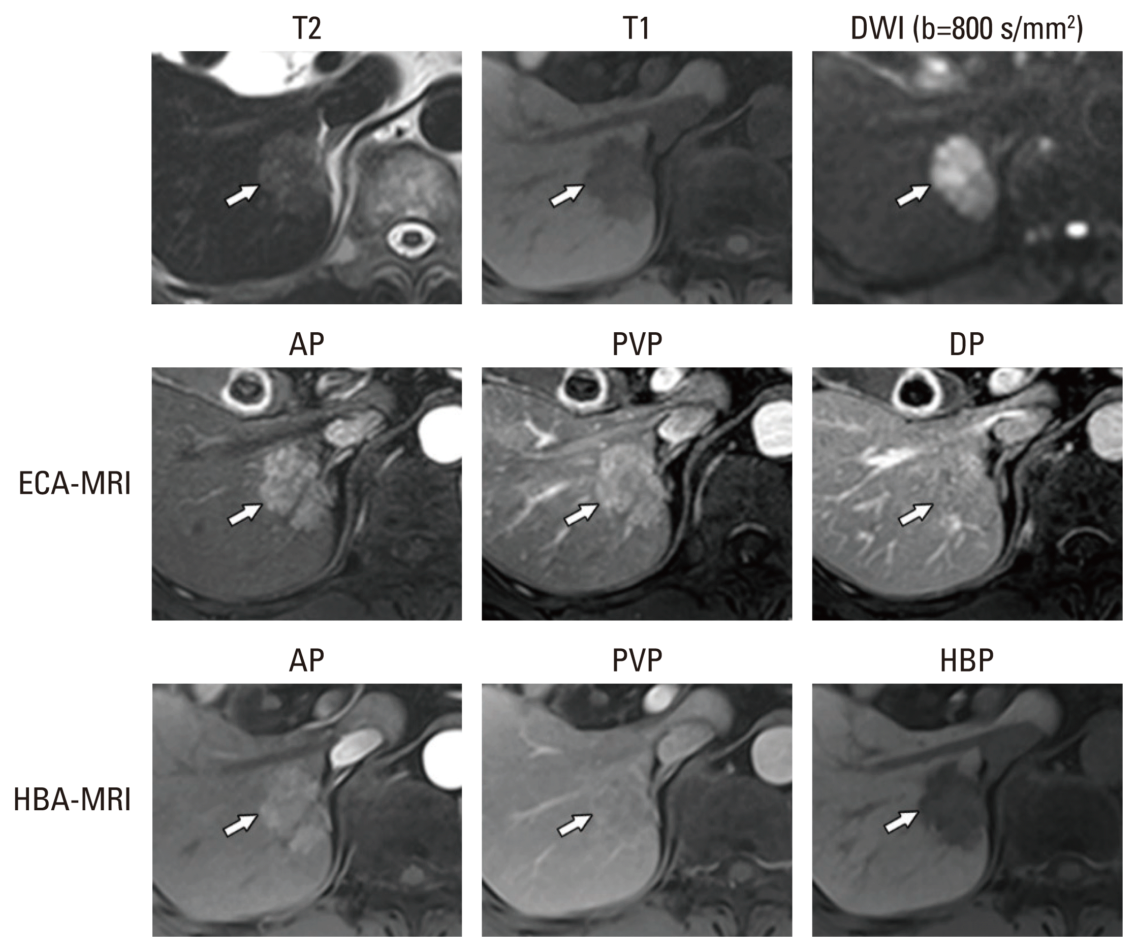
Fig. 1
Flowchart of the inclusion and exclusion criteria of the study population. ECA-MRI, magnetic resonance imaging with an extracellular agent; HBA-MRI, magnetic resonance imaging with a hepatobiliary agent; HCC, hepatocellular carcinoma.
crt-2022-1645f1.gif
Fig. 2
Categorization of a pathologically confirmed HCC on ECA-MRI and HBA-MRI according to the KLCA-NCC 2022 criteria. A 3.2-cm sized mass in the right posterior section of the liver (arrows) shows moderate T2 hyperintensity and T1 hypointensity. The lesion did not show targetoid appearance on DWI nor enhanced sequences. On ECA-MRI, the mass showed APHE without demonstrable washout appearance during the PVP or DP, and was categorized as “probable” HCC. On HBA-MRI, the mass showed APHE without washout appearance on PVP, but with washout appearance on HBP, and was categorized as “definite” HCC. AP, arterial phase; APHE, arterial phase hyperenhancement; DP, delayed phase; DWI, diffusion-weighted images; ECA-MRI, magnetic resonance imaging with an extracellular agent; HBA-MRI, magnetic resonance imaging with a hepatobiliary agent; HBP, hepatobiliary phase; HCC, hepatocellular carcinoma; KLCA-NCC, Korean Liver Cancer Association-National Cancer Center; PVP, portal venous phase.
crt-2022-1645f2.gif
Table 1
Baseline demographics of included patients and lesions
Characteristic No. (%)
Sex 91
 Male 76 (83.5)
 Female 15 (16.5)
Age (yr), mean±SD 58.1±10.6
Etiology of liver disease
 Hepatitis B 74 (81.3)
 Hepatitis C 4 (4.4)
 Alcoholic 3 (3.3)
 Others 10 (11.0)
Liver cirrhosis 40 (44.0)
MELD score, median (IQR) 7 (6–8)
Lesions 118
Size (mm)a) 25.6±15.9
Final diagnosis
 HCC 93 (78.8)
 Non-HCC malignancy
  cHCC-CCA 3 (2.5)
  Intrahepatic cholangiocarcinoma 1 (0.9)
 Benign lesion
  Hemangioma 9 (7.6)
  Regenerative nodule or dysplastic nodule 12 (10.2)

Values are presented as number (%) unless otherwise specified. cHCC-CCA, combined hepatocellular-cholangiocarcinoma; HCC, hepatocellular carcinoma; IQR, interquartile range; MELD, Model of End-Stage Liver Disease; SD, standard deviation.

Table 2
Categorization of lesions according to KLCA-NCC 2022 on ECA-MRI and HBA-MRI
Categorization according to KLCA-NCC 2022 ECA-MRI HBA-MRI
Benign category 9 (7.6) 9 (7.6)
“Indeterminate” nodule 12 (10.2) 14 (11.9)
“Probable” HCC 27 (22.9) 7 (5.9)
“Definite” HCC 56 (47.4) 75 (63.6)
Targetoid appearance 14 (11.9) 13 (11.0)

Values are presented as number (%). ECA-MRI, magnetic resonance imaging with an extracellular agent; HBA-MRI, magnetic resonance imaging with a hepatobiliary agent; HCC, hepatocellular carcinoma; KLCA-NCC, Korean Liver Cancer Association–National Cancer Center.

Table 3
Diagnostic performances of “definite” HCC category based on the KLCA-NCC 2022 criteria
Categorization of KLCA-NCC 2022a) ECA-MRI HBA-MRI p-valueb)


No. % (95% CI) No. % (95% CI)
“Definite” HCC

 All lesions (n=118)

  Sensitivity 54/93 58.1 (47.8–67.6) 73/93 78.5 (69.0–85.7) < 0.001

  Specificity 23/25 92.0 (73.1–98.0) 23/25 92.0 (73.1–98.0) > 0.999

  PPV - 96.4 (87.6–99.0) - 97.3 (90.6–99.3) -

  NPV - 37.1 (31.1–43.5) - 53.5 (43.4–63.3) -

 Lesions 10–19 mm (n=53)

  Sensitivity 12/33 36.4 (21.9–53.7) 24/33 72.7 (53.5–83.4) 0.005

  Specificity 19/20 95.0 (71.1–99.3) 19/20 95.0 (71.8–99.3) > 0.999

  PPV - 92.3 (62.8–98.8) - 96.0 (77.8–99.4) -

  NPV - 47.5 (40.7–54.4) - 67.9 (54.5–78.8) -

 Lesions ≥ 20 mm (n=65)

  Sensitivity 42/60 70.0 (57.3–80.2) 49/60 81.7 (69.8–89.5) 0.020

  Specificity 4/5 80.0 (30.9–97.3) 4/5 80.0 (30.9–97.3) > 0.999

  PPV - 97.7 (87.8–99.6) - 98.0 (89.4–99.6) -

  NPV - 18.2 (11.0–28.5) - 26.7 (15.4–42.0) -

CI, confidence interval; ECA-MRI, magnetic resonance imaging with an extracellular agent; HBA-MRI, magnetic resonance imaging with a hepatobiliary agent; HCC, hepatocellular carcinoma; KLCA-NCC, Korean Liver Cancer Association-National Cancer Center; NPV, negative predictive value; PPV, positive predictive value.

a) Consensus data by the two readers were used,

b) McNemar’s test was used.

Table 4
Diagnostic performances of “probable” or “definite” HCC categories based on the KLCA-NCC 2022 criteria
Categorization of KLCA-NCC 2022a) ECA-MRI HBA-MRI p-valueb)


No. % (95% CI) No. % (95% CI)
“Probable” or “definite” HCC
 All lesions (n=118)
  Sensitivity 79/93 84.9 (76.2–90.9) 79/93 84.9 (76.2–90.9) > 0.999
  Specificity 21/25 84.0 (64.3–93.9) 21/25 84.0 (64.3–93.9) > 0.999
  PPV - 95.2 (88.9–98.0) - 95.2 (88.9–98.0) -
  NPV - 60.0 (47.3–71.5) - 60.0 (47.3–71.5) -
 Lesions 10–19 mm (n=53)
  Sensitivity 28/33 84.8 (68.4–93.5) 28/33 84.8 (68.4–93.5) > 0.999
  Specificity 17/20 85.0 (62.4–95.1) 17/20 85.0 (62.4–95.1) > 0.999
  PPV - 90.3 (76.5–96.4) - 90.3 (76.5–96.4) -
  NPV - 77.3 (59.8–88.6) - 77.3 (59.8–88.6) -
 Lesions ≥ 20 mm (n=65)
  Sensitivity 51/60 85.0 (73.6–92.0) 51/60 85.0 (73.6–92.0) > 0.999
  Specificity 4/5 80.0 (30.9–97.3) 4/5 80.0 (30.9–97.3) > 0.999
  PPV - 98.1 (98.8–99.7) - 98.1 (98.8–99.7) -
  NPV - 30.8 (17.4–48.4) - 30.8 (17.4–48.4) -

CI, confidence interval; ECA-MRI, magnetic resonance imaging with an extracellular agent; HBA-MRI, magnetic resonance imaging with a hepatobiliary agent; HCC, hepatocellular carcinoma; KLCA-NCC, Korean Liver Cancer Association-National Cancer Center; NPV, negative predictive value; PPV, positive predictive value.

a) Consensus data by the two readers were used,

b) McNemar’s test was used.

Table 5
Comparison of radiologic hallmarks on ECA-MRI and HBA-MRI
HCC (n=93) ECA-MRI HBA-MRI p-value
All lesions (n=93)
 APHE 78 (83.9) 74 (79.6) 0.219
 Washouta) 67 (72.0) 92 (98.9) < 0.001
Lesions 10–19 mm (n=33)
 APHE 28 (84.8) 23 (69.7) 0.063
 Washouta) 16 (48.5) 33 (100) < 0.001
Lesions ≥ 20 mm (n=60)
 APHE 50 (83.3) 51 (85.0) > 0.999
 Washouta) 51 (85.0) 59 (98.3) 0.008

Values are presented as number (%). APHE, arterial phase hyperenhancement; ECA-MRI, magnetic resonance imaging with an extracellular agent; HBA-MRI, magnetic resonance imaging with a hepatobiliary agent; HCC, hepatocellular carcinoma.

a) Washout on portal venous, delayed, and hepatobiliary phases (for HBA-MRI).

TOOLS
Similar articles