

 PDF
PDF Citation
 Print
Print


Brain metastases (BM) are common in patients with solid malignancies and are the most common intracranial tumor in adults. Compared to primary malignant brain tumors, BM occurs approximately 10 times more frequently [1]. Although the actual incidence of BM is difficult to determine, estimates of BM incidence range from 8.3 to 14.3 per 100,000 [2,3]. As the use of magnetic resonance imaging (MRI) expands, cancer is not only detected early, but systemic therapy is gradually improved, which increases the survival rate of cancer patients and consequently increases the incidence of BM [4].
Imaging modalities such as computed tomography and MRI have increased the surgical resection of BM, and neurosurgical management has been shown to increase patient survival and enhance neurological performance [5,6]. At 2019 Congress of Neurological Surgeons Guidelines on the Role of Surgery in the Management of Adults with Metastatic Brain Tumors, the adult patients newly diagnosed with metastatic brain tumors, except for radiosensitive tumors, surgery plus whole brain radiation therapy are recommended as first-line treatments if the following three conditions are satisfied: (1) single BM, (2) favorable performance status, and (3) limited extracranial disease [7].
According to increased neurosurgical operation for metastatic brain tumor, the number of surgical pathologic specimens has also recently increased. Molecular pathology analyses of these BM tissues can offer additional clinical gains. To date, some studies on epidemiology and pathology of patients with BM have been conducted. In the epidemiologic research of BM, some studies have been conducted based on patients with BM, but most molecular pathological studies were performed targeting primary cancer patients such as lung cancer and breast cancer, and studies based on BM cases nearly have not been conducted. We performed an analysis encompassing the epidemiological and molecular pathological characteristics of metastatic brain tumors based on BM patients. Therefore, this is the first study of patients with BM in South Korea in the past decade.
In this retrospectively collected data of patients with BM, we studied epidemiology, pathologic and molecular features of BM. We specifically address metastatic brain tumors from the three major primary cancers in which BM predominates: lung cancer, breast cancer, and colon cancer, with a focus on the therapy-associated molecular alterations and their clinicopathological implications. We collected extensive data using next-generation sequencing (NGS), immunohistochemistry (IHC), in situ hybridization, and sequencing. In this study, some specific data, which are emphasized in clinical importance among them, were selected and analyzed for comparison. We addressed epidermal growth factor receptor (EGFR), anaplastic lymphoma kinase (ALK), and KRAS status in lung cancer, human epidermal growth factor receptor 2 (HER2) and hormone receptor conversion in breast cancer, and KRAS and NRAS status in colorectal cancer.
MATERIALS AND METHODS
Patients
From January 2010 to March 2020, patients diagnosed with BM were identified from our institutional database. A total of 269 patients underwent surgical resection for intra-axial lesion of the BM. We retrospectively reviewed the electronic medical records for these patients, including demographic information, clinical characteristics, and surgical pathology reports. Among these, the most frequent primary tumors, that is, lung cancer (n=125), breast cancer (n=42), and colorectal cancer (n=27), were reviewed their protein expression and molecular pathological status. Due to the molecular pathology studies for primary tumor and BM were performed at the physician’s request for treatment decision, the tests were not uniform between patients.
Immunohistochemistry
IHC for estrogen receptor (ER), progesterone receptor (PR), HER2, Ki-67 index, and ALK was performed on formalin-fixed paraffin-embedded (FFPE) tumor specimens. Primary antibodies used for immunohistochemical staining were ER (prediluted, SP1, Roche Tissue Diagnostics, Tucson, AZ, USA), PR (prediluted, 1E2, Roche Tissue Diagnostics), HER2 (prediluted, 4B5, Roche Tissue Diagnostics), Ki67 (prediluted, MIB-1, Roche Tissue Diagnostics), and ALK (prediluted, D5F3, Roche Tissue Diagnostics). Immunohistochemical staining was performed using an automated Ventana Benchmark XT slide stainer (Roche Tissue Diagnostics). The staining results of ER and PR were interpreted using the Allred scoring system [8]. The proportion score was evaluated according to the proportion of positive-stained tumor cells as follows: 0 (0% positive), 1 (<1% positive), 2 (1% to 10% positive), 3 (11% to 33% positive), 4 (34% to 66% positive), and 5 (≥67% positive). Intensity was scored according to the average staining intensity of tumor cells, which is as follows: 0 (none), 1 (weak), 2 (intermediate), and 3 (strong). Positive and negative were evaluated by the sum of proportion and intensity scores, and a sum score >2 was interpreted as positive.
HER2 staining results were interpreted according to the American Society of Clinical Oncology (ASCO)/College of American Pathologists (CAP) guidelines [9,10]. The criteria for reporting HER2 test results are as follows: score 0 (negative, no staining observed or membrane staining that is incomplete and is faint/barely perceptible and within ≤ 10% of tumor cells), score 1+(negative, incomplete membrane staining that is faint/barely perceptible and within >10% of tumor cells), score 2+ (equivocal, weak to moderate complete membrane staining in >10% of tumor cells or complete membrane staining that is intense but within ≤10% of tumor cells), and score 3+ (positive, complete membrane staining that is intense and >10% of tumor cells). HER2 results were scored from 0 to 3+ according to the criteria presented in the guideline, and in the case of 2+, subsequent analysis of HER2 amplification by silver in situ hybridization was performed.
Ki-67 proliferation index was evaluated either by manual counting with ‘eyeballing’ or using automated digital image analysis. All stained invasive tumor cells were included for evaluation, regardless of staining intensity. Slides were scanned with an iScan Coreo slide scanner (Roche Tissue Diagnostics) and analyzed with Virtuoso software (Roche Tissue Diagnostics). Ki-67 was counted from more than 1,000 invasive tumor cells in at least three high-power fields (400×), including one hotspot area and two average areas, according to the recommendations of the International Ki67 in Breast Cancer Working Group [11].
In situ hybridization
ALK rearrangement was detected from FFPE by fluorescence in situ hybridization (FISH) using ALK (2p23) Break Apart Probe Kit (Leica Biosystems, Melbourne, Australia) or LSI ALK Dual Color Probe Kit (Abbott, Chicago, IL, USA). Since 2015, Vysis ALK Dual Color Break Apart FISH Probe Kit (Abbott) has been used for ALK analysis. HER2 amplification status was determined by using HER2 Dual ISH Probe Cocktail Assay (Roche Diagnostics, Risch-Rotkreuz, Switzerland).
Sequence analysis
Sanger direct sequencing, peptide nucleic acid (PNA)–mediated real-time polymerase chain reaction (PCR), and pyrosequencing were used for sequence analysis, and the DNA extraction method was the same for each. DNA was extracted from FFPE tumor specimen using Maxwell 16 FFPE purification kit (Promega, Madison, WI, USA), according to manufacturer’s protocol. DNA concentration was measured by Nano drop (Thermo Fisher Scientific, Waltham, MA, USA). Sanger direct sequencing was used to analyze EGFR exons 18, 19, 20, 21 and KRAS. After DNA extraction, PCR was performed using HotStar Taq Plus DNA Polymerase (Qiagen, Hilden, Germany). As forward primers, 5'-ACTGCTTTCCAGCATGGTGAGG-3' for EGFR exon 18, 5'-GTGGCACCATCTCACAATTGCC-3' for EGFR exon 19, 5'-ATGCGTCTTCACCTGGAAGG-3' for EGFR exon 20, 5'-CCTGAA TTCGGATGCAGAGCTTC-3' for EGFR exon 21, 5'-GGTGAGTTTGTATTAAAAGG-3' for KRAS exon 2, and 5'-GGTGCACTGTAATAATCCAGAC-3' for KRAS exon 3 were used. After initial denaturation at 95°C for 5 minutes, the following process was performed for 40 cycles. 94°C for 30 seconds, 60°C (EGFR exon 18) or 57°C (EGFR exon 19, 20, and 21) or 50°C (KRAS exon 2 and 3) for 30 seconds, and 72°C for 30 seconds. A final extension was performed at 72°C for 7 minutes. Sequencing was performed using Applied Biosystems 3730XL (Thermo Fisher Scientific) with the BigDye Terminator v.3.1 Cycle Sequencing kit (Applied Biosystems, Foster City, CA, USA). Since 2013, Applied Biosystems 3500 XL (Thermo Fisher Scientific) has been used. Pyrosequencing of NRAS codon 12, 13 and 61 region was performed using Threascreen NRAS Pyro kit (Qiagen). NRAS PCR conditions were as follows: initial denaturation at 95°C for 15 min, 42 cycles at 95°C for 30 seconds, 53°C for 30 seconds, and 72°C for 30 seconds, and final extension was performed at 72°C for 10 minutes. Streptavidin sepharose beads (GE Healthcare, Pittsburgh, PA, USA) and PCR product were dispensed into PyroMark Q24 plate wells, and the pyrosequencing results were analyzed using PyroMark Q24 software (ver. 2.0.6). PNA-mediated real-time PCR clamping method was used to analyze EGFR exons 18, 19, 20, 21 and NRAS codons 12, 13, 59, 61, 117, and 146. The extracted DNA was amplified using the PANAMutyper R EGFR kit (PANAGENE, Daejeon, Korea). PCR was performed using the CFX96 Real-Time PCR Detection System (Bio-Rad, Hercules, CA, USA) for EGFR and the QuantStudio 5 Real-Time PCR Instrument for NRAS. The results were analyzed using the PANAMutyper Analyzer (PANAGENE).
Next-generation sequencing
Genomic DNA was extracted from dissected FFPE samples using the RecoverAll Total Nucleic Acid Isolation Kit (Thermo Fisher Scientific) according to the manufacturer’s instructions. DNA concentration was measured by Nano drop and Qubit dsDNA HS Assay Kit (Thermo Fisher Scientific). DNA library construction was performed using the Ion AmpliSeq Library kit 2.0 (Thermo Fisher Scientific) according to the manufacturer’s protocol and quantified by quantitative polymerase chain reaction. IonTorrent S5 XL Sequencer (Thermo Fisher Scientific) was used for sequencing, and sequencing data were analyzed by Ion Reporter v5.4 (Thermo Fisher Scientific). Since December 2019, Oncomine Comprehensive Assay v3 panel and Ion Reporter v5.10 (Thermo Fisher Scientific) have been used.
Breast cancer subtype classification
Breast cancer subtype were classified as follows: Luminal A (ER and/or PR positive, HER2 negative, Ki-67 ≤20%), luminal B (HER2 negative type: ER and/or PR positive, HER2 negative, Ki-67 >20%, HER2 positive type: ER and/or PR positive, HER2 positive, any Ki-67), HER2-enriched (ER and PR negative, HER2 positive), and triple-negative (ER, PR, and HER2 negative) [12]. In our institution, the Ki-67 cutoff value is 20% [13].
Statistical analysis
Difference comparisons between groups were performed using the chi-square test. A p-value of <0.05 was set as the level of statistical significance. Statistical analyses were performed utilizing R software (ver. 4.2.1, R core team 2022, R Foundation for Statistical Computing, Vienna, Austria).
RESULTS
Clinical characteristics
Two hundred sixty-nine patients were diagnosed with BM for the surgically resected brain specimen, including 139 men and 130 women (Table 1). The median age at diagnosis of primary tumor was 58 years (range, 13 to 87 years). Of these patients, 86 (32.0%) had synchronous BM at initial presentation. The median interval between diagnosis of primary tumor and metastatic brain tumor was 28 months (range, 1 to 286 months). Most metastatic brain tumors (74.4%) were located in the supratentorial region. The most frequent primary tumors were lung cancers (46.5%), followed by breast cancer (15.6%), and colon cancer (10.0%).
Lung was the most frequent site of origin of BM, occupying 125 out of 269 cases (46.5%). The median age at diagnosis of primary tumor was 62 years (range, 38 to 87 years). Many of these patients were male (81/125, 64.8%). More than half of the cases (66/125, 52.8%) were diagnosed with BM at initial presentation and the median BM-free interval was 20 months (range, 4 to 217 months). The location of BM was supratentorial in 79.2% (99/125).
For breast cancer (15.6%), which had the second highest incidence of BM, all 42 patients were female. The median age at diagnosis of primary cancer was 46 years (range, 28 to 68 years), and most of them (41/42, 97.6%) developed BM over time. There was a median interval of 39 months between diagnosis of BM and primary cancer (range, 1 to 215 months). Twenty-six of the 42 patients (61.9%) had BM in the supratentorial region.
Colorectal cancer was the third most common primary cancer, accounting for 10.0% (n=27). The median age at diagnosis of colorectal cancer was 58 years (range, 40 to 76 years). There was no significant difference in the sex, with males and females accounting for 48.1% (13/27) and 51.9% (14/27), respectively. Most of them (24/27, 88.9%) had metachronous BM, with a median interval of 41 months from primary cancer diagnosis to BM diagnosis (range, 12 to 146 months). Fifty-one point nine percent (14/27) were supratentorial metastases.
Other additional clinical characteristics according to the primary tumor are shown in Table 1. Overall, among patients with BM, patients with primary lung cancer, gallbladder and extrahepatic bile duct cancer were older than patients with the other types of primary cancer, and breast and skin cancer patients were younger. Lung, kidney, and liver cancers showed male predominance, 64.8%, 88.9%, and 85.7% in males, respectively. On the other hand, skin cancers showed female predominance, 80% in females.
The histological types of the primary tumors with brain metastasis
The histological types of the primary tumors are summarized in Table 2. In the case of lung cancer, most were non–small cell lung cancers (NSCLC, 92.0%) and small cell lung cancers were 8.0%. Among NSCLC, the most frequent histologic type was adenocarcinoma, followed by squamous cell carcinoma and adenosquamous carcinoma. In breast cancer, invasive breast carcinoma of no special type (90.4%) was the most common, followed by invasive lobular carcinoma (4.8%) and mucinous carcinoma (4.8%) in equal proportions. Most of colon cancer were adenocarcinoma. Concerning kidney, clear cell renal cell carcinomas (RCC) were most frequent (94.4%) and chromophobe RCC is in one case. In liver, all cases were hepatocellular carcinoma. All of ovary cancer were high grade serous carcinoma, and all of skin cancer were malignant melanoma. In uterine cancer, endometrioid carcinoma was four cases (80.0%) and undifferentiated carcinoma was one case (20.0%). In five cases of unknown of primary tumor, the histologic types were adenocarcinoma in three cases, small cell neuroendocrine carcinoma and poorly differentiated carcinoma in one case each. The other diverse primary sites and histologic types are described in Table 2.
Molecular pathology of lung cancer with brain metastasis
Molecular pathology studies were applied to 71 metastatic brain lesions and 63 primary lung lesions. This study was not totally paired with primary and metastatic lesions in all cases, but was performed on paired lesions in some cases. EGFR mutation or amplification, KRAS mutations, and ALK rearrangement status were evaluated. In primary tumors, EGFR mutations were observed in 50.8% of cases (Table 3). Exon19 deletion was the most frequent genotype (17 cases), followed by exon21 p.L858R (8 cases). In metastatic brain tumors, 58.0% of cases were EGFR-mutant types. As with primary tumors, exon19 deletion and exon21 p.L858R mutations were the most frequent, 19 cases and eight cases, respectively. Multiple mutation sites were detected in four cases of primary tumors and eight cases of metastatic brain tumors. Exon20 p.T790M mutations were much more detected in BM (6 cases) than lung primary (1 cases). Detailed mutation sites of EGFR are shown in Table 3. Correlations between EGFR status and clinicopathological characteristics are listed in Table 4. There was no significant correlation between EGFR status and age, timing of BM, BM location, and histologic type except sex. EGFR-mutant type was observed more frequently in females (p=.002). Of the 125 cases, 33 cases were studied for EGFR mutations in both primary lung cancer and paired metastatic brain tissues (Table 5). Eight cases of EGFR-wild type and 25 cases of EGFR-mutant type were observed. EGFR mutation sites were changed in nine cases. In six cases, exon20 p.T790M was additionally found in the existing mutation. In one of the three cases, EGFR amplification was additionally confirmed in addition to the existing mutation, in one case, exon18 E709G disappeared from the existing mutation, and in the other case, exon19 del and exon21 L833S disappeared from the existing mutations and EGFR amplification appeared.
Four cases of primary lung tumors and three cases of metastatic brain tumors were KRAS-mutant, and the detected mutation sites were codon 12 (3 cases) and codon 13 (1 case) in primary brain tumors and codon 12 in metastatic brain tumors (Table 3).
ALK rearrangement was detected in five cases of lung and six cases of brain, respectively, and one case was examined by IHC in the brain, and all other cases were confirmed by FISH (Table 3).
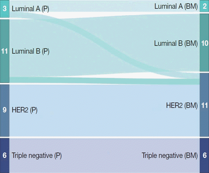
Protein expression profile and molecular subtypes of breast cancer with brain metastasis
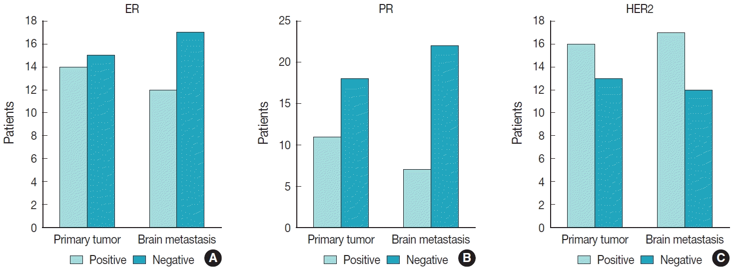
In 42 breast cancer BM cases, 29 primary tumors and 42 metastatic brain tumors were studied for protein expression status. Detailed data and resultant molecular subtypes are shown in Table 6. In both breast and brain, the most frequent subtype was luminal B type (37.9% and 42.9%), followed by HER2-enriched type (31.0% and 33.3%) and triple-negative type (20.7% and 16.7%). Among 42 cases of breast cancer BM, protein expression in both primary and paired metastatic brain tumors was studied in 29 cases. In primary tumors, ER, PR, and HER2 positive cases were 14/29 (48.3%), 11/29 (37.9%), and 16/29 (55.2%), respectively, and in metastatic brain tumors, 12/29 (41.4%), 7/29 (24.1%), and 17/29 (58.6%), respectively (Fig. 1). Receptor status discordances between primary and paired metastatic brain tumors were observed in a total of 11 out of 29 cases. There were two cases of ER status conversion, both of which were positive to negative conversion. PR status conversion occurred in a total of eight cases, with positive to negative conversion in six cases and negative to positive conversion in two cases. HER2 conversion was negative to positive in one case (Table 7). Accordingly, one case each of luminal A type and luminal B type was converted to HER2-enriched type (Fig. 2).
Molecular pathology of colorectal cancer with brain metastasis
Among 27 cases of colorectal cancer BM, 14 cases of primary tumors and three cases of metastatic brain tumors were studied for the molecular status. In primary tumors and metastatic brain tumors, 13 cases and three cases were analyzed for KRAS mutation and eight cases and two cases for NRAS mutation, respectively. In primary tumors, KRAS mutations were observed in 76.9% (10/13) and no NRAS mutations were detected. In metastatic brain tumors, KRAS mutations were observed in 66.7% (2/3) and no NRAS mutations were detected (Table 8). In primary tumors, KRAS mutations were most frequently observed in codon 12 of exon 2 (6 cases), and the other detected mutation sites are described in Table 8. Multiple mutations in KRAS were not observed. Two cases of KRAS mutations in metastatic brain tumors were observed at codons 12 and 13 of exon 2, respectively, and both mutations were consistent with mutations in the primary tumors. Correlations between KRAS status and clinicopathological characteristics are listed in Table 9. There was no significant correlation between EGFR status and age, sex, and BM location except timing of BM. Most of the wild type were diagnosed with BM at initial presentation (2/3), while mutant types were mostly metachronous (9/10) (p=.041).
DISCUSSION
This study analyzed the 10-year epidemiology and molecular pathological characteristics of BM patients. In particular, molecular pathological analysis was performed on metastatic brain tumor tissues of lung cancer, breast cancer, and colorectal cancer with high BM incidence. As stated in several studies, lung cancer (43%–51%), breast cancer (15%–16%), and melanoma (7%–16%) are known as the three most common primary cancers that metastasize to the brain, followed by renal cancer (7%–9%) and colorectal cancer (0.6%–9%) [2,14-17]. In this study, the most common primary lesions of BM were lung (46.5%), breast (15.6%), and colorectum (10.0%), followed by kidney (6.7%), liver (5.2%), and ovary (2.6%), uterus (1.9%), skin (1.9%), and unknown (1.9%) in the order, and 8.1% in other lesions. This difference appears to be due to the incidence of primary cancer. In the United States and Europe, the incidence of melanoma is frequent at 3.7% to 5.2% but in South Korea it is only 0.3%, while the incidence of liver cancer is higher at 5.2% than in the United States and Europe (2.2%) [18-20]. The BM incidence of colorectal cancer in this study is relatively higher than the results of western countries’ previous studies. The reasons for this are that the oncologic outcome has improved with the development of the surgical technique and adjuvant chemotherapy in colon cancer, and the detection rate of small brain lesions has increased due to increased number of performed brain imaging procedure, including brain MRI, which has been covered by medical insurance in South Korea.
Among the three BM-prone primary sites, the median age at diagnosis of primary cancer was 62 years old for lung cancer, 46 years old for breast cancer, and 58 years old for colorectal cancer, and, with lung cancer patients being the oldest and breast cancer patients being the youngest. These results showed similar tendencies to the results of previous studies. In a study of 309 patients with BM, the median age at diagnosis of primary cancer was reported as 60 years for NSCLC, 61 years for small cell lung cancer, 57 years for colon cancer, and 50 years for breast cancer [15]. Of the 269 patients with BM, 51.7% were male. In the case of lung cancer patients with BM, 64.8% were male and 35.2% were female. All patients with BM from breast cancer were female, while there was minimal sex disparity among patients with colorectal cancer BM. These results are similar to study that analyzed patients diagnosed with BM from 1973 to 2001 [2]. According to this study, 7,167 patients (60.9%) were male and 4,596 (39.1%) female patients with BM from lung cancer, with a high proportion of males. The majority of patients with BM from breast cancer were female, with only 19 males and 2,616 females. There were 414 males and 365 females with BM from colorectal cancer patients, and no significant difference was observed by sex. According to the incidence of major carcinomas by sex in South Korea, lung cancer was more common in males, and breast cancer was far more common in females [20]. Colorectal cancer has a slight male predominance, and there is no significant difference in the sex ratio. Considering this, the difference in the proportion of BM patients by sex in our study also seems to depend on the incidence of primary cancer, as in the above study.
The natural history of progression from primary cancer to BM varies depending on the site of the primary tumor. According to a study analyzing the incidence and mortality of synchronous BM in the United States, the most common primary tumor site in patients with synchronous BM was the lung (80.3%) [21]. Likewise, in our study, lung cancer was the most frequently diagnosed at the same time as the primary tumor at 52.8%. In a retrospective cohort study of 2419 patients [14], median time interval from diagnosis of primary cancer to diagnosis of BM was as follows; lung cancer (11 months), breast cancer (44 months), and colorectal cancer (33 months). In contrast, our results demonstrate the median time interval was lung cancer (20 months), breast cancer (39 months), and colorectal cancer (41 months). Lung cancer had the shortest free interval, showing a similar result to previous studies, but the primary cancer with the longest free interval was colorectal cancer, not breast cancer.
Berghoff et al. [14] reported a supratentorial predominance for the location at diagnosis of BM. Our result shows also supratentorial predominance (79.2%).
As described in Table 2, we summarized the histological types of primary tumors in 269 patients with BM. NSCLC was the most common type with BM at 92.0% in lung cancer. While previous studies reported that NSCLC accounts for 66.4 to 86.3% of lung cancer cases [14,16], the results in our study showed a slightly higher rate. Among NSCLC, adenocarcinoma (67.8%) was the most frequent, followed by squamous cell carcinoma (10.4%) and adenosquamous carcinoma (8.7%). A study of 975 patients with early-stage NSCLC found that 48% of patients with BM had adenocarcinoma and 32% had squamous cell carcinoma [22]. According to a population-based study, invasive ductal carcinoma (IDC, now changed to invasive breast carcinoma of no special type) was the most common histological type in 64.6% of patients with BM from breast cancer [23]. Another study reported that 81.1% of cases were IDC [4]. In our study, invasive breast carcinoma of no special type was the majority at 90.4%. Yang et al. [24] reported that 84.5% of 401 patients with colorectal cancer BM were adenocarcinoma, 6.7% were mucinous adenocarcinoma, 5.5% were other types, and 3.2% were unknown. As a result of our study, adenocarcinoma, NOS accounted for 88.9% of colorectal cancer BM, followed by mucinous adenocarcinoma in 7.4% and signet-ring cell carcinoma in 3.7%.
Metastatic lung cancer
Molecular pathology analysis provides information on NSCLC oncogenes that are responsive to targeted therapy, such as EFGR mutations, ALK rearrangements, and KRAS mutations. EGFR mutations in NSCLC patients occur in about 17.4% of Caucasians and 38.8% of Asians [25], and EGFR mutations in BM were found in 3.9%–6.2% and 44.4%–61.2% of Caucasians and Asians, respectively [26-30]. In this study, EGFR mutations were observed in 50.8% of primary tumors and 58.0% of BM. These results, similar to previous studies, indicate that EGFR mutations in NSCLC and BM have clear ethnic and geographical differences.
Discordances in EGFR mutations between NSCLC and BM have been reported at 22.5% to 32.0% [29-31]. In our study, the EGFR mutation was altered in nine patients with BM. Exon20 p.T790M and EGFR amplification were additionally found in the existing mutation, and in two patients, some of the mutations present in primary lung cancer were disappeared. One patient had exon18 E709G and exon21 L858R in primary lung cancer and only exon21 L858R in metastatic cancer. The other patient had exon19 del, exon21 L858R, and exon21 L833S in primary lung cancer and exon21 L858R and EGFR amplification in metastatic cancer. These findings can be explained by tumor heterogeneity associated with genetic changes such as cancer stem cell theory, clonal evolution, and chromosomal instability. In addition to genetic heterogeneity, epigenetic and microenvironmental changes can contribute to tumor heterogeneity [32,33].
Most NSCLC patients with EGFR mutations respond to treatment with first-generation EGFR tyrosine kinase inhibitors (TKIs) [34] but develop drug resistance within 1 to 2 years, approximately 50%–60% due to acquired EGFR T790M mutation [35,36]. For patients who have progressed after treatment with an EGFR TKI, the molecular pathology analyses of the BM tissue should be considered to determine whether to continue treatment with an EGFR inhibitor or switch to another TKI, such as the T790M mutation-specific brain penetration inhibitor osimertinib.
EML4-ALK is a tyrosine kinase generated by gene fusion and appears in about 3%–5% of NSCLC patients [37-39]. ALK-positive NSCLC patients have been reported to have a higher risk of developing BM than ALK-negative patients [40]. Rangachari et al. [41] analyzed NSCLC patients and found that 23.8% of NSCLC patients with ALK rearrangements had BM at initial diagnosis. In this study, ALK rearrangement was detected in 9.3% of primary tumors and 13.5% of BM, respectively. One case was examined by IHC in the brain, and all other cases were confirmed by FISH. A highly reactive ALK inhibitor, crizotinib is the treatment of choice for NSCLC patients with ALK rearrangement. However, crizotinib has a problem of poor blood-brain barrier penetration [42]. In contrast, alectinib and brigatinib have been proven to penetrate the central nervous system and show excellent efficacy [43,44], so it is important to analyze the ALK mutation status of BM in establishing a treatment plan for BM of NSCLC.
KRAS mutations are prevalent mutations in human cancers, and occur in about 20%–40% of lung adenocarcinomas [45,46]. According to Kalikaki et al. [47], in paired primary tumor and BM specimens, KRAS mutations were found in 20% and 8% of primary tumors and BM, respectively. In this study, KRAS mutations were found in 12.5% and 9.1% of primary tumors and BM, respectively. Considering that the incidence of KRAS mutations in lung adenocarcinoma is 25%–33% in Europe and the United States, which is higher than in Asia (5%–8%) [48], the lower incidence of primary tumors’ KRAS mutations in our study seems to be due to geographical or racial differences. On the other hand, the incidence of KRAS mutations in lung cancer was higher in our study (12.5%) than in Asia (5%–8%), which seems to be due to the analysis of lung cancer tissues with BM. Evaluation of KRAS mutations in BM paired with primary lung carcinoma using NGS revealed a higher incidence of KRAS mutations in lung cancer with BM [49]. In both lung cancer and BM, KRAS mutations have been reported to be most prevalent in the G12C subtype [48,49], and sotorasib targeting G12C was approved by the U.S. Food and Drug Administration in 2021 for patients with locally advanced or metastatic NSCLC. Thus, KRAS profile analysis can serve as a basis for suggesting customized treatment for NSCLC and NSCLC BM patients with KRAS mutations.
Molecular alterations in EGFR, ALK, and KRAS in patients with NSCLC BM are significant information for planning treatment including drug selection. In this way, as molecular pathology develops and its influence gradually expands, pathologists should focus on molecular study in addition to histological examination and diagnosis.
Metastatic breast cancer
Hormone receptor and HER2 status in breast cancer play an important role in determining the direction of treatment. Several studies have reported discordance in receptor (ER, PR, and HER2) status between primary tumors and paired metastatic tumors. The conversion rates for each receptor were broad, ranging from 7.3% to 29.2% for ER, 2.4% to 38.1% for PR, and 2.3% to 23.8% for HER2 [4,50-58]. In this study, the conversion rates of receptors were 6.9% for ER, 27.6% for PR, and 3.4% for HER2. The conversion rates of PR and HER2 were within the range of previous studies, while the conversion rates of ER were comparatively lower. The reason for these results is not clear. Receptor conversion reported in another study conducted in South Korea was different from our study (ER, 9.5%, PR, 38.1%, HER2, 23.8%) [4]. In addition, no clear racial or geographic trends were found associated with a wide range of receptor conversion rates reported in previous studies. Although the exact reason is not known, it is thought that the receptor conversion may vary according to the unknown characteristics of the patients included in the study. A multicenter analysis of subtype switching reported that subtype conversion occurred due to changes in the receptor status, and the HER2 enriched and triple-negative types increased [58]. In this study, ERs were converted from positive to negative (2/2), PRs were predominantly converted from positive to negative (6/8), and HER2 was converted from negative to positive. Accordingly, one case each of luminal A type and luminal B type was converted to HER2-enriched type. Although the exact mechanisms by which discordance in receptor expression occurs have not been elucidated, several explanations have been made. Turner et al. [59] introduced three explanations, including the selection of existing clones that may have been obscured by bulk tumors, changes in molecular expression of hormone receptors and HER2, or both of these possibilities occurring. In addition, inadequate fixation of the tumor tissue may also contribute to receptor conversion. Yildiz-Aktas et al. [60] reported that a delayed fixation time contributed to reduced immunostaining of hormone receptors and HER2 receptors. The ASCO/CAP guidelines recommend that cold ischemia time must be recorded and samples should be fixed in 10% neutral buffered formalin for 6 to 72 hours [61].
Receptor discrepancy between the primary tumor and BM may affect treatment decisions, so receptor status testing in BM tissue should be performed. ASCO Clinical Practice Guidelines recommend biopsy to determine ER, PR, and HER2 status in patients with newly diagnosed metastases, as receptor discrepancies can be found in the primary tumor and metastases [62].
Metastatic colon cancer
The RAS gene family consists of the proto-oncogenes KRAS, NRAS, and HRAS, and RAS mutations appear in various cancers. According to a study analyzing the COSMIC (The Catalog of Somatic Mutations in Cancer) database, the KRAS, NRAS, and HRAS mutation rates in the large intestine were 33%, 3%, and <1%, respectively [63]. Roussille et al. [64] reported that KRAS mutations were observed in 56% of primary tumor and 74% of BM, respectively, and NRAS mutations were observed in 6% and 11%, respectively. In primary tumors and BM, KRAS mutations were observed at exon 2 codon 12 and codon 13, and the most frequent were G12D and G12V [64]. Our results show that KRAS mutations were observed in 76.9% of primary tumors and 66.7% of BM, and NRAS mutations were not detected in either. In primary tumors, KRAS mutations were most frequently observed in exon 2 codon 12. In two cases of BM, KRAS mutations were observed in exon 2 codons 12 and 13, respectively. In our study, the incidence of KRAS mutations in primary tumors was 76.9%, which was higher than the previous study result of 56%. Differences in KRAS mutation rates in primary cancers are probably due to regional and ethnic differences. In a study researching KRAS mutation frequencies in Caucasian colorectal cancer patients, KRAS mutations were found in 38.3% [65]. A study of colorectal cancer patients in South Korea reported that KRAS mutations were found in 45.9% [66]. Meanwhile, the KRAS mutation rate of primary cancer was higher in this study compared to 30%–40% in previous studies [65,66], which seems to be because the study was conducted on BM patients. RAS mutation analysis in colorectal cancer patients reported that RAS mutations affect the increased incidence of lung, bone and BM [67]. As a result of our study, KRAS status in metastatic brain tumors was analyzed in three patients and found 66.7% of mutations, but this is too small a number to discuss.
The incidence of NRAS mutations is low, as reported in 4% or 3.2% of colorectal cancer patients [65,68]. In our study, NRAS status was analyzed in eight cases of primary cancer and two cases of BM, so the results of the study do not represent the incidence of NRAS mutations. Therefore, additional studies with more patients should be conducted.
Molecular alterations different from the information obtained in the primary tumor can occur in the BM, and these differences can be detected by analysis of the molecular profile, providing clinical advantages in the selection of candidate targeted therapies. As the NGS platform is covered by medical insurance in South Korea, it has recently become a standard procedure in many institutions. NGS analysis of resected metastatic brain tumor tissue specimens provided treatment-associated mutations, contributing to the development of drug resistance.
A multidisciplinary approach is often required to discuss treatment plans for BM, and the molecular tumor board plays an important role in discussing the opinions of multiple medical experts, including pathologists. Molecular tumor board helps in the understanding of the results of molecular analysis and is of benefit in establishing treatment plans and clinical trial enrollment in patients with genetic alterations available to targeted therapy [69]. As the field of molecular medicine develops, the role of the pathologist as an important member of multidisciplinary teams is increasing. In particular, molecular studies are becoming more important in diagnosis and management due to discordance in target molecular mutations between BM and primary tumors. Therefore, in addition to the pathological diagnosis, pathologists will have to provide information on the molecular biomarkers.
For the first time, our study provided clinicopathological and molecular features of patients with BM in South Korea. The main limitation of this study is it’s the single-center, retrospective design and molecular analysis results in the unpaired samples since most patients which were performed surgery of BM did not have analyzed molecular status for primary cancer. Moreover, we only included patients who underwent surgical resection of the BM tumor, so population selection bias occurred. Therefore, further studies on comparative analysis targeting primary cancer paired with BM should be conducted.
 XML Download