

 PDF
PDF Citation
 Print
Print


Introduction
Double-lumen endobronchial tube (DLT) intubation is the method of choice for lung separation and one-lung ventilation during thoracic procedures [1]. However, displacement of a well-positioned DLT frequently occurs and may adversely affect patient safety by inducing severe hypoxemia and hindering clear surgical visualization of the operative field [1].
Lateral decubitus positioning (LDP) can predispose the patient to DLT displacement [2]; however, to our knowledge, evidence regarding predictors for DLT displacement after this maneuver is lacking. Although a recent study proposed that the American Society of Anesthesiologists classification 3 status, emergency surgery, procedures on the left lung parenchyma, and the use of a right-sided DLT increased the risk of DLT malposition [3], the authors described DLT displacement for the entire intraoperative period, and the effect of surgical manipulation on DLT placement could not be excluded. Another study reported a history of previous thoracotomy as a risk factor for DLT displacement [4], but this does not adequately explain the frequent occurrence of displaced DLT observed in patients without a previous surgical history.
Previous literature has described how the gravitational shift of the mediastinum in the lateral decubitus position may affect the DLT position [5,6]. It is also known that obesity substantially distorts mediastinal anatomy by inducing a considerable mechanical load on the diaphragm [7]. However, no comprehensive data concerning the impact of obesity as measured by body mass index (BMI) on DLT displacement after LDP exist. Therefore, we evaluated the predictive value of preoperative risk factors, including obesity, for DLT displacement > 15 mm after LDP in patients scheduled for pulmonary resection.
Materials and Methods
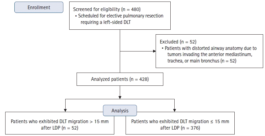
The present study was approved by the Institutional Review Board (no. 4-2021-0694) of Yonsei University Health System (Seoul, Republic of Korea), and the requirement for written informed consent was waived. The study was a retrospective review of 480 patients who underwent elective pulmonary resection requiring placement of a left-sided DLT between July 2020 and July 2021 in Severance Hospital, Seoul, Republic of Korea. Patients with centrally positioned tumors invading the anterior mediastinum, trachea, or main bronchus, which may distort the tracheobronchial anatomy were excluded (Fig. 1). The study adhered to the Strengthening the Reporting of Observational Studies in Epidemiology guidelines and was conducted in accordance with the Ethical Principles for Medical Research Involving Human Subjects as outlined in the Helsinki Declaration of 1975 (revised 2013).
All patients received standardized anesthetic management according to our departmental protocols. Lung isolation was performed using a left-sided DLT (VentiBroncTM Anchor; Flexicare Medical Ltd., UK). DLT size (33, 35, 37, or 39 Fr) was selected according to the inner diameter of the left main bronchus, as determined by the coronal view of two-dimensional chest computed tomography (< 11 mm, 33 Fr; ≥ 11 and < 13 mm, 35 Fr; ≥ 13 and < 15 mm, 37 Fr; and ≥ 15 mm, 39 Fr) [8]. The diameter of the left main bronchus was measured 2 cm below the tracheal carina, where the bronchial cuff of the left-sided DLT is conventionally placed.
After placing the DLT, a bronchoscope was inserted into the tracheal lumen to adjust it to its optimal position, where the proximal margin of the inflated bronchial cuff was immediately below the tracheal carina [9]. Subsequently, the bronchoscope was withdrawn and inserted into the bronchial lumen to ensure a clear view of the left upper and lower lobe bronchi [9]. The DLT depth was measured at the level of the central incisor, and the DLT was firmly fixed with tape.
Before turning the patient laterally, a protractor was used to facilitate cervical spine neutrality, according to our institutional protocol. We assessed the neutral neck posture in the supine position by measuring the angle formed at the intersection of lines from the tragus of the ear to the lateral canthus of the eye and the spinous process of C7. The patient was then placed in the lateral decubitus position with the neck and DLT firmly held by the anesthesiologist. Subsequently, the head and neck postures were adjusted according to the assessed cervical angle. A higher headrest was placed to prevent the lateral flexion of the neck. Thereafter, the DLT was optimally adjusted under bronchoscopic guidance in the same manner as in the supine position, and the depth of the DLT in the lateral decubitus position was recorded.
The assessed data included age, sex, height, weight, BMI, left main bronchus diameter (as assessed using preoperative chest computed tomography), DLT size, direction of the lateral decubitus position, and bronchoscope-adjusted DLT depths in the supine and lateral decubitus positions.
Obesity was defined as BMI ≥ 25 kg/m2, according to the Asia-Pacific BMI classification [10]. Clinically significant DLT displacement was defined as the migration of the DLT by > 15 mm from its optimal position, regardless of the direction. The displacement was calculated by subtracting the bronchoscope-adjusted DLT depth in the supine position from that in the lateral decubitus position. To express the DLT depth relative to each patient’s height, we divided the patient’s height by the optimal DLT depths measured in the supine and lateral decubitus positions (H/Dsupine and H/Dlateral, respectively).
The primary endpoint of this study was to assess the predictive value of preoperative risk factors, including obesity (BMI ≥ 25 kg/m2), for DLT displacement (> 15 mm) after LDP.
Statistical analyses were performed using SPSS® 23.0 (SPSS Inc., USA). The results are expressed as mean ± standard deviation, median (Q1, Q3), or the number of patients (percentage). Patients who exhibited DLT migration ≤ 15 mm after LDP were allocated to the optimal group, and those who exhibited DLT migration > 15 mm after LDP were allocated to the displacement group. Continuous variables were first assessed for normality using the Kolmogorov–Smirnov test. Intergroup comparisons were performed using the t-test, Mann–Whitney U test, or χ2/Fisher’s exact test, as appropriate. Multivariable logistic regression analysis was performed to assess the endpoint of the current study. The optimal cutoff value for continuous variables was determined using receiver operating characteristic (ROC) curve analysis. Pearson correlation analysis was performed to assess the relationship between continuous variables. Additionally, a comparison of the baseline and intraoperative variables between patients with and without obesity was performed to further illustrate the effect of obesity on DLT depths and the extent of DLT migration. Statistical significance was set at P < 0.05.
Results
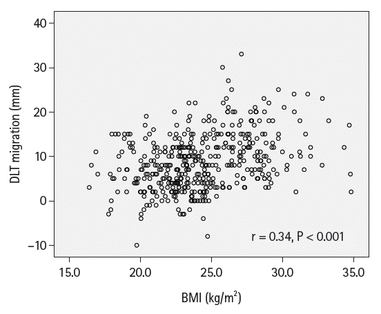
Of the 480 patients, 52 were excluded owing to the distorted airway anatomy caused by tumors invading the anterior mediastinum, trachea, or main bronchus. Among the remaining 428 patients, a DLT displacement > 15 mm was observed in 52 patients (12.1%) after LDP (Fig. 1 and Table 1). A scatterplot of BMI and the extent of DLT migration are displayed in Fig. 2. Pearson correlation coefficient (r) of the two variables was 0.34 (P < 0.001). Table 2 compares the demographic data of the optimal group and displacement group. The displacement group exhibited significantly higher weights and BMI and a larger proportion of patients with obesity than that in the optimal group. Age, sex, and height were comparable between the groups.
Intergroup comparisons of clinical variables are shown in Table 3. The left main bronchus was narrower in the displacement group. The displacement group had a shallower supine DLT depth, which induced a higher H/Dsupine. The lateral DLT depth and H/Dlateral ratio were comparable between the groups. The extent of DLT migration in the displacement group was 20.3 (18.5, 22.4) mm and in the optimal group was 7.1 (4.0, 11.3) mm. All patients in the displacement group exhibited proximal DLT migration after LDP, which required further advancement of the DLT to locate the tube in an accurate position. There was no difference in the distribution of the DLT size and direction of the lateral decubitus position between the groups. When patients were divided by obesity (BMI ≥ 25 kg/m2 and BMI < 25 kg/m2), patients with obesity exhibited a shallower supine DLT depth, higher H/Dsupine, and greater extent of DLT migration than patients without obesity did (Supplementary Table 1).
Table 4 shows the results of the logistic regression analysis. In the univariate logistic regression analysis, BMI, obesity, left main bronchus diameter, supine DLT depth, and H/Dsupine were identified as predictors for DLT displacement. Because multicollinearity was detected between BMI and obesity, BMI was removed from the multivariable analysis, which indicated obesity (odds ratio [OR]: 5.69, 95% CI [2.89, 11.23], P < 0.001) and H/Dsupine (OR: 8.28, 95% CI [2.92, 23.48], P < 0.001) as independent predictors for DLT displacement. In the multivariable analysis, when BMI was used instead of obesity, BMI was also identified as an independent predictor for DLT displacement (OR: 1.26, 95% CI [1.15, 1.38], P < 0.001; Supplementary Table 2).
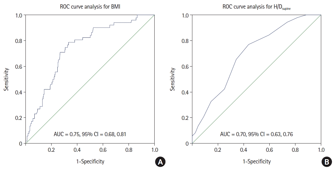
The cutoff value of BMI for predicting DLT displacement was 24.8 kg/m2, with a sensitivity of 78.8% and specificity of 66.8% (area under the ROC curve [AUROC]: 0.75, 95% CI [0.68, 0.81] P < 0.001; Fig. 3A), and that of H/Dsupine was 6.1, with a sensitivity of 76.9% and specificity of 56.1% (AUROC: 0.70, 95% CI [0.63, 0.76], P < 0.001; Fig. 3B).
When BMI and H/Dsupine were introduced as dichotomous variables divided by their cutoff values, BMI ≥ 25 kg/m2 exhibited an OR of 6.08 with a 95% CI of 3.09, 11.95 (P < 0.001), and H/Dsupine > 6 exhibited an OR of 2.91 with a 95% CI of 1.42, 6.02 (P = 0.004, Supplementary Table 3).
Discussion
In this study, we demonstrated that obesity and H/Dsupine were significantly associated with left-sided DLT displacement of > 15 mm after LDP in patients scheduled for elective pulmonary resection. The direction of displacement was predominantly proximal, and obesity was associated with a six-fold increased risk of DLT displacement.
The extent of displacement regarded as clinically significant varies among studies [2,4,11,12]. We defined displacement as the tube having to be moved by more than 15 mm to correct its position. Although some clinicians believe that even a 5 mm deviation from an optimal placement could be dangerous [2,11], no data have proven the clinical relevance of minor misplacements [13]. In this study, we employed a safety margin in positioning DLT, as suggested by Benumof et al. [14]. The authors demonstrated that the average margin of safety in positioning left-sided double-lumen tubes ranged from 16 to 19 mm, and exceeding this safety margin would induce hypoxemia and inadequate lung isolation [14]. However, as shown in Table 1, more than 20-mm displacement was extremely rare in our study (17 patients, 4.0%), and considering the short stature of Asian patients, we assumed that DLT migration of more than 15 mm was clinically relevant and in need of correction.
In this study, obesity was defined as BMI ≥ 25 kg/m2, according to the Asia-Pacific BMI classification [10]. Although the cutoff value for obesity differs from that proposed in the international guideline issued by the World Health Organization in 2000 [15], recent investigations have demonstrated that the Asia-Pacific BMI classification more appropriately reflects the correlation of obesity and disease manifestation in Asian patients [16,17], and the cutoff values of this criteria are the consensus among the Asia-Pacific region [16–18]. Since our study encompassed the Korean population only, we considered it more appropriate to define obesity according to the Asia-Pacific BMI classification.
Despite BMI and the extent of DLT migration exhibiting a weak linear correlation when assessed as continuous variables, BMI was identified as a significant predictor for DLT displacement > 15 mm. In addition, the BMI cutoff value from the ROC curve analysis coincided with the upper threshold of the normal BMI range [10], further supporting our finding that patients with obesity are likely to develop clinically significant DLT displacement after LDP. H/Dsupine was also associated with DLT displacement with a cutoff value of 6, providing a simple formula for assessing the risk of DLT displacement before changing the patient position. For example, when the bronchoscope-adjusted DLT depth in the supine position is 27 cm in a patient with a height of 180 cm, our findings suggest that clinicians should be aware of displaced DLT after turning the patient laterally. Considering that the direction of the DLT migration was proximal in the displacement group, pre-emptively advancing the DLT from the initial correct location before LDP may be advantageous in high-risk patients, which is consistent with the recommendations of the previous studies [4,6].
The optimal DLT depth is well known to significantly correlate with height [19], and considering that the two groups exhibited similar heights, we expected similar supine DLT depths in the two groups. However, the displacement group exhibited a shallower supine DLT depth than did the optimal group. As compression on the diaphragm in the supine posture is increased in obesity [20,21], we speculate that the cranial shift of the tracheobronchial structures was more pronounced in patients with obesity, resulting in a shallow supine DLT depth. However, in the lateral decubitus position, the anterior displacement of the pannus significantly relieves the compressed diaphragm [22], which is reflected in our results as comparable lateral DLT depths between the groups. Comprehensively, the extensive alteration of mechanical load on the mediastinum between body postures seems to have contributed to the significant displacement of DLT in patients with obesity.
In previous literature, DLT displacement after LDP has been described as a result of a downward shift of the carina or upward movement of the DLT inside the carina [6]. Many clinicians suspected that vigorous neck movements after LDP are a major cause of the upward movement of DLT inside the carina [23]. Indeed, efforts to minimize cervical movement using a neck brace [24] or to intentionally induce neck extension in the supine position [25] significantly reduced the incidence of DLT displacement; however, displacement could not be prevented entirely. A cadaver study suggested a similar result, as significant migration of the DLT was observed even when the movement of the neck was strictly restricted and the tip of the DLT was fixed with forceps at the bronchus level [26], implying that the impact of carinal movement on DLT position was not negligible. However, to our knowledge, no previous study has assessed factors that may affect significant carinal movement after LDP, and the current study provides evidence in this regard.
Some studies have suggested that small DLTs are frequently related to positioning problems [27,28]; however, the DLT size was not associated with displacement in our study. This discrepancy may be attributable to the different preferences of clinicians with respect to the appropriate DLT size for each patient. We selected the DLT size on the basis of the inner diameter of the left mainstem bronchus, whereas previous studies considered patient height, sex, and tracheal diameter to select the DLT size [27,28]. We presume that our selection method, in which the outer diameter of the endobronchial tip fits the inner diameter of the left mainstem bronchus, may have partly contributed to preventing significant DLT displacement.
This study has an inherent limitation owing to its retrospective design. First, although we suggested plausible mechanisms for DLT displacement in patients with obesity, they need to be clarified in further studies. Second, our definition of obesity according to the Asia-Pacific BMI classification limits the generalizability of our findings for other ethnic groups such as Caucasians. Third, due to the retrospective nature of this study, we could not clearly address the incidence of ramp or reverse Trendelenburg position for airway management of patients with morbid obesity, which may have affected the extent of DLT migration. Finally, although we used a protractor to preserve cervical spine neutrality, some cases of minor alterations in the cervical posture may have been undetected, affecting the DLT position [29].
In conclusion, obesity was significantly associated with DLT displacement after LDP. Our findings indicate that preemptively advancing the DLT from its optimal position before LDP may be advantageous in patients with obesity.
 XML Download