Journal List > J Neurocrit Care > v.16(1) > 1516083161

Ryu, Lee, Kwon, and Chang: Delayed post-ischemic leukoencephalopathy in emergent large-vessel occlusive stroke after mechanical thrombectomy: case reports

Abstract

Background

Delayed post-ischemic leukoencephalopathy (DPIL) is a rare complication after mechanical thrombectomy, with no well-established clinical characteristics and pathomechanism. We explored the characteristics and possible mechanisms in three patients with DPIL.

Case Report

Based on the clinical manifestations and laboratory findings including magnetic resonance imaging, magnetic resonance spectroscopy, electroencephalography, and lumbar puncture, DPIL was diagnosed in three patients. We administered antiplatelet agents and conservative treatment. Cardioembolism, successful recanalization using a balloon guiding catheter, and fluctuating or gradually worsening neurological symptoms in delayed phase between 13 and 70 days were common features of DPIL. Diffusion-weighted imaging and fluid-attenuated inversion recovery showed high-signal intensity in the affected subcortical white matter. Laboratory findings provided no evidence of an epileptic disorder, inflammatory demyelination, or tumorous conditions.

Conclusion

This report shows the characteristics and neuroradiologic images of DPIL. Among the various hypotheses, regional hypoxic-ischemic leukoencephalopathy and delayed reperfusion injury might be the pathomechanism underlying DPIL.

INTRODUCTION

Delayed post-ischemic leukoencephalopathy (DPIL) is a rare complication after mechanical thrombectomy (MT) characterized by delayed-onset neurological deterioration, with subcortical white matter hyperintensities, sparing the cortex and basal ganglia [1-3]. The clinical manifestations of DPIL were heterogenous, including aphasia, dysarthria, apathy, and executive dysfunction. Despite several hypotheses, the pathomechanism underlying DPIL remains unclear. We encountered four cases of DPIL from May 2021 to February 2022. Herein, we report the cases of three patients who underwent further evaluation, which is expected to contribute to a better understanding of the clinical characteristics and pathomechanism of DPIL for improved diagnosis in future.

CASE REPORTS

Case 1

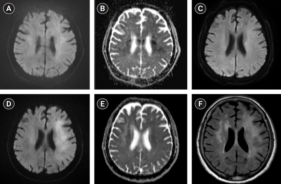
A 79-year-old woman with a history of hypertension and atrial fibrillation presented with right hemiparesis and global aphasia. Her National Institutes of Health Stroke Scale (NIHSS) score was 21. Diffusion-weighted imaging (DWI) showed no lesions (Fig. 1); however, magnetic resonance angiography (MRA) revealed a left cavernous internal carotid artery (ICA) occlusion. The onset-to-puncture time was 270 minutes. After one aspiration thrombectomy attempt with a balloon guiding catheter (BGC), we achieved modified thrombolysis in cerebral infarction (mTICI) grade 3. The puncture-to-recanalization time was 40 minutes. At discharge on day 6, her NIHSS had decreased to 1. On day 28, she returned with fluctuating motor aphasia, from mild to mute. Additionally, she had a fever (38 ℃) with Escherichia coli in the blood culture, suggesting a urinary tract infection and uncontrolled blood pressure of up to 160/110 mm Hg. DWI and fluid-attenuated inversion recovery (FLAIR) showed high-signal intensities in the entire subcortex of left middle cerebral artery (MCA) territory, without signal changes in the left basal ganglia and gray matter. She was discharged with mild to moderate motor aphasia, urinary tract infection improvement, and controlled blood pressure.

Case 2

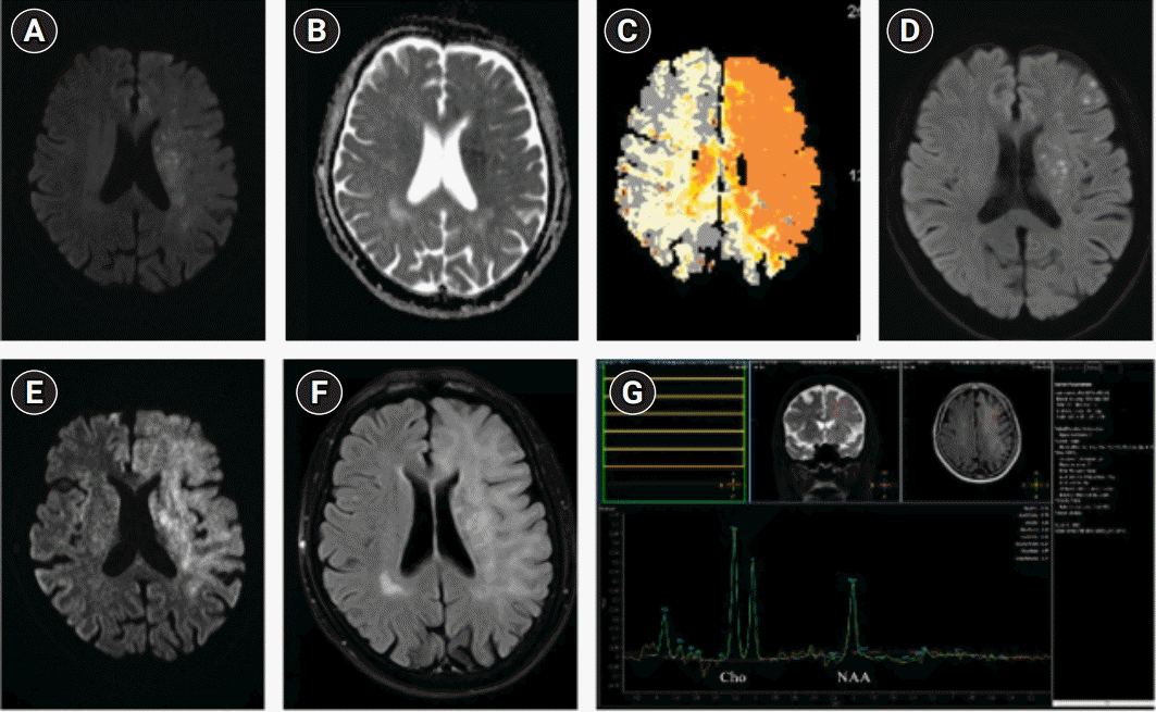
A 77-year-old woman with a history of hypertension and atrial fibrillation visited our hospital. Her NIHSS score was 16. DWI showed hyperintense lesions in the left basal ganglia, and severe diffusion-perfusion mismatch was observed (Fig. 2). MRA revealed a left cavernous ICA occlusion. The onset-to-puncture time was 1,125 minutes. After two attempts with a stent retriever and aspiration thrombectomy with a BGC, we obtained mTICI grade 3. The puncture-to-recanalization time was 65 minutes. The patient was transferred to another hospital on day 6 with an NIHSS score of 5. On day 30, she was readmitted for fluctuating motor aphasia. DWI and FLAIR revealed diffuse hyperintensities in the left MCA territory subcortex, with partial low-signal changes in the apparent diffusion coefficient (ADC). There were no newly developed steno-occlusive lesions in the left ICA or MCA. She was transferred to a rehabilitation hospital on day 38 with improved motor aphasia.

Case 3

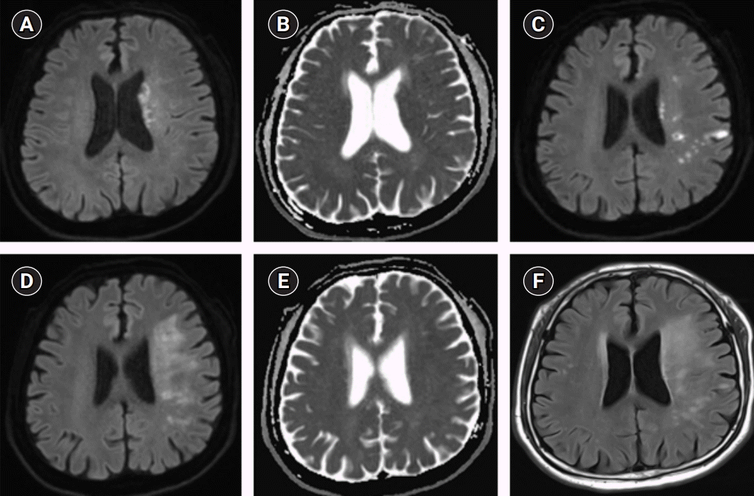
A 78-year-old man with a history of hypertension and atrial fibrillation visited a tertiary hospital. His NIHSS score was 16. DWI demonstrated new infarcts in the left basal ganglia and corona radiata (Fig. 3), with severely decreased perfusion in the left MCA territory. MRA revealed occlusion of the left MCA M1 segment. The onset-to-puncture time was 733 minutes. After an attempt with a stent retriever and simultaneous aspiration thrombectomy with BGC, we achieved mTICI grade 2b. The puncture-to-recanalization time was 47 minutes. On day 8, the patient was discharged with an NIHSS score of 1. On day 13, he visited our stroke center because his aphasia had gradually aggravated. He underwent magnetic resonance imaging on day 29. DWI and FLAIR showed diffuse white matter hyperintensities in the left MCA territory, without signal changes in the ADC. He was discharged with moderate motor aphasia.
No patient showed pleocytosis, albuminocytologic dissociation, or oligoclonal bands on cerebrospinal fluid analysis. In the first case, the immunoglobulin G (IgG) index was normal at 0.49. Moreover, magnetic resonance spectroscopy demonstrated mildly increased choline/N-acetyl aspartate ratios of 1.19, 1.57, and 1.38, respectively, which indicate nonspecific findings with no tumorous condition. Electroencephalography showed no epileptiform discharge in the patients.

DISCUSSION

The present case series reported the clinical and neuroradiological feature of three patients with DPIL that developed after MT due to large vessel occlusion. Table 1 summarizes the clinical characteristics of the patients with DPIL that developed after MT due to large vessel occlusion from the current study and previous ones. (1) Cardioembolism, (2) fluctuating or gradually worsening neurologic symptoms in the delayed phase, (3) hyperintensities in the subcortical territory of the occluded vessel, and (4) successful recanalization using BGC during the procedure were common features in all cases. Diffuse subcortical white matter hyperintensities on T2 weighted images of the previously occluded vessel territory were observed in all cases, and impairment of remyelination by oligodendrocytes could be a plausible pathomechanism of DPIL [4]. There was no evidence of neuro-infection, seizures, inflammatory demyelinating disease, or tumorous conditions.
Various hypothetical pathomechanisms have been suggested for DPIL [1-3]. The neuroradiologic features of this condition are similar to those of delayed hypoxic leukoencephalopathy that are reported after carbon monoxide intoxication, cardiac arrest, and opioid or benzodiazepine overdose [5]. Impairment of repair mechanisms by oligodendrocytes during the subacute to chronic period of ischemic insult might attribute to DPIL. Oligodendrocytes in the white matter may be more vulnerable to ischemic insult than neurons in the cerebral cortex since leptomeningeal collateral circulation is more prominent in the cortex than in the deep white matter [4]. Additionally, oligodendrocytes may be more severely injured in cardioembolic stroke, which is associated with poor collateral circulation [6]. Impairment of myelin regeneration by oligodendrocytes after ischemic insult leads to delayed neurological deterioration with subcortical white matter hyperintensities in the affected vessel territory. Moreover, the use of a BGC to prevent distal embolization during MT, might be one of the hypothetical causes of damage in the subcortical oligodendrocytes and accelerate the impairment of compensatory mechanisms for myelin loss. The absence of albuminocytologic dissociation and normal range of IgG index could rule out active demyelination as a histopathologic process.
Another potential cause of DPIL is reperfusion injury after recanalization of the large-vessel occlusion. Notably, all patients, including those from previous reports, presented with cardioembolism, which is more likely to cause reperfusion injury [7]. Similar manifestations have been observed in cerebral hyperperfusion syndrome after carotid stenting, caused by disrupted cerebral autoregulation. Moreover, delayed cerebral hyperperfusion syndrome, occurring a few weeks after carotid stenting, has also been reported [8]. Hypertension, chronic kidney disease, poor collateral circulation, and recent stroke are associated with the occurrence of cerebral hyperperfusion syndrome [9]. As the optimal range of blood pressure to maintain cerebral autoregulation varies according to the extent of baseline ischemia or degree of recanalization, delayed reperfusion injury can occur in the absence of high blood pressure [10].
Based on clinical manifestations and neuroradiologic findings, we speculated on the pathomechanism of DPIL. Early recognition of the disease may guide proper management and help avoid unnecessary diagnostic evaluation. Further investigation regarding the histopathologic mechanism, predictors, and long-term prognosis of patients with DPIL is needed.

Notes

Ethics statement

The local Ethics Committee of Asan Medical Center provided approval for this study (No. 2022-0357). The need for written informed consent was waived because this was a retrospective study.

Conflict of interest

Jun Young Chang is an editorial board member of the journal, but he was not involved in the peer reviewer selection, evaluation, or decision process of this article. No other potential conflicts of interest relevant to this article were reported.

Funding

This research was supported by a grant from the Korea Health Technology R&D Project through the Korea Health Industry Development Institute, funded by the Ministry of Health and Welfare, Republic of Korea (grant no. HR18C0016).

Author contributions

Conceptualization: all authors. Data curation: JCR, DL. Formal analysis: JCR. Funding acquisition: JYC. Investigation: JCR, JYC. Methodology: all authors. Project administration: JYC. Resources: all authors. Software: JCR, JYC. Supervision: SUK, JYC. Validation: JCR, JYC. Visualization: JCR. Writing–original draft: JCR, JYC. Writing–review & editing: all authors.

REFERENCES

1. Sasaki T, Tomura N, Okada H, Tsuji E, Hayashi N, Kuwata T. A case of delayed white matter lesion after mechanical thrombectomy for middle cerebral artery occlusion. Jpn J Stroke. 2017; 40:270–4.
2. Singu T, Inatomi Y, Yonehara T, Ando Y. Delayed leukoencephalopathy after recanalized cardioembolic stroke: two case reports. J Neurol Sci. 2017; 379:81–3.
3. Nehme A, Pistono AA, Guilbert F, Stumpf É. Post-ischemic leukoencephalopathy after endovascular treatment for acute ischemic stroke. Can J Neurol Sci. 2019; 46:363–5.
4. Iadecola C, Park L, Capone C. Threats to the mind: aging, amyloid, and hypertension. Stroke. 2009; 40(3 Suppl):S40–4.
5. Zamora CA, Nauen D, Hynecek R, Ilica AT, Izbudak I, Sair HI, et al. Delayed posthypoxic leukoencephalopathy: a case series and review of the literature. Brain Behav. 2015; 5:e00364.
6. Guglielmi V, LeCouffe NE, Zinkstok SM, Compagne KC, Eker R, Treurniet KM, et al. Collateral circulation and outcome in atherosclerotic versus cardioembolic cerebral large vessel occlusion. Stroke. 2019; 50:3360–8.
7. Ferro JM. Cardioembolic stroke: an update. Lancet Neurol. 2003; 2:177–88.
8. Lieb M, Shah U, Hines GL. Cerebral hyperperfusion syndrome after carotid intervention: a review. Cardiol Rev. 2012; 20:84–9.
9. Lin YH, Liu HM. Update on cerebral hyperperfusion syndrome. J Neurointerv Surg. 2020; 12:788–93.
10. Petersen NH, Silverman A, Strander SM, Kodali S, Wang A, Sansing LH, et al. Fixed compared with autoregulation-oriented blood pressure thresholds after mechanical thrombectomy for ischemic stroke. Stroke. 2020; 51:914–21.

Fig. 1.
Magnetic resonance imaging findings in case 1. Diffusion-weighted imaging (DWI) and apparent diffusion coefficient (A, B) on admission revealed no definite acute lesions. (C) DWI after mechanical thrombectomy showed some infarcts in the left corona radiata. On day 28, DWI (D) revealed high-signal intensities in the entire subcortex of left middle cerebral artery territory without signal changes on apparent diffusion coefficient (E). Fluid-attenuated inversion recovery (F) showed diffuse subcortical white matter lesions in the same territory.
jnc-230011f1.tif
Fig. 2.
Magnetic resonance imaging findings in case 2. Diffusion-weighted imaging (DWI) and apparent diffusion coefficient (A, B) on admission revealed new infarcts in the left corona radiata and subcortical area. (C) Magnetic resonance perfusion showed severe diffusion-perfusion mismatch. (D) DWI on day 6 showed new infarcts after mechanical thrombectomy. (E, F) On day 30, DWI and fluid-attenuated inversion recovery demonstrated the presence of diffuse subcortical white matter lesions. (G) Magnetic resonance spectroscopy showed a mildly increased choline/N-acetyl aspartate ratio.
jnc-230011f2.tif
Fig. 3.
Magnetic resonance imaging findings in case 3. (A, B) Diffusion-weighted imaging (DWI) and apparent diffusion coefficient on admission showed diffusion-restriction lesions in the left basal ganglia and corona radiata. (C) DWI after mechanical thrombectomy revealed new scattered infarcts in the left middle cerebral artery territory. On day 29, DWI and fluid-attenuated inversion recovery (D, F) demonstrated the presence of diffuse subcortical white matter lesions, without signal changes on apparent diffusion coefficient (E).
jnc-230011f3.tif
Table 1.
Clinical characteristics of the patients with delayed post-ischemic leukoencephalopathy
Study Sex/age (yr) Risk factor TOAST Occlusion location NIHSS (pre → post) DWI ASPECTS mTICI grade HTf Duration from stroke to delayed symptom (day) Suspected trigger factor
Current study (case 1) F/79 HT, AF, previous stroke CE Lt cavernous ICA 21 → 6 9 3 No 28 Fever, UTI, high BP
Current study (case 2) F/77 HT, HL, AF, CHF CE Lt cavernous ICA 16 → 5 6 3 HI-1 20 None
Current study (case 3) M/78 HT, HL, AF, CAD, smoking CE Lt MCA M1 16 → 1 7 2b No 13 None
Current study (case 4a)) F/71 HT, HL, AF, renal infarct CE Rt proximal CCA 18 → 8 6 3 HI-1 48 High BP
Sasaki et al. (2017) [1] F/79 AF, CKD CE Rt MCA M1 9 → 0 10 3 No 70 None
Singu et al. (2017) [2] M/66 HT, DM, HL, CAD, HF CE Lt MCA M1 NA NA 2b or 3 No 35 None
Nehme et al. (2019) [3] F/71 HT, DM, HL, CAD CE Lt terminal ICA 18 → 2 10 (CT) 3 No 18 None

TOAST, Trial of Org 10172 in Acute Stroke Treatment; NIHSS, National Institutes of Health Stroke Scale; DWI, diffusion-weighted imaging; ASPECTS, Alberta Stroke Program Early CT score; mTICI, modified thrombolysis in cerebral infarction; HTf, hemorrhagic transformation; HT, hypertension; AF, atrial fibrillation; CE, cardioembolism; Lt, left; ICA, internal carotid artery; UTI, urinary tract infection; BP, blood pressure; HL, hyperlipidemia; CHF, congestive heart failure; HI-1, hemorrhagic infarction type 1; CAD, coronary artery disease; MCA, middle cerebral artery; Rt, right; CCA, common carotid artery; CKD, chronic kidney disease; DM, diabetes mellitus; NA, not available; CT, computed tomography.

a)Not included in the current case reports.

TOOLS
Similar articles