

 PDF
PDF Citation
 Print
Print


INTRODUCTION
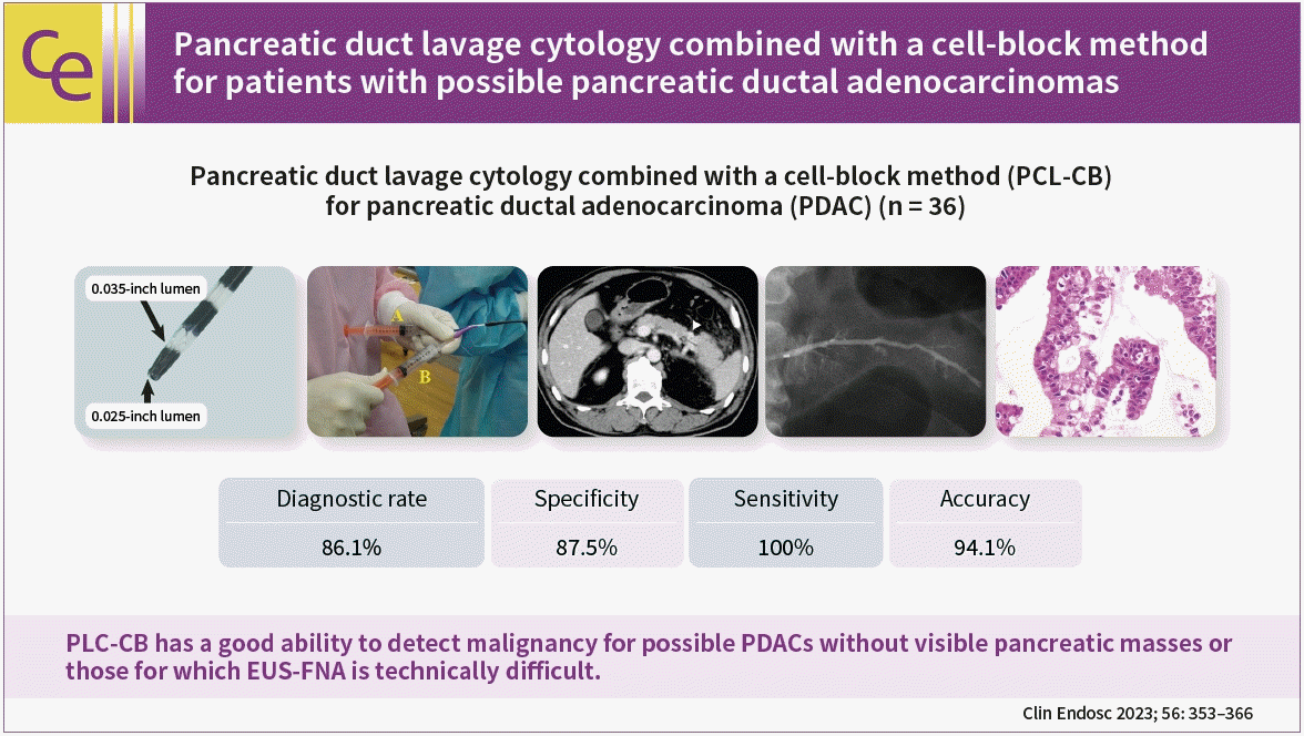
METHODS
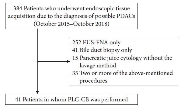
Study population
Methods
1) Outcome measurement
2) Endoscopic procedures
 | Fig. 2.Tip of a double lumen sampling catheter used in this study (Uneven Double Lumen Cannula; Piolax Medical Devices Inc.).
|
 | Fig. 3.Endoscopic procedure for pancreatic duct lavage cytology by using a double lumen catheter (endoscopy assistant). The injection of saline into the main pancreatic duct (A) and the suction of fluid in the main pancreatic duct with negative pressure by using a syringe (B) were simultaneously performed through two separate lumens (0.025- and 0.035-inch lumen, respectively).
|
3) Histocytological evaluations
4) Determination of definitive benignancy and malignancy
5) Procedure-related adverse events
Ethical statements
RESULTS
Baseline characteristics of 41 subjects
Table 1.
Values are presented as median (interquartile range) or number (%). MPD, main pancreatic duct; Ph, pancreatic head; Pb, pancreatic body; Pt, pancreatic tail; PLC-CB, pancreatic duct lavage cytology combined with a cell block method; EUS-FNA, endoscopic ultrasound-guided fine needle aspiration; PDAC, pancreatic ductal adenocarcinomas; PCIS, pancreatic carcinoma in situ; PanIN, pancreatic intraepithelial neoplasia; TNM, tumor-node-metastasis; UICC, Union for International Cancer Control.
![]()
Table 2.
MPD, main pancreatic duct; EUS-FNA, endoscopic ultrasound-guided fine needle aspiration; PLC-CB, pancreatic duct lavage cytology combined with a cell-block method; PDAC, pancreatic ductal adenocarcinoma; UICC, Union for International Cancer Control; PEP, post-endoscopic retrograde cholangiopancreatography pancreatitis; NSAIDs, non-steroidal anti-inflammatory drugs; F, female; M, male; EUS, endoscopic ultrasonography; CT, computed tomography; MRI, magnetic resonance imaging; US, ultrasonography; Ph, pancreatic head; Pt, pancreatic tail; Pb, pancreatic body; N/A, not available; PCIS, pancreatic carcinoma in situ; PanIN, pancreatic intraepithelial neoplasia; MFP, mass forming pancreatitis; AIP, autoimmune pancreatitis.
a)Indeternimate final diagnosis due to lack of surveillance periods (<12 months) after undergoing PLC-CB.
![]()
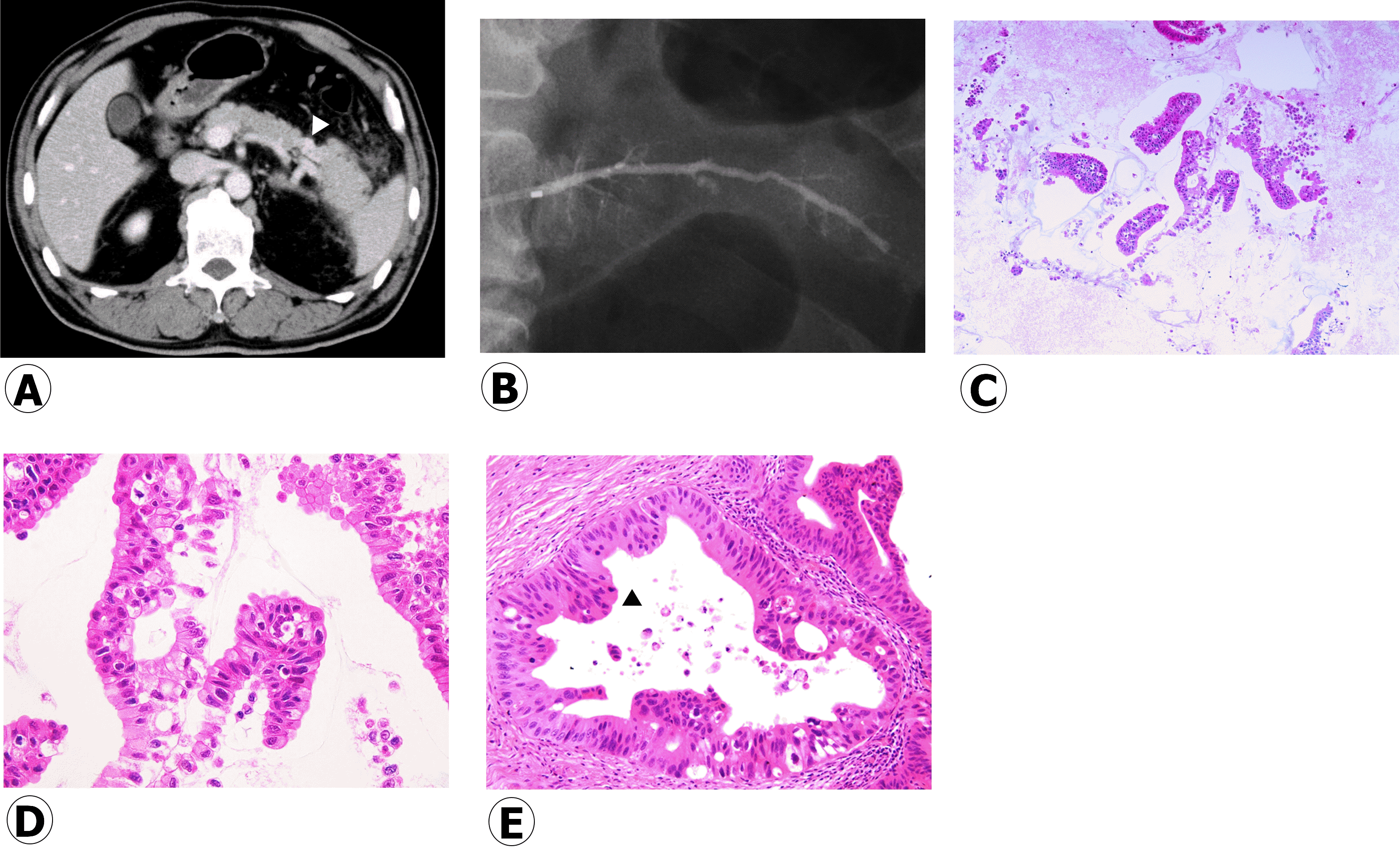 | Fig. 4.A 64-year-old patient with an initial diagnosis of acute pancreatitis (patient no. 14 in Table 2). The enlargement of the pancreatic tail with mild pancreatitis and localized atrophy of the pancreatic parenchyma at the proximal site of the enlarged pancreatic tail were detected (A, contrast-enhanced computed tomography, arrowhead). Endoscopic ultrasonography did not detect mass lesions, and magnetic resonance cholangiopancreatography did not show the main pancreatic duct stenosis at the pancreatic tail. We suspected pancreatic carcinoma in situ and performed pancreatic duct lavage cytology combined with a cell-block method using a double lumen catheter. Endoscopic retrograde pancreatography did not show clear stenosis of the main pancreatic duct at the pancreatic tail (B, pancreatography). Cell-block specimens obtained were firstly evaluated by using hematoxylin and eosin staining (C, ×10; D, ×40). Histocytological diagnosis was shown to be adenocarcinoma (Class V), and distal pancreatectomy was performed. For the histological findings of resected specimens, the atypical ductal epithelium was continuously observed from the main pancreatic duct (E, ×20; arrowhead) to branch pancreatic duct, and invasive components derived from this atypical epithelium were not found. Thus, this patient was diagnosed with pancreatic carcinoma in situ (high-grade pancreatic intraepithelial neoplasm).
|
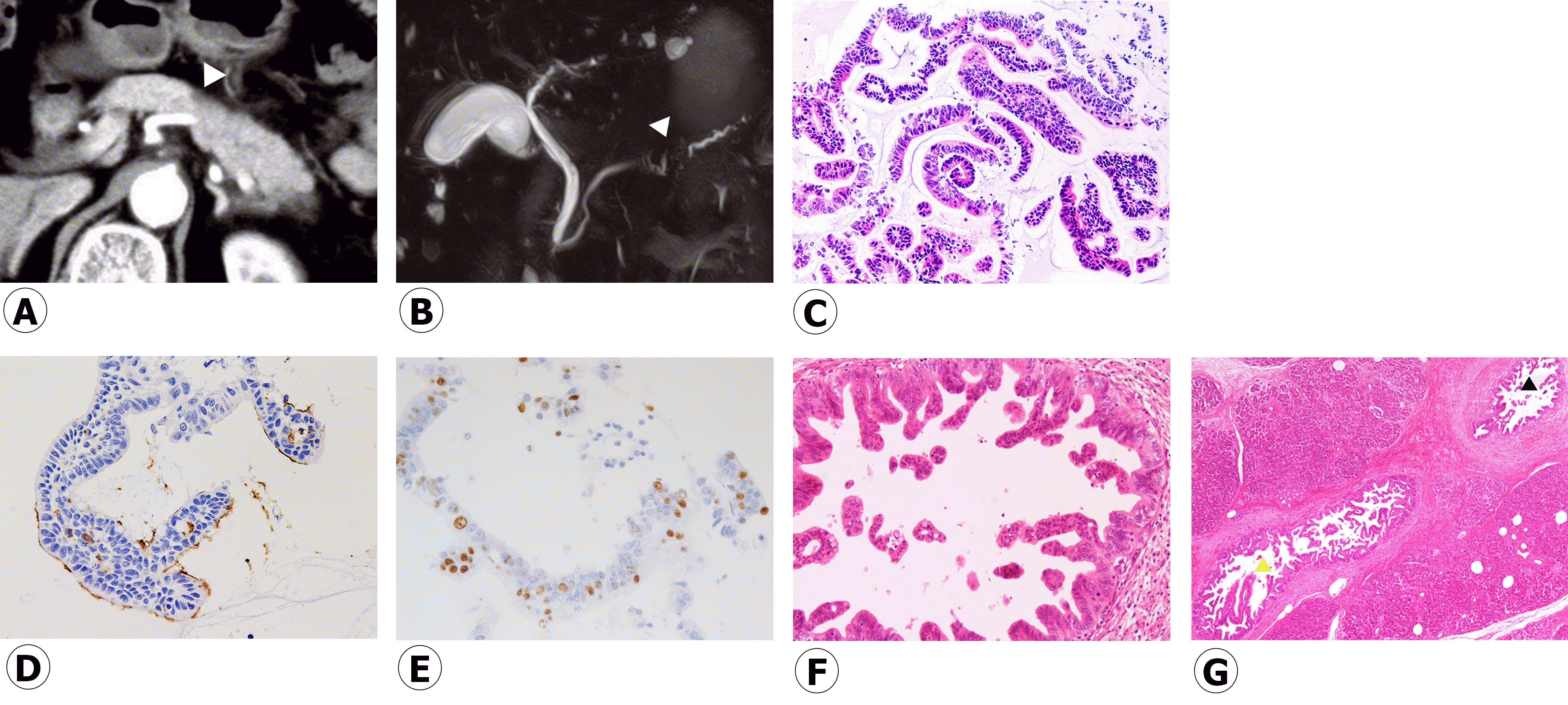 | Fig. 5.An 82-year-old patient (patient no. 17 in Table 2) with recurrent acute pancreatitis had a localized parenchymal atrophy of the pancreatic body (A, contrast-enhanced computed tomography; arrowhead, localized atrophic parenchyma in the pancreatic body). Although magnetic resonance cholangiopancreatography (B) showed a stenosis of the main pancreatic duct (MPD) in the pancreatic body (arrowhead), endoscopic ultrasonography did not detect mass lesions in this MPD stenosis. Pancreatic duct lavage cytology combined with a cell-block method using a double lumen catheter was conducted due to the diagnosis of suspected pancreatic carcinoma in situ. For histocytology of the specimens obtained through pancreatic duct lavage cytology combined with a cell-block method, some clusters of atypical cells were observed by using hematoxylin and eosin staining (C, ×20). MUC1 staining (D, ×40) was focally positive, a Ki-67 labeling index (E, ×40) was approximately 20% and p53 staining was negative, indicating adenocarcinoma (Class IV). She underwent distal pancreatectomy, and histology (hematoxylin and eosin staining) of resected specimens showed an atypical epithelium mainly located in the MPD and branch ducts (F, ×4; G, ×20; black arrowhead: MPD, yellow arrowhead: branch duct), resulting in the final diagnosis of pancreatic carcinoma in situ (high-grade pancreatic intraepithelial neoplasm).
|
Results of PLC-CB and clinical courses after PLC-CB
Accuracy of PLC-CB to detect malignancy for patients with possible PDACs
Table 3.
![]()
Post-ERCP adverse events
Table 4.
PEP, post-endoscopic retrograde cholangiopancreatography pancreatitis; BMI, body mass index; NSAIDs, non-steroidal anti-inflammatory drugs, EST, endoscopic sphincterotomy; EPST, endoscopic pancreatic sphincterotomy; IDUS, intraductal ultrasonography; F, female; M, male; TGRY, total gastrectomy with Roux-en-Y reconstruction; DGBI, distal gastrectomy with Billroth I reconstruction.
![]()
 XML Download