

 PDF
PDF Citation
 Print
Print


INTRODUCTION
Leptomeningeal metastasis (LM) is a dissemination or invasion of tumor cells into the cerebrospinal fluid (CSF) space or the leptomeninges and usually represents a dismal prognosis [7]. Nevertheless, in glioma patients the clinical characteristics and risk factors for LM have not yet been revealed due to the rare occurence [10,16]. Recently however, owing to prolongation of survival, technical developments such as image resolution, and more frequent testing, an increase in LM diagnoses in glioma patients has been reported [2].
Regarding magnetic resonance imaging (MRI) findings, different patterns of LM in glioma patients have been reported [2,21], such as disseminated or subependymal spread. However, it has not been described whether such different patterns reflect different clinical characteristics. Moreover, risk factors for LM based on clinical characteristics and interventions remain unknown. In the parenchymal brain metastasis of systemic cancer, it has been suggested that the proximity of brain metastasis or CSF contact during the operation increases the risk of LM development [1,27]. Thus, the location of gliomas in reference to the CSF circulation pathway might be related to the development of LM, but this question has not yet been investigated. Moreover, with the development of molecular diagnostics, a new classification of diffuse midline glioma (DMG) was created but the relationship with the development of LM has not been identified yet [15].
As the treatment of choice for gliomas is primarily maximal safe resection [8], numerous surgical methods are used to achieve the goal. Nevertheless, surgical spillage is one of the concerns during the surgical removal of cancer, including gliomas. Although there are controversial reports regarding whether the seeding of tumor cells is enhanced, ultrasonic aspirators are a common armamentarium used in the field [9,22]. Ventricular opening during tumor removal surgery is also reported to carry the risk of tumor seeding [1,27,28].
In this study, we first investigated the proportional occurrence of LM in glioma patients according to both clinical and radiological characteristics and analyzed the risk factors for LM among glioma patients, especially those who were surgically treated. Second, we reviewed the clinical characteristics of LM in gliomas according to different metastasis patterns of nodular and linear spread.
Go to : 
MATERIALS AND METHODS
The present study was approved by the Institutional Review Board of National Cancer Center of Korea (IRB No. NCC2021-0236).
Patient selection
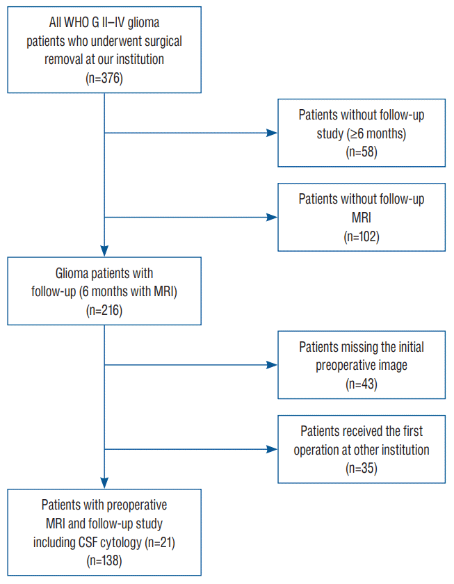
We retrospectively reviewed the medical charts and radiological records of adult glioma patients who underwent surgical removal between January 2001 and December 2020 in our institution. A total of 376 World Health Organization (WHO) grade II to IV glioma patients underwent surgery for gliomas during the study period, and patients without a follow-up period of more than 6 months, those who underwent surgery at other institutions, or those whose initial images were missing were excluded (Fig. 1).
Data collection
The data for age, sex, the Karnofsky performance scale (KPS) score [18], MRI findings, CSF cytology, the WHO grade, the time from surgery to LM diagnosis, LM-related symptoms, the extent of tumor resection, and adjuvant therapy such as chemotherapy or radiation therapy were collected and categorized for analysis. In addition, surgical factors, including the use of ultrasonic aspirators and the opening of ventricles during surgery, were reviewed as risk factors for LM, which was previously evaluated in metastatic brain tumor or glioma patients [1,24].
MRI findings
Brain MRI was performed on all patients prior to surgery and images were reviewed by a neuroradiologist (S.H.L.). Spinal MRI was not routinely performed due to the low occurrence of spinal LM for gliomas [4]. Only patients manifesting symptoms related to spinal LM, such as urinary difficulty or low extremity pain, were selected for spinal MRI. The MRI sequence included T1-, T2- and T1- with gadolinium enhancement. Axial, coronal and sagittal images were reviewed. The tumor proximity to the CSF circulation pathway was defined with the same criteria as a previous study [1] : 1) involved : the tumor was in contact with the pial surface or ventricular wall without intervening brain parenchyma, and/or was accompanied by pial or ependymal enhancement or asymmetrical cortical vessel enhancement and 2) separated : the tumor was not in contact with the pial surface or ventricular wall and was separated by brain parenchyma, as shown in Fig. 2. The location of the tumor was dichotomized as midline for tumors residing in the thalamus, basal ganglia and brainstem, or lateral for tumors residing in the cerebral and cerebellar hemispheres. Tumor volume was calculated using the diameter method ABC/2, which are greatest diameter of the tumor in axial plane (A), diameter of the tumor perpendicular in the axial plane (B), and multiplication value of the numbers of MRI slices and thickness of each slice (C) [26]. The extent of resection (EOR) was analyzed by MRI 48 hours postoperatively and was categorized as gross total resection (GTR) or non-GTR.
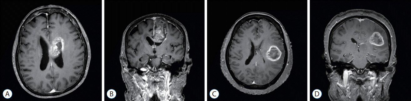 | Fig. 2.The tumor proximity to the cerebrospinal fluid pathway. Axial (A) and coronal (B) images of “involved” proximity. “Involved” proximity was defined as when the tumor was in contact with the pial surface or ventricular wall without intervening brain parenchyma and/or accompanied by pial or ependymal enhancement over the tumor. Axial (C) and coronal (D) images of “separated” proximity. “Separated” proximity was defined as when the tumor was not in contact with the pial surface or ventricular wall and was separated by brain parenchyma.
|
Diagnosis of LM
The diagnosis of LM was made by gadolinium-enhanced MRI [12] or CSF cytology. The date of LM diagnosis was calculated from the earliest date that was noted positive by either MRI or CSF cytology. If the LM diagnosis date preceded the surgery, the date of surgery was defined as the date of LM diagnosis. The extent of LM was categorized as nodular or linear by enhancement pattern (Fig. 3). Nodular LM was defined as dot-shaped enhancement, whereas linear LM was defined as disseminated enhancement.
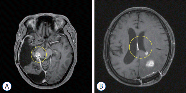 | Fig. 3.Different leptomeningeal metastasis patterns of gliomas on magnetic resonance imaging. “Nodular” leptomeningeal metastasis shows dot-shaped enhancement (A), whereas (B) “linear” leptomeningeal metastasis shows disseminated or diffuse enhancement along the leptomeningeal surface. Lesions are circled in yellow.
|
LM-related symptoms were defined as symptoms that were solely caused by LM and not by other anatomical or pathological causes. Such symptoms included manifestations of increased cranial pressure caused by communicating hydrocephalus, cranial neuropathy without other space-occupying lesions, or radiculopathy that was not related to other primary spine diseases.
Statistical analysis
SPSS Statistics version 18 (SPSS Inc., Chicago, IL, USA) was used for statistical analyses. Comparisons were made according to the presence and extent pattern of LM. The chi-square test or Fisher’s exact test was used to compare categorical and ordinal variables. Student’s t test or the Wilcoxon rank sum test was used for continuous variables. Kaplan-Meier analysis was used to estimate cumulative survival. The Cox proportional hazard model was used for univariate analysis and multivariate analysis to determine risk factors for LM. All analyses were two-sided, and a p-value <0.05 was considered significant. Factors with a p-value <0.05 in univariate analyses were selected for multivariate analyses.
Go to :
RESULTS
Patients’ characteristics according to LM development
A total of 138 patients met the eligibility criteria (Fig. 1). The overall demographic characteristics of these patients are shown in Table 1. The median age at surgery was 56.5 years (range, 23–88). Eighty patients (58%) were male, and 58 patients (42%) were female. The median follow-up duration was 19 months (range, 6–216). Eighty-one patients (58%) were diagnosed with WHO grade IV gliomas (79 glioblastomas, two gliosarcomas), while 57 patients (42%) were diagnosed with WHO grade II (eight oligodendrogliomas, 11 oligoastrocytomas) or III gliomas (18 anaplastic astrocytomas, nine anaplastic oligodendrogliomas, 11 anaplastic oligoastrocytomas). Spinal LM was found in four patients, and all the patients had WHO grade IV gliomas (glioblastoma).
Table 1.
Incidence of glioma leptomeningeal metastases according to various clinical and radiological factors
![]()
We analyzed whether there was any significant difference in clinical and radiological variables between the non-LM and LM groups. During the follow up period, 94 patients (68%) did not develop LM, whereas 44 patients (32%) showed MRI finding compatible with LM. Among the patients with LM, nine patients (20%) had LM preoperatively at the time of glioma diagnosis. The median age of the patients without LM was 59 years (range, 26–88), which was significantly older than that of patients with LM (53 years; range, 23–82; p=0.039). The number of patients with KPS of 70 or higher was not significantly different between non-LM and LM patients (91 vs. 39, p=0.110). Pathological grading was significantly different between the two groups, as 49 out of 94 patients (52%) in the non-LM group had WHO grade IV gliomas, and 31 out of 44 (70%) patients with LM were WHO grade IV glioma patients (p=0.042).
Regarding MRI findings, both the tumor proximity to the CSF space and location of the tumor differed significantly between the two groups. Twenty-three percent of the patients without LM had a tumor involving the CSF space prior to surgery, whereas 52% of the 44 patients with LM had this finding (p=0.001). There were a total of 23 midline located tumors, and LM was found in 78% (18 patients) of the patients whereas 23% of patients with lateral located tumors developed LM (p<0.001). On the other hand, the tumor volume was not significantly different between the two groups.
Variables such as the EOR, usage of the cavitron ultrasonic surgical aspirator (CUSA) or opening of ventricles during surgery were not significantly different between the non-LM and LM groups. GTR was achieved in 31 patients (23%), of which 24 patients (26%) developed LM and seven patients (16%) did not. The CUSA was used in 87 of overall patients (63%) : 62% (58 patients) of the patients who did not have LM and 66% (29 patients) of the patients who had LM. Among the patients without LM, 20 of the 94 patients (21%) had their ventricle opened during the surgery, whereas 15 patients (34%) with LM had their ventricle opened.
Comparison of clinical and radiological variables according to LM pattern
For the next step, we investigated whether there was any clinical difference according to LM patterns (nodular vs. linear) in the LM group (Table 2). Among the LM patients, 25 (57%) had a nodular pattern, and 19 (43%) had a linear pattern. Patients with nodular LM were younger than the patients with linear LM (median age, 49 years vs. 57 years; p=0.048). In addition, more gliomas with midline location showed linear LM (61%) whereas lateral located gliomas showed nodular pattern of LM more (69%, p=0.046). However, presence of LM-related symptoms, the WHO grades (IV vs. II and III) and MRI findings such as tumor proximity to the CSF circulation pathway were not significantly different according to the metastasis pattern. LM-related symptoms were present in 15 of overall LM patients (34%), and no significant difference was found between the two groups of metastasis patterns. Common LM symptoms were cognitive impairment, headache, nausea, vomiting and gait disturbance. Other symptoms were urinary incontinence (two patients) and diplopia (one patient). Patients usually manifest multiple symptoms at presentation.
Table 2.
Clinical and radiological characteristics according to LM patterns on MRI
![]()
Relative risk for LM development
Progression-free survival for LM was calculated by the time from the initial surgery to diagnosis of LM on MRI, and relative risk was compared by log rank test (Fig. 4). Among the clinical factors investigated, CSF proximity, location of the tumor and the WHO grade were significant factors for LM development in both univariate analysis (Table 3). In multivariate analysis, location of the tumor was the most significant factor affecting the LM development followed by WHO grade (95% confidence interval [CI], 1.384–4.974; p=0.003). The midline located gliomas increased the development of LM with a hazard ratio (HR) of 2.624 (95% CI, 1.384–4.974, p=0.003) compared with lateral located gliomas. The HR was 3.008 for WHO grade IV gliomas (95% CI, 1.379–6.561; p=0.006).
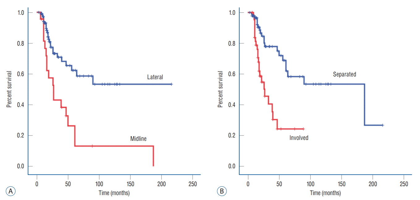 | Fig. 4.Kaplan-Meier survival curve of all patients for the development of leptomeningeal metastasis. Development of leptomeningeal metastasis was significantly different according to the location (A) and tumor involvement of cerebrospinal fluid space (B).
|
Table 3.
Overall relative risks for leptomeningeal metastasis in all gliomas (n=138)
![]()
Surgical factors such as the EOR and the use of the CUSA, did not significantly affect the risk of LM development. On the other hand, opening of the ventricle during surgery was significant in the univariate analysis (p=0.023) but not in the multivariate analysis.
Overall survival (OS) according to LM development and MRI patterns
The median OS after the surgery (the time of gliomas diagnosis) of the enrolled patients was 47 months (95% CI, 28.141– 65.859). Patients without LM had a median survival of 62 months (95% CI, 27.572–96.428), while patients with LM had a median survival of 26 months (95% CI, 12.743–39.257), but this apparent difference failed to reach statistical significance (p=0.130). According to the LM pattern, the median survival after LM diagnosis was 8 months for both nodular and linear metastases. However, OS after the surgery was shorter in patients with a linear metastasis pattern (26 months; 95% CI, 22.076–49.924) than in those with a nodular metastasis pattern (33 months; 95% CI, 9.124–56.876), although the difference did not reach statistical significance. In contrast, a tendency of shorter time to LM development was observed in linear metastasis pattern than nodular metastasis pattern (p=0.190).
Considering the heterogeneity of our cohort for the WHO grade, which is known to be one of greatest factors affecting OS, we performed subgroup analysis for WHO grade IV gliomas. The median OS for WHO grade IV gliomas was 22 months (95% CI, 18.221–25.779), with 22 months for patients with LM (95% CI, 16.185–27.815) and 24 months (95% CI, 19.737–28.263) for patients without LM. The median survival after LM diagnosis was 4 months.
Go to :
DISCUSSION
Clinical significance of LM in glioma patients
The occurrence of LM in glioma patents in the literature varies according to the definition of LM or the histological subtype/grade. Additionally, as most previous studies were a single institute retrospective studies, it is difficult to have a population-based or prospectively collected LM occurrence of gliomas. The only population-based LM incidence of oligodendroglioma was reported by Roldán et al. [25] and based on a population of 1.8 million over 19 years. They used the LM criteria of 2 or more of the following : 1) LM-related symptoms or signs; 2) MRI findings; and 3) CSF cytology, and 9/204 (3.9%) were diagnosed with LM from oligodendrogliomas. Recently, Andersen et al. [2] reported the incidence of LM in glioma patients and different prognoses according to the histological subtype of gliomas in a single institution over 15 years period. The incidence of LM was 4.6% (188/4082 patients) using the diagnostic criteria of the clinical description of LM with either MRI findings or positive CSF cytology. Despite the fact that the interventions after LM diagnosis in these patients were not controlled, it seems obvious that the OS of the LM patients varied by original histology, as the median OS of glioblastoma patients was 3.8 months vs. that of oligodendroglioma patients, which was 10.8 months. In our study, the apparent LM occurrence was much higher than those in previous studies, as 44/138 patients (32%) were diagnosed with LM and considering the excluded patients, which mostly lacked information, the possible lowest proportional occurrence was 44/376 (12%). Another reason for our higher occurrence of LM was probably due to more generous diagnostic criteria of positive MRI findings regardless of LM-related symptoms and CSF cytology.
It has been reported that LM manifestation on MRI can be divided into linear spread (diffuse), nodular spread or mixed types. Bae et al. [4] observed different MRI findings of spinal LM patients, with eight diffuse, one nodular and two mixed types, but they did not evaluate clinical differences according to MRI manifestation. Andersen et al. [2] divided cerebral LM patients into two subgroups, disseminated and subependymal LM, as well as groups based on the original histology. As a result, the overall clinical characteristics, such as the time to development of LM, survival from LM diagnosis, OS after surgery and occurrence of related symptoms, did not differ between the subgroups [2]. In contrast, they reported that the most important determining factor for the time to development of LM and survival of LM patients is the original histology. This is in line with our results, which showed no difference according to the metastasis pattern. The study did not analyze risk factors for LM, which was the goal of our study.
For spinal LM dissemination, the median OS after LM diagnosis has not increased much over the decades. Awad et al. [3] reported a median survival after LM diagnosis of 3 months in 13 patients diagnosed with LM by myelogram or CSF cytology in 1986. Later, Bae et al. [4] reported a median OS of 2.7±1.3 months in 11 patients with spinal leptomeningeal dissemination from various gliomas in 2011. In contrast to cerebral LM manifestations, these spinal LM patients all had LM-related symptoms, dissemination after the initial surgery for supratentorial gliomas, and dismal prognoses regardless of glioma subtype. In our study, the medial OS of 3 months after diagnosis was apparently longer than those of previous studies. The difference probably came from our diagnostic criteria, and most (134/138) of our patients did not have spinal dissemination.
Prognostic factors for OS after LM diagnosis have not yet been determined. However, previous studies reported objective responses to chemotherapy or radiation in selected patients, and some of these responses lasted for quite a long period [5,13,17,19].Thus, we expected that proper treatment, including recently developed target inhibitors or immune therapy, could prolong patient OS and lessen patient suffering through future clinical trials.
Risk factors for developing LM in glioma patients
We were able to analyze the clinical characteristics according to LM and their risk factors in glioma patients. Thirty-two percent of the patients with WHO grade II–IV gliomas developed LM in our cohort. Patients with LM were younger than non-LM patients, whereas the performance scale was equal. The younger age of LM patients compared to non-LM patients is a common feature reported in previous papers [3,11,20].
Andersen et al. [2] tried to evaluate whether any clinical or molecular characteristics affect the risk for LM. However, they failed to obtain the HR of each variable for LM development and described that no variables were identified as a risk factor. Our study is the first attempt to evaluate clinical variables for the development of LM using HRs, although we could not balance these variables for comparison in this retrospective study. With regard to preoperative MRI, midline location and higher histological grade (IV vs. II/III) showed a significantly higher risk for LM. In addition, gliomas involving the CSF circulation pathway and opening of ventricles during surgery were another important factor for LM.
Previous studies have verified that surgical spillage caused by piecemeal resection, ultrasonic aspiration or ventricular opening to be a risk factor for LM development [1,27,28]. Roelz et al. [24] reported that ventricular opening during surgery is a risk factor for LM. Similarly, Bae et al. [4] reported that all 11 patients who were enrolled in the study had their ventricle opened during surgery showed spinal LM. On the other hand, to see the surgical effect, surgical procedures such as those using CUSA should be included. A previous study with brain metastasis showed that surgery increased the risk of LM compared with radiosurgery [14], and the use of CUSA increased the risk for LM [1]. However, in our study, surgical spillage-related factors of the use of the CUSA failed to show a significant influence on LM development (Table 3).
Qiu et al. [23] reported MRI characteristics of H3K27M-mutant DMG. Although our study could not perform molecular analysis to verify H3-K27M mutation, non-midline location of H3K27M-mutant DMGs are reported to be rare, implying high proportion of H3-K27M mutation in our cohort [23]. In the study, 11/66 (17%) of the patients showed CSF dissemination, which is lower than that of our cohort. This is probably due to the short time period of the study. Nevertheless, the proportion of LM in H3-K27M mutant DMG was higher compare to previous studies which is in line with our study [2,6]. Further study regarding the role of H3-K27M in LM development is mandatory.
Ventricular opening was significant only in the univariate analysis, but not in multivariate analysis, suggesting that the LM that were thought to be caused by ventricular opening is in fact a secondary effect by tumor proximity to CSF space. It is more likely for the ventricle to be opened if the tumor lies more closely to the ventricle. Although we cannot deny the possibility of mechanical spillage contributing to the development of LM, our present data implies that the tumor involvement to the CSF space has more influence on the development of LM than ventricular opening during surgery. Hence, based on our results, maximal safe resection of the tumor should not be avoided due to fear of LM occurrence. In this context, CSF proximity was in accordance with an increased chance of cancer cell spillage in the CSF space. Roelz et al. [24] verified that the distance of the tumor to the ventricle was also a significant risk factor in the univariate analysis of a previous report. This is in line with our results in that the relation of the tumor to the CSF space is important for LM progression.
Limitations
The limitations of the current study are that it was based on a retrospective analysis in a single institution. However, the demographic characteristics in our study were similar to the trends of other reports, which supports the reliability of our results. Another limitation is that our analysis did not include molecular studies. Because majority of the patients underwent surgery before the revision of the WHO grade and the rise of molecular analysis, molecular study was not performed on routine basis. Nevertheless, because MRI is a more common and simple examination that can be performed, screening patients with a higher risk of LM by MRI findings would be more helpful in the clinical field compare to molecular studies.
Go to :
CONCLUSION
LM in glioma patients is no longer a rare complication, owing to prolonged survival. Histological grading is the most important factor for LM. The midline location, WHO grade IV gliomas, and proximity to the CSF circulation pathway and is the most important factor for the development of LM in WHO grade IV glioma patients. Clinicians should be aware of and closely follow up patients who carry a high risk for LM.
Go to :
 XML Download