Journal List > Korean J Pain > v.36(3) > 1516083093

Wang, Karri, Tontisirin, and Cohen: Antimicrobial therapies for chronic pain (part 1): analgesic mechanisms

Abstract

There is increasing evidence that the relationship between chronic pain and infections is complex and intertwined. Bacterial and viral infections can cause pain through numerous mechanisms such as direct tissue damage and inflammation, the induction of excessive immunologic activity, and the development of peripheral or central sensitization. Treating infections might relieve pain by attenuating these processes, but a growing body of literature suggests that some antimicrobial therapies confer analgesic effects, including for nociceptive and neuropathic pain symptoms, and affective components of pain. The analgesic mechanisms of antimicrobials are indirect, but might be conceptualized into two broad categories: 1) the reduction of the infectious burden and associated pro-inflammatory processes; and 2) the inhibition of signaling processes (e.g., enzymatic and cytokine activity) necessary for nociception and maladaptive neuroplastic changes via off-target effects (unintended binding sites). For the former, there is evidence that symptoms of chronic low back pain (when associated with Modic type 1 changes), irritable bowel syndrome, inflammatory bowel disease, chronic pelvic pain, and functional dyspepsia might be improved after antibiotic treatment, though significant questions remain regarding specific regimens and dose, and which subpopulations are most likely to benefit. For the latter, there is evidence that several antimicrobial classes and medications exert analgesic effects independent of their reduction of infectious burden, and these include cephalosporins, ribavirin, chloroquine derivatives, rapalogues, minocycline, dapsone, and piscidin-1. This article aims to comprehensively review the existing literature for antimicrobial agents that have demonstrated analgesic efficacy in preclinical or clinical studies.

INTRODUCTION

Pain is a cardinal sign of infection, so it stands to reason that antimicrobial agents that treat infection may alleviate pain. Yet, in clinical practice the relationship between pain, infection and antimicrobial agents is more complex. Many individuals whose infection is eradicated may continue to experience chronic pain via mechanisms that include autoimmune reactions and peripheral and central sensitization (e.g., Lyme disease, myalgic encephalomyelitis, “long COVID”), in which case antimicrobial treatment may be less effective or ineffective [1]. Low-grade infection may cause neuropathic pain via demyelination, neuronal damage, and deafferentation, and predispose to somatic (e.g., discogenic back or neck pain, periodontitis) or visceral nociceptive pain (e.g., some cases of bladder pain syndrome/interstitial cystitis, inflammatory bowel disease [IBD]) through mucosal injury, chronic inflammation, or accelerated degenerative processes [25]. Recent evidence also points to variations in gut and other organ system microbiomes as sources of acute pain and chronic neuropathic, nociceptive, or nociplastic pain [6].
All organisms, including humans, share similarities in cellular machinery with microbes, and alterations in the microbiomes of the gut and other organ systems (e.g., respiratory, skin, genitourinary) can have a profound impact on pain. Therefore, the connections among infections, the treatment of infections, and chronic pain are not surprising. Over the past few decades, a growing body of literature has been devoted to these complex relationships [1,4,6], but there has been no systematic attempt to review antimicrobial therapy as a treatment for chronic pain in general, which is more clinically relevant to frontline pain practitioners. The aims of this two-part series are to outline the direct and indirect mechanisms by which antimicrobial therapies can alleviate nociception and pain, categorize the preclinical and clinical evidence supporting antimicrobial therapies in pain conditions, and provide a framework for future directions in this important, but hitherto underrecognized area.

MAIN BODY

1. Search strategy and study selection

The search strategy was the same for parts 1 and 2 of this series. From December 2022 to April 2023, we searched the following databases: PubMed, Embase, and Google Scholar, without language or date restrictions. We cross-referenced the major search terms “chronic pain,” “pain,” “antimicrobial,” “antibiotic,” “antiviral,” “antifungal,” “mechanism,” and “infection” with various iterations and subcategories of these keywords to correspond with various pathogens, medications, mechanisms, and chronic pain conditions. We prioritized peer-reviewed pooled analyses (e.g., meta-analyses and systematic reviews) and randomized controlled trials (RCTs), but also included preclinical studies, narrative reviews, case series, and retrospective studies as indicated. In addition to primary sources, we searched reference lists of retrieved articles.

2. Mechanisms of analgesia by antimicrobial agents

The aim of an antimicrobial agent is to eradicate its target pathogen before resistance develops [7]. Bacterial and viral infections can cause pain via numerous mechanisms including direct tissue damage, the induction of injurious immune responses, and the development of peripheral or central sensitization [1]. Although some antibiotics and antivirals might cause painful adverse effects (e.g., arthralgia with fluoroquinolones, peripheral neuropathy with antiretroviral therapy) [8], others may confer analgesia in the setting of infections. The analgesic mechanisms of antimicrobials are via indirect actions which might be conceptualized into two broad categories (Fig. 1): 1) the reduction of infectious burden and associated pro-inflammatory processes; and 2) the inhibition of signaling processes (e.g., enzymatic and cytokine activity) necessary for nociception and maladaptive neuroplastic changes via off-target effects (unintended binding sites). Altogether, a variety of acute and chronic pain conditions may be alleviated by antimicrobial agents (Fig. 2).
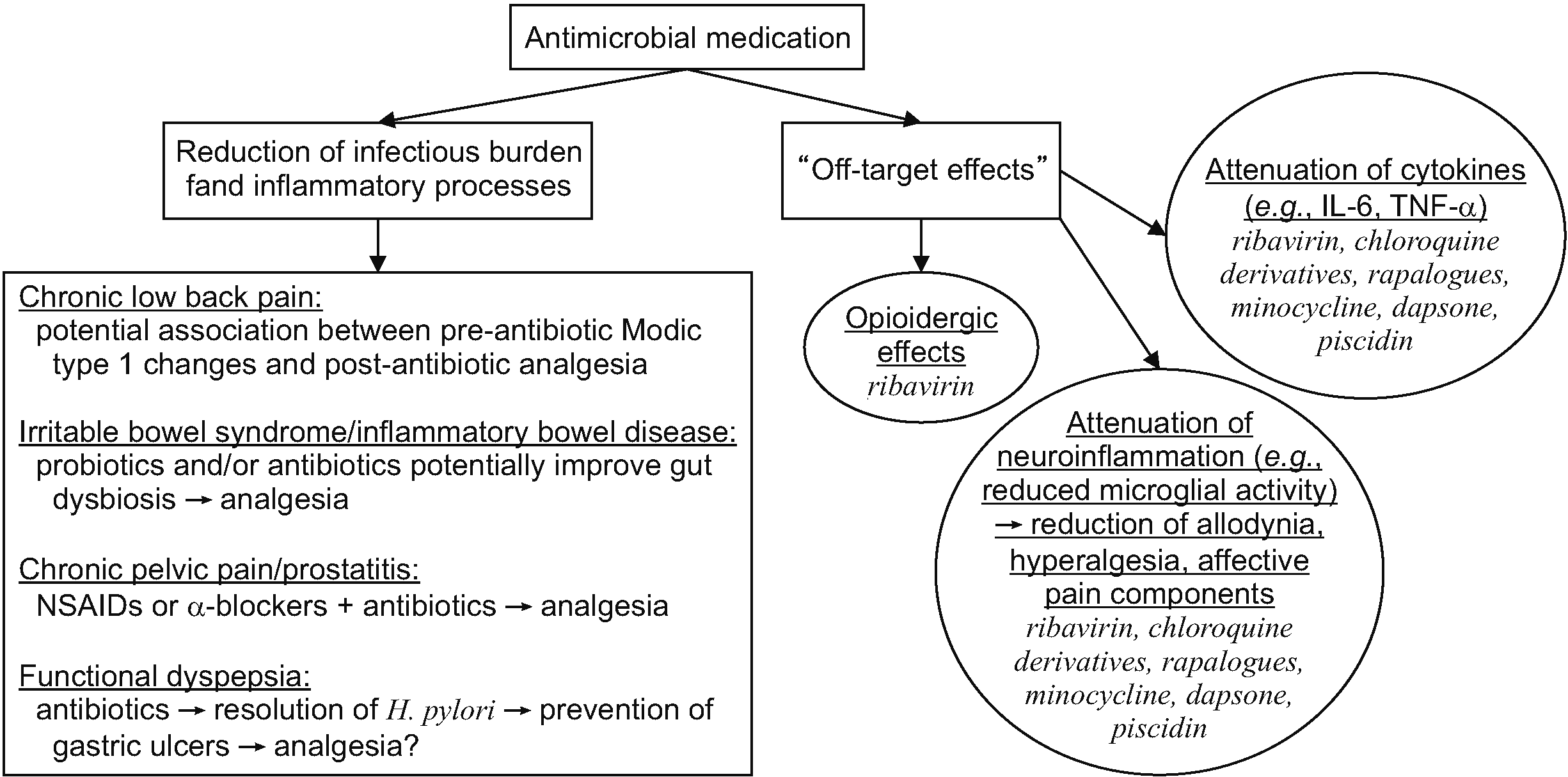
3. Reduction of infectious burden and inflammation

The etiology of Modic type 1 vertebral endplate changes remains controversial, but infection remains one of several plausible causes [9]. Positive cultures (especially for anaerobic microorganisms such as Propionibacterium acnes) from biopsies of herniated disc material have been associated with new Modic changes at adjacent vertebrae [10]. In a recent literature review assessing the use of antibiotics for chronic low back pain, Gilligan and colleagues [4] identified only two randomized placebo-controlled trials [11,12]. In the trial by Albert et al. [11], patients with magnetic resonance imaging (MRI) findings of Modic type 1 changes who received 100 days of amoxicillin-clavulanic acid reported statistically significant improvements in pain and function. Patients in the antibiotic group also had a greater likelihood of MRI-confirmed resolution of their Modic changes. However, in the trial by Bråten et al. [12], patients with MRI-confirmed Modic type 1 or type 2 changes reported no benefit in pain or functional outcomes after 100 days of amoxicillin, though post-intervention MRIs were not assessed. Given this limited and mixed evidence, it is uncertain whether antibiotics can reliably treat pain in the context of Modic type 1 changes, which might be associated with low-grade infection, pro-inflammatory processes, or both [9,10]. Nonetheless, the association between Modic type 1 resolution and analgesia with antibiotic use suggests that the reduction of microbial and pro-inflammatory burden is a potential mechanism.
A meta-analysis of patients with irritable bowel syndrome (IBS) found potential benefit for probiotics and rifaximin for abdominal pain symptoms via potential alterations to the gut microbiota, but high heterogeneity among the included studies precluded estimates of effect size [13]. A systematic review of patients with IBD also found analgesic benefit from antibiotics by treating small bowel bacterial overgrowth [14], although the data for this conclusion were based on one RCT [15]. A meta-analysis of patients with chronic prostatitis/chronic pelvic pain syndrome found that antibiotics (e.g., fluoroquinolones) and non-steroidal anti-inflammatory drugs reduced pain scores compared to a placebo, with a combination of α-blockers and antibiotics being the most efficacious regimen for analgesia [16].
In functional dyspepsia, although a meta-analysis of patients with known Helicobacter pylori infection failed to identify short-term (< 1 year) symptomatic improvement with antibiotics, there was significant improvement noted on long-term (≥ 1 year) follow-up [17]. These deferred benefits might be attributable to a greater likelihood of antibiotic-treated patients to have histologic resolution of chronic gastritis and the prevention of peptic ulcer disease [17]. In one placebo-controlled RCT that excluded patients with H. pylori, rifaximin significantly reduced dyspeptic symptoms, though follow-up was limited to only 8 weeks and the putative mechanism (alteration of the duodenal microbiota) has yet to be confirmed [18]. The precise relationship between H. pylori and functional dyspepsia remains unclear. H. pylori is recognized as an organic cause of dyspeptic symptoms, but consensus guidelines have suggested with some controversy [19] that functional dyspepsia is a specific diagnosis that is distinct from H. pylori-associated dyspepsia [20] and better reserved for describing symptoms that persist despite successful antibiotic treatment.
In summary, when microbiome dysbiosis (e.g., IBS) or an active infection (whether symptomatic or subclinical) potentially mediates pain symptoms, antibiotics might confer analgesia by reducing pathogen burden or altering microbiome compositions, leading to the attenuation of further tissue injury and pro-inflammatory processes. The available evidence is limited, and more studies are needed to assess how strongly analgesia is associated with radiologic or histologic evidence of infection eradication.

4. Inhibition of nociception and pain signaling via off-target effects

Antimicrobials can confer analgesia via mechanisms independent of reducing infectious burden and associated inflammation. Several antimicrobial agents are known to inhibit enzymes, proteins, or neurotransmitters necessary for pain signaling (e.g., protein kinase expression, pro-inflammatory cytokine activity) and maladaptive neuroplastic changes (e.g., dorsal horn remodeling, induction of hyperalgesia). These disruptions to pain processing are generally the consequence of unintended binding sites, and may be described as beneficial off-target effects [21]. Antimicrobial agents with known off-target analgesic effects are summarized below and in Table 1. It is important to note that because analgesia is not the primary therapeutic intent for antimicrobials, relatively few studies specifically assess the effect of antimicrobials on nociception, and this relationship might be underrecognized.

1) Cephalosporins

Cephalosporins are beta-lactam antimicrobials first discovered and isolated from the mold Cephalosporin acremonium (also named Acremonium chrysogenum) in 1945, and have since become one of the most prescribed antibiotics [22]. Five generations of cephalosporins have been developed and are collectively utilized against a variety of gram-positive and gram-negative bacteria. Cephalosporins exert bactericidal activity via their beta-lactam rings, which inhibit penicillin-binding proteins essential for bacterial cell wall synthesis [23].
In preclinical studies, there is evidence that ceftriaxone increases the expression of glial glutamate transporter-1 (GLT-1), which might confer neuroprotective effects by preventing neurotoxicity from excessive glutamate levels [24]. In a murine chronic constriction injury (CCI) model, hyperalgesia and allodynia were associated with downregulation of GLT-1 in the spinal dorsal horn; when intraperitoneal or intrathecal ceftriaxone was administered, GLT-1 expression and glutamate uptake increased, and thermal hyperalgesia and mechanical allodynia were reversed [25]. Moreover, when a GLT-1 inhibitor was administered following ceftriaxone administration, these beneficial effects were blunted [25]. In another murine study comparing the effects of ceftriaxone and gabapentin on neuropathic pain, both medications produced a similar effect size on the reduction of allodynia and hyperalgesia [26]. Ceftriaxone might also inhibit pro-inflammatory cytokine production (e.g., tumor necrosis factor [TNF]-α, interleukin [IL]-1β) in response to neuropathic injury [27], potentially via a poorly understood relationship between GLT-1 and inflammatory mediators [28].
Limited clinical data exist pertaining to cephalosporins and analgesia. In one case report, cephalexin following a course of minocycline provided a near-resolution of spasticity and pain in a patient with neurosarcoidosis [29]. In one comparative-effectiveness trial in 45 patients undergoing median or ulnar nerve decompression, participants were randomized to receive a single pre-incisional infusion of saline, saline with ceftriaxone (2 grams), or saline with cefazolin (2 grams). The patients in the ceftriaxone group reported a significant increase in pain thresholds up to 6 hours after surgery, whereas the patients in the other groups reported no significant difference [30]. The same investigators also tested a murine model of postsurgical pain, and the mice that received intraperitoneal ceftriaxone had greater dorsal horn GLT-1 expression and a greater reduction in nocifensive behavior than those that received saline or cefazolin [30]. This study did not evaluate why cefazolin, also a cephalosporin, appeared to lack analgesic efficacy. Although cefazolin is a first-generation cephalosporin whereas ceftriaxone is a third-generation cephalosporin [23], cefazolin has also demonstrated the ability to upregulate GLT-1 expression [31]. Further studies are needed to clarify whether the different generations of cephalosporins involve differential analgesic mechanisms or confer varying levels of analgesia.

2) Ribavirin

Ribavirin is a guanosine analog that is used to treat respiratory syncytial virus and Lassa virus, but is perhaps most recognized as a treatment for chronic hepatitis C when co-administered with interferon-alpha [32]. Ribavirin has several putative mechanisms of action, such as facilitating viral RNA chain termination, increasing the sensitivity of target cells to interferon, and inhibiting viral enzymes (e.g., inosine monophosphate dehydrogenase) necessary for energy production or genetic replication [3234].
In one murine study, ribavirin decreased nociceptive responses to noxious stimuli (e.g., formalin, capsaicin) and provided analgesia for visceral pain (e.g., intra-abdominal acetic acid injections) [34]. Because these analgesic effects were attenuated with the administration of naloxone and enhanced with dopamine D2 receptor activity (regardless of agonism or antagonism), endogenous opioid or dopaminergic pathways might be involved, though the exact mechanisms remain uncertain [34]. In two other murine studies, ribavirin reduced histologic signs of neuroinflammation, such as microglial infiltration and demyelination [35], as well as astrocyte proliferation and glial scarring [36]. Ribavirin has also demonstrated the ability to reduce levels of pro-inflammatory cytokines (e.g., IL-1β, IL-6, and TNF-α) [37], but the degree to which this confers analgesia remains unclear.
Although ribavirin is not widely used for the purpose of analgesia, at least one proprietary medication using ribavirin has been developed and tested in a peripheral nerve injury mouse model, which appeared to facilitate axonal regeneration and increase thresholds for hyperalgesia and allodynia [38].

3) Chloroquine derivatives

Chloroquine and hydroxychloroquine are antimalarial medications used as disease-modifying antirheumatic drugs (DMARDs) in several autoimmune conditions, such as systemic lupus erythematosus (SLE) [39] and rheumatoid arthritis (RA) [40], though more recent guidelines [41] have deemphasized the use of hydroxychloroquine as monotherapy or a first-line DMARD in RA due to concerns of modest benefits and substantial risks of adverse effects (e.g., nausea and vomiting [42], myopathy [43], retinopathy [44]). Chloroquine derivatives inhibit lysosomes [45] and several pathways in the immune cascade, including Toll-like receptor (TLR) activity [46], pro-inflammatory cytokine production (e.g., IL-1, TNF) [47], and T-cell antigen presentation [48]. The inhibition of lysosomal and T-cell activity, in conjunction with the reduction of TLR and proinflammatory cytokine signaling, is likely the means through which autoimmune activation and associated tissue injury is attenuated, but the mechanisms of action for chloroquine derivatives are numerous and incompletely understood [49]. Although outside the scope of this review, the immunologic (e.g., inhibition of autophagy) and anti-inflammatory effects of chloroquine derivatives might inhibit tumor growth and increase the efficacy of chemotherapy drugs [50].
The numerous immunomodulatory and anti-inflammatory effects of chloroquine derivatives provide a mechanistic basis for conferring analgesia. Additionally, murine studies have demonstrated a local anesthetic effect when chloroquines are administered intrathecally [51] or subcutaneously [52,53], possibly via their ability to antagonize potassium, sodium, and calcium channels [54]. However, clinical studies have mostly been negative, with large multicenter RCTs in osteoarthritis hand pain demonstrating no benefit of hydroxychloroquine over placebo [55,56]. Similarly, a recent cost-utility analysis found that hydroxychloroquine does not provide cost-effective benefits for pain and quality of life in hand osteoarthritis [57]. Although early data suggested that hydroxychloroquine might benefit arthritis pain in the context of SLE [58], a large placebo-controlled multicenter RCT demonstrated no benefit for hydroxychloroquine in inflammatory arthritis pain [59]. A systematic review of treatments for chikungunya virus-associated pain found that chloroquine was superior to placebo for chronic pain, though not for acute pain, with only five trials comprising 402 total patients included [60]. A more recent systematic review that included 11 studies pertaining to chikungunya-associated joint pain found no benefit from chloroquine or hydroxychloroquine compared to placebo [61].
Smaller studies continue to suggest that chloroquine derivatives might be of benefit in other pain syndromes with potential autoimmune-mediated mechanisms. Two case series of oral lichen planus [62,63] and a retrospective analysis in perineal lichen planus [64] suggest that hydroxychloroquine might facilitate the healing of painful erosions, but prospective placebo-controlled trials have yet to be completed. A small case series in seven patients with complex regional pain syndrome (CRPS) combined with a CRPS murine model demonstrated a decrease in self-reported pain scores with daily hydroxychloroquine, and reduced spinal cord dorsal horn microglial and cytokine activity in the mice [65]. Allodynia, paw edema, and temperature discrepancies (signs associated with CRPS) were also decreased in the mice receiving hydroxychloroquine, suggesting a reduction in neuroinflammation [65].
Although chloroquine derivatives do not appear to confer a large magnitude of analgesia, and controlled studies and pooled analyses have so far been negative, their inhibition of numerous inflammatory and immunologic processes involved with pain signaling is well-described. It is possible that the pain mechanisms (and pain diagnoses) most amenable to treatment from chloroquine derivatives have simply yet to be clarified.

4) Rapamycin (sirolimus) and rapalogues

Rapamycin (sirolimus) is a macrolide produced by the bacteria Streptomyces hygroscopicus that was initially utilized for its antifungal and immunosuppressive properties [66,67]. The molecular target of rapamycin was identified as a protein kinase that regulates intracellular anabolic and catabolic signaling in mammals, and this kinase was subsequently named “mTOR” (initially an abbreviation for “mammalian target of rapamycin,” later revised to “mechanistic target of rapamycin”) [67].
The mTOR kinase is a component of numerous intracellular functions and is implicated in various diseases. The inhibition of mTOR by rapamycin or its analogues (“rapalogues”) has been studied as a treatment strategy for various cancers [68,69] (e.g., pancreatic [70], renal cell [71]), myopathies (e.g., mitochondrial myopathy [72], inflammatory myopathy [73], inclusion body myositis [74]), sickle cell disease [75], fragile X syndrome [76], viral prophylaxis for transplant recipients [77], and possible anti-aging effects [78,79].
Rapalogue inhibition of mTOR disrupts several maladaptive processes associated with the chronification of pain. In murine neuropathic pain models, intrathecal [80,81] or intraperitoneal [82,83] rapamycin inhibits astrocyte and microglial cell activation, as well as levels of pro-inflammatory neuropeptides including calcitonin gene-related peptide, substance P, and several cytokines (e.g., IL-1β, IL-6, TNF-α [83]). Significant mTOR activity has been identified in the insular cortex, dorsal root ganglia, and laminae I–III of the dorsal horn in murine studies [8486], with mTOR blockade by rapalogues attenuating wind-up and mechanically-evoked potentials [85], thereby reducing allodynia [84], increasing the activation thresholds of nociceptive Aδ fibers [86], and inhibiting hyperalgesic priming [87]. There is also data supporting the role of mTOR in mediating affective components of pain, with murine models demonstrating improvement of pain behaviors with mTOR blockade [88,89].
It is noteworthy that mTOR upregulates intracellular signaling pathways (e.g., PI3K/Akt/mTOR) and the expression of protein kinases (e.g., PKCγ, neuronal NOS) associated with opioid-induced tolerance and opioid-induced hyperalgesia (OIH) [90,91]. In murine models, rapamycin and other mTOR inhibitors (e.g., metformin) have shown efficacy for reducing morphine tolerance and hyperalgesia [90,92,93]. The use of rapalogues for treating opioid-induced tolerance and OIH has not been described in a clinical setting, but their utility for this purpose might be limited by potentially serious adverse effects associated with mTOR inhibition (e.g., metabolic dysfunction, anemia, renal failure [94]).

5) Minocycline

Minocycline is a second-generation tetracycline that has demonstrated the ability to reduce neuroinflammation, neuropathic pain, and nociceptive pain in preclinical studies [95] via mechanisms independent of its antimicrobial effects [96,97]. Minocycline inhibits central [98101] and peripheral [102,103] glial cell activity and attenuates the release of pro-inflammatory cytokines such as IL-1β [104,105] and TNF [106]. There is limited data suggesting that minocycline might also confer analgesia by binding to the NR2B subunit of N-methyl-D-aspartate (NMDA) receptors [107]. Because minocycline crosses the blood-brain barrier, recent focus has been on its potential neuroprotective effects [108,109], but clinical efficacy for this indication remains uncertain [110].
Numerous murine studies have demonstrated analgesic benefit in a variety of mechanistic pain categories, including neuropathic pain (e.g., mechanical allodynia and hyperalgesia [105,111,112], chemotherapy-induced peripheral neuropathy [CIPN] [113,114], and painful diabetic neuropathy [107,115117]), nociceptive pain [96,97,118,119] (including visceral pain [120122]), and mixed pain conditions (e.g., endotoxin-induced hyperalgesia and arthralgia [123], and cancer-associated bone pain [124,125]). Minocycline potentially improves affective components of pain, such as depression, anxiety, and fear [126128], and older studies suggested that minocycline might also prevent the development of opioid tolerance via glutaminergic or anti-microglial mechanisms [129,130].
However, clinical studies are few and have shown mixed results. A recent literature review identified only nine prospective trials assessing the analgesic efficacy of minocycline [131]. Three small RCTs [132134] studied the use of minocycline for CIPN, with only one [134] demonstrating clinically meaningful benefit. Two RCTs [135,136] assessed the efficacy of minocycline for lumbar radicular pain, and there was no significant benefit over a placebo [135] or amitriptyline [136]. One RCT of 50 participants found benefit for painful diabetic neuropathy symptoms [137], and in a small pilot study in patients with leprosy-associated neuropathy, 9 of 11 participants reported improvements in sensory and motor function tests [138]. Minocycline did not accelerate the resolution of postsurgical symptoms after hand surgery in an RCT of 131 patients [139] and did not yield clinically meaningful analgesia in a small open-label trial of 20 patients with neuropathic pain from heterogeneous etiologies (e.g., phantom limb pain, CIPN, and brachial plexopathy) [128]. Although minocycline appears to be safe [131], headaches and vestibular symptoms are common [140], and more studies are necessary to demonstrate whether minocycline has clinical utility for analgesia.

6) Dapsone

Dapsone is a sulfonamide antibiotic that was initially synthesized in 1908 [141] and has both antimicrobial and anti-inflammatory properties [142]. Dapsone is bacteriostatic rather than bactericidal [141], and impedes bacterial replication by inhibiting dihydrofolic acid synthesis [143]. Numerous potential anti-inflammatory mechanisms for dapsone have been proposed [141], such as the inhibition of reactive oxidants and proteases [144], attenuation of mast cell activity [145], and the suppression of pro-inflammatory cytokines (e.g., IL-8 [146] and TNF-α [147]). Although dapsone has been a treatment for leprosy and malaria for decades [142], recent interest has focused on its utility for non-infectious, inflammatory dermatologic conditions (e.g., dermatitis herpetiformis) [141,143].
Murine models have provided evidence for a neuroprotective effect from dapsone, which might have relevance for neurodegenerative and neuropathic pain conditions. Dapsone has demonstrated the ability to attenuate the development of striatal necrosis [148] and the depletion of gamma-aminobutyric acid levels [149] after the injection of quinolinic acid, a neurotoxic NMDA receptor agonist. Dapsone might prevent excessive lipid peroxidation [148] or glutamate agonism [149], which was corroborated by a spinal cord injury (SCI) murine model in which dapsone appeared to antagonize lipid peroxidase and normalize glutathione concentrations [150]. Notably, tactile allodynia and mechanical hyperalgesia were similarly improved with either early (3 hours post-injury) or delayed administration (15 days post-injury) of dapsone [150]. Other SCI murine models have shown that dapsone can improve neurological function by reducing cell apoptosis [151] and inhibiting myeloperoxidase [152], limiting the extent of neurological tissue damage. The evidence for dapsone’s effects on the peripheral nervous system is limited, but it appears to similarly inhibit proinflammatory cytokines and maintain glutathione activity, improving thermal and mechanical pain thresholds in a CIPN murine model [153].
Clinical studies have used dapsone for nociceptive pain conditions rather than for neuropathic pain. A placebo-controlled trial [154] and two small comparative-effectiveness trials pitting dapsone against chloroquine derivatives [155,156] found dapsone to be superior to a placebo but not chloroquine in reducing inflammatory biomarkers and pain in RA. Hemolysis and hemolytic anemia were adverse events in all three studies, demonstrating a relatively poor risk-to-benefit profile. These studies were completed several decades ago and newer studies have not re-assessed the efficacy of dapsone for arthritis pain. More recently, dapsone has been recognized for its efficacy in neutrophilic urticarial dermatosis [157] and cutaneous lupus erythematosus (CLE) [158,159], with case reports demonstrating significant benefit in subtypes including lupus erythematosus profundus [160] and bullous lupus erythematosus [161]. Although more placebo-controlled or comparative-effectiveness studies are needed, dapsone is now recognized as a second-line therapy in CLE treatment guidelines [162].
Given its antioxidant and antiapoptotic properties, dapsone has also demonstrated efficacy in animal models and controlled and uncontrolled human studies for neurodegenerative diseases frequently associated with central neuropathic pain and spasticity, such as Parkinson’s disease, Alzheimer’s disease, stroke, and epilepsy [163165]. Whereas these studies have not focused on pain as a primary outcome, given the correlation between disease burden and pain symptoms for these conditions, future studies evaluating dapsone should consider assessing pain and related quality of life measures.
It is important to recognize that dapsone is used in inflammatory dermatoses primarily for its immunomodulating and disease-modifying effects [166] rather than for analgesia. In addition to hemolysis, chronic dapsone use is associated with peripheral neuropathy [167] (potentially via paradoxical axonal toxicity [168]), methemoglobinemia, agranulocytosis, and dapsone hypersensitivity syndrome [142], a condition that can lead to fatal liver dysfunction [169]. Although dapsone can be safely used, its significant risks of toxicity require regular serum monitoring and likely preclude its use as an analgesic medication.

7) Piscidin-1

Piscidin is an antimicrobial peptide named for its natural occurrence in the skin and gills of various fish species [170]. Piscidin is produced in mucosal and epithelial cells [171] as well as mast cells and eosinophils [170], and likely plays an important role in the immune systems of fish by preventing microbial colonization of the skin. While at least seven piscidin isoforms have been identified, all of which have antimicrobial and immunological functions [172], only piscidin-1 (PCD-1) has been reported to have analgesic properties [173].
In a murine CCI model, PCD-1 was found to inhibit the upregulation of inducible nitric oxide synthase and cyclooxygenase-2 in response to lipopolysaccharide antigen, which suggests potential efficacy for nociceptive pain symptoms [173]. In addition, PCD-1 improved thermal hyperalgesia (increased paw withdrawal latency) at a magnitude of effect similar to that of gabapentin and increased paw withdrawal thresholds (e.g., mechanical allodynia). Immunohistologic examination also demonstrated decreased dorsal horn microglial activity in the CCI rats that had been treated with PCD-1 compared to those in the control group [173]. In preclinical studies, PCD-1 facilitates mitochondrial dysfunction and apoptosis in osteosarcoma cells [174], and piscidin-4 induces tumor necrosis in triple-negative breast cancer cells [175].
No clinical studies to date have assessed the analgesic effects of PCD-1, and there is a lack of additional preclinical studies confirming potential mechanisms. Given the limited but encouraging data available suggesting efficacy for nociceptive pain, neuropathic pain, and certain cancers, further study of PCD-1 and other piscidin isoforms is warranted.

5. Future research

The analgesic effects of many medications (e.g., antidepressants) besides antimicrobial agents have been discovered serendipitously, and the exploration of these off-target effects in the quest to develop antimicrobial agents has led to the development of medications used for non-infectious conditions that share significant overlap with chronic pain (e.g., meprobamate for anxiety and sleep disorders, chlorpromazine for sleep, anxiety and psychosis) [176]. However, there are several unique challenges in repurposing antimicrobial agents for analgesic and other purposes, including antibiotic stewardship (e.g., preventing future resistance in non-infected individuals) and unintended effects on the microbiome, which can have myriad unintended effects on the development of chronic pain conditions [6].
Identifying mechanisms requires preclinical studies, of which a substantial proportion (greater than 50% in some estimates) involves indirect-acting mechanisms such as phages or phage-derived peptides, virulence factors, antibiotic-drug conjugates, microbiome-modulating therapies, immunomodulators, drug potentiators, and a host of other non-traditional targets [177]. Unique characteristics that undermine translation from animals to humans for antimicrobial therapy and pain should be addressed. For the former, these include differences in antimicrobial effectiveness between preclinical and clinical contexts, differences in genomics, proteomics and metabolism between species, the need for evaluating disease-modulating properties which typically take longer to realize than detecting reductions in microbial populations, and difficulties in detecting long-term cytotoxic effects in non-humans, amongst others. For the latter, they might include finding ways to concomitantly measure the effects of therapy on nociception (for neuropathic and non-neuropathic pain) and antimicrobial activity, and the inherent difficulties outlined elsewhere in translating preclinical pain studies to humans. These include differences in the physiological properties of nociception and the subjective phenomenon of pain that includes affective and cognitive components, designing studies that account for the diversity of humans including vulnerable populations often excluded from clinical trials, and addressing common design flaws such as blinding, randomization, and small sample sizes which may fail to detect modest signals for analgesic properties [178].

CONCLUSIONS

Antimicrobials are not expected to have mechanistic effects on several key processes implicated in the initiation and propagation of chronic pain syndromes, many of which are autoimmune-mediated (e.g., epitope spreading, molecular mimicry) [1]. Moreover, antimicrobials cannot reverse tissue or nerve damage that has already occurred from infection (e.g., vaccination remains the best intervention to prevent postherpetic neuralgia [179]). Antimicrobials should be used judiciously, and should not be utilized to treat pain conditions when there is no physiologic or mechanistic basis for efficacy. Nonetheless, antimicrobials have shown surprising analgesic effects, including via numerous off-target effects on pain signaling. These mechanisms require further investigation in order to optimize any potential analgesic benefits and to understand which patient populations may most likely benefit.

Notes

DATA AVAILABILITY

Data sharing is not applicable to this article as no datasets were generated or analyzed for this paper.

CONFLICT OF INTEREST

No potential conflict of interest relevant to this article was reported.

AUTHOR CONTRIBUTIONS

Eric J. Wang: Writing/manuscript preparation; Jay Karri: Writing/manuscript preparation; Nuj Tontisirin: Writing/manuscript preparation; Steven P. Cohen: Writing/manuscript preparation.

REFERENCES

1. Cohen SP, Wang EJ, Doshi TL, Vase L, Cawcutt KA, Tontisirin N. 2022; Chronic pain and infection: mechanisms, causes, conditions, treatments, and controversies. BMJ Med. 1:e000108. DOI: 10.1136/bmjmed-2021-000108. PMID: 36936554. PMCID: PMC10012866.
crossref
2. Brizzi KT, Lyons JL. 2014; Peripheral nervous system manifestations of infectious diseases. Neurohospitalist. 4:230–40. DOI: 10.1177/1941874414535215. PMID: 25360209. PMCID: PMC4212417.
crossref
3. Kramer S, Baeumler P, Geber C, Fleckenstein J, Simang M, Haas L, et al. 2019; Somatosensory profiles in acute herpes zoster and predictors of postherpetic neuralgia. Pain. 160:882–94. DOI: 10.1097/j.pain.0000000000001467. PMID: 30585985.
crossref
4. Gilligan CJ, Cohen SP, Fischetti VA, Hirsch JA, Czaplewski LG. 2021; Chronic low back pain, bacterial infection and treatment with antibiotics. Spine J. 21:903–14. DOI: 10.1016/j.spinee.2021.02.013. PMID: 33610802.
crossref
5. Cai Z, Zhu T, Liu F, Zhuang Z, Zhao L. 2021; Co-pathogens in periodontitis and inflammatory bowel disease. Front Med (Lausanne). 8:723719. DOI: 10.3389/fmed.2021.723719. PMID: 34616755. PMCID: PMC8488124. PMID: b651136bdaec4a04808c734b2be38365.
crossref
6. Minerbi A, Shen S. 2022; Gut microbiome in anesthesiology and pain medicine. Anesthesiology. 137:93–108. DOI: 10.1097/ALN.0000000000004204. PMID: 35486831. PMCID: PMC9183187.
crossref
7. Song JH. 2003; Introduction: the goals of antimicrobial therapy. Int J Infect Dis. 7 Suppl 1:S1–4. DOI: 10.1016/S1201-9712(03)90064-6. PMID: 12839701.
crossref
8. Gilbert DN, Chambers HF, Saag MS, Pavia AT, Boucher HW.
9. Crockett MT, Kelly BS, van Baarsel S, Kavanagh EC. 2017; Modic type 1 vertebral endplate changes: injury, inflammation, or infection? AJR Am J Roentgenol. 209:167–70. DOI: 10.2214/AJR.16.17403. PMID: 28402132.
crossref
10. Albert HB, Lambert P, Rollason J, Sorensen JS, Worthington T, Pedersen MB, et al. 2013; Does nuclear tissue infected with bacteria following disc herniations lead to Modic changes in the adjacent vertebrae? Eur Spine J. 22:690–6. DOI: 10.1007/s00586-013-2674-z. PMID: 23397187. PMCID: PMC3631023.
crossref
11. Albert HB, Sorensen JS, Christensen BS, Manniche C. 2013; Antibiotic treatment in patients with chronic low back pain and vertebral bone edema (Modic type 1 changes): a double-blind randomized clinical controlled trial of efficacy. Eur Spine J. 22:697–707. DOI: 10.1007/s00586-013-2675-y. PMID: 23404353. PMCID: PMC3631045.
crossref
12. Bråten LCH, Rolfsen MP, Espeland A, Wigemyr M, Aßmus J, Froholdt A, et al. AIM study group. 2019; Efficacy of antibiotic treatment in patients with chronic low back pain and Modic changes (the AIM study): double blind, randomised, placebo controlled, multicentre trial. BMJ. 367:l5654. DOI: 10.1136/bmj.l5654. PMID: 31619437. PMCID: PMC6812614.
crossref
13. Ford AC, Harris LA, Lacy BE, Quigley EMM, Moayyedi P. 2018; Systematic review with meta-analysis: the efficacy of prebiotics, probiotics, synbiotics and antibiotics in irritable bowel syndrome. Aliment Pharmacol Ther. 48:1044–60. DOI: 10.1111/apt.15001. PMID: 30294792.
crossref
14. Norton C, Czuber-Dochan W, Artom M, Sweeney L, Hart A. 2017; Systematic review: interventions for abdominal pain management in inflammatory bowel disease. Aliment Pharmacol Ther. 46:115–25. DOI: 10.1111/apt.14108. PMID: 28470846.
crossref
15. Castiglione F, Rispo A, Di Girolamo E, Cozzolino A, Manguso F, Grassia R, et al. 2003; Antibiotic treatment of small bowel bacterial overgrowth in patients with Crohn's disease. Aliment Pharmacol Ther. 18:1107–12. DOI: 10.1046/j.1365-2036.2003.01800.x. PMID: 14653830.
crossref
16. Anothaisintawee T, Attia J, Nickel JC, Thammakraisorn S, Numthavaj P, McEvoy M, et al. 2011; Management of chronic prostatitis/chronic pelvic pain syndrome: a systematic review and network meta-analysis. JAMA. 305:78–86. DOI: 10.1001/jama.2010.1913. PMID: 21205969.
crossref
17. Du LJ, Chen BR, Kim JJ, Kim S, Shen JH, Dai N. 2016; Helicobacter pylori eradication therapy for functional dyspepsia: systematic review and meta-analysis. World J Gastroenterol. 22:3486–95. DOI: 10.3748/wjg.v22.i12.3486. PMID: 27022230. PMCID: PMC4806206.
18. Tan VP, Liu KS, Lam FY, Hung IF, Yuen MF, Leung WK. 2017; Randomised clinical trial: rifaximin versus placebo for the treatment of functional dyspepsia. Aliment Pharmacol Ther. 45:767–76. DOI: 10.1111/apt.13945. PMID: 28112426.
crossref
19. Ford AC, Mahadeva S, Carbone MF, Lacy BE, Talley NJ. 2020; Functional dyspepsia. Lancet. 396:1689–702. DOI: 10.1016/S0140-6736(20)30469-4. PMID: 33049222.
crossref
20. Sugano K, Tack J, Kuipers EJ, Graham DY, El-Omar EM, Miura S, et al. faculty members of Kyoto Global Consensus Conference. 2015; Kyoto global consensus report on Helicobacter pylori gastritis. Gut. 64:1353–67. DOI: 10.1136/gutjnl-2015-309252. PMID: 26187502. PMCID: PMC4552923.
crossref
21. National Cancer Institute. 2011. NCI dictionary of cancer terms [Internet]. National Cancer Institute;Bethesda (MD): https://www.cancer.gov/publications/dictionaries/cancer-terms.
22. Bennett JE, Dolin R, Blaser MJ. 2015. Mandell, Douglas, and Bennett's principles and practice of infectious diseases. 8th ed. Saunders;p. 278–92.e4. DOI: 10.1016/c2012-1-00075-6.
23. Bui T, Preuss CV. 2023. Cephalosporins. StatPearls [Internet]. StatPearls Publishing.
crossref
24. Rothstein JD, Patel S, Regan MR, Haenggeli C, Huang YH, Bergles DE, et al. 2005; Beta-lactam antibiotics offer neuroprotection by increasing glutamate transporter expression. Nature. 433:73–7. DOI: 10.1038/nature03180. PMID: 15635412.
crossref
25. Hu Y, Li W, Lu L, Cai J, Xian X, Zhang M, et al. 2010; An anti-nociceptive role for ceftriaxone in chronic neuropathic pain in rats. Pain. 148:284–301. DOI: 10.1016/j.pain.2009.11.014. PMID: 20022427.
crossref
26. Hajhashemi V, Hosseinzadeh H, Amin B. 2013; Antiallodynia and antihyperalgesia effects of ceftriaxone in treatment of chronic neuropathic pain in rats. Acta Neuropsychiatr. 25:27–32. DOI: 10.1111/j.1601-5215.2012.00656.x. PMID: 26953071.
crossref
27. Amin B, Hajhashemi V, Hosseinzadeh H. Abnous Kh. 2012; Antinociceptive evaluation of ceftriaxone and minocycline alone and in combination in a neuropathic pain model in rat. Neuroscience. 224:15–25. DOI: 10.1016/j.neuroscience.2012.07.058. PMID: 22871519.
crossref
28. Chu K, Lee ST, Sinn DI, Ko SY, Kim EH, Kim JM, et al. 2007; Pharmacological induction of ischemic tolerance by glutamate transporter-1 (EAAT2) upregulation. Stroke. 38:177–82. DOI: 10.1161/01.STR.0000252091.36912.65. PMID: 17122424.
crossref
29. Mohan A, Lefstein KM, Chang E. 2021; Minocycline and cephalexin in a patient with spastic neuropathic pain secondary to neurosarcoidosis. Pain Med. 22:2767–79. DOI: 10.1093/pm/pnab044. PMID: 33560414.
crossref
30. Macaluso A, Bernabucci M, Trabucco A, Ciolli L, Troisi F, Baldini R, et al. 2013; Analgesic effect of a single preoperative dose of the antibiotic ceftriaxone in humans. J Pain. 14:604–12. DOI: 10.1016/j.jpain.2013.01.774. PMID: 23725677.
crossref
31. Rao PS, Goodwani S, Bell RL, Wei Y, Boddu SH, Sari Y. 2015; Effects of ampicillin, cefazolin and cefoperazone treatments on GLT-1 expressions in the mesocorticolimbic system and ethanol intake in alcohol-preferring rats. Neuroscience. 295:164–74. DOI: 10.1016/j.neuroscience.2015.03.038. PMID: 25813713. PMCID: PMC4408259.
crossref
32. Loustaud-Ratti V, Debette-Gratien M, Jacques J, Alain S, Marquet P, Sautereau D, et al. 2016; Ribavirin: past, present and future. World J Hepatol. 8:123–30. DOI: 10.4254/wjh.v8.i2.123. PMID: 26807208. PMCID: PMC4716528.
crossref
33. Dixit NM, Perelson AS. 2006; The metabolism, pharmacokinetics and mechanisms of antiviral activity of ribavirin against hepatitis C virus. Cell Mol Life Sci. 63:832–42. DOI: 10.1007/s00018-005-5455-y. PMID: 16501888.
crossref
34. Abdel-Salam OM. 2006; Antinociceptive and behavioral effects of ribavirin in mice. Pharmacol Biochem Behav. 83:230–8. DOI: 10.1016/j.pbb.2006.01.010. PMID: 16563475.
crossref
35. Milicevic I, Pekovic S, Subasic S, Mostarica-Stojkovic M, Stosic-Grujicic S, Medic-Mijacevic L, et al. 2003; Ribavirin reduces clinical signs and pathological changes of experimental autoimmune encephalomyelitis in Dark Agouti rats. J Neurosci Res. 72:268–78. DOI: 10.1002/jnr.10552. PMID: 12672002.
crossref
36. Lavrnja I, Savic D, Bjelobaba I, Dacic S, Bozic I, Parabucki A, et al. 2012; The effect of ribavirin on reactive astrogliosis in experimental autoimmune encephalomyelitis. J Pharmacol Sci. 119:221–32. DOI: 10.1254/jphs.12004FP. PMID: 22785017.
crossref
37. Liao SH, Li Y, Lai YN, Liu N, Zhang FX, Xu PP. 2017; Ribavirin attenuates the respiratory immune responses to influenza viral infection in mice. Arch Virol. 162:1661–9. DOI: 10.1007/s00705-017-3291-7. PMID: 28243801.
crossref
38. Romeo-Guitart D, Casas C. 2020; NeuroHeal treatment alleviates neuropathic pain and enhances sensory axon regeneration. Cells. 9:808. DOI: 10.3390/cells9040808. PMID: 32230770. PMCID: PMC7226810.
crossref
39. Fanouriakis A, Kostopoulou M, Alunno A, Aringer M, Bajema I, Boletis JN, et al. 2019; 2019 update of the EULAR recommendations for the management of systemic lupus erythematosus. Ann Rheum Dis. 78:736–45. DOI: 10.1136/annrheumdis-2019-215089. PMID: 30926722.
crossref
40. Rempenault C, Combe B, Barnetche T, Gaujoux-Viala C, Lukas C, Morel J, et al. 2020; Clinical and structural efficacy of hydroxychloroquine in rheumatoid arthritis: a systematic review. Arthritis Care Res (Hoboken). 72:36–40. DOI: 10.1002/acr.23826. PMID: 30629341.
crossref
41. Smolen JS, Landewé RBM, Bergstra SA, Kerschbaumer A, Sepriano A, Aletaha D, et al. 2023; EULAR recommendations for the management of rheumatoid arthritis with synthetic and biological disease-modifying antirheumatic drugs: 2022 update. Ann Rheum Dis. 82:3–18. Erratum in: Ann Rheum Dis 2023; 82: e76. DOI: 10.1136/ard-2022-223356corr1. PMID: 36764818.
42. inivasa A Sr, Tosounidou S, Gordon C. 2017; Increased incidence of gastrointestinal side effects in patients taking hydroxychloroquine: a brand-related issue? J Rheumatol. 44:398. DOI: 10.3899/jrheum.161063. PMID: 28250164.
crossref
43. Khosa S, Khanlou N, Khosa GS, Mishra SK. 2018; Hydroxychloroquine-induced autophagic vacuolar myopathy with mitochondrial abnormalities. Neuropathology. 38:646–52. DOI: 10.1111/neup.12520. PMID: 30411412.
crossref
44. Jorge A, Ung C, Young LH, Melles RB, Choi HK. 2018; Hydroxychloroquine retinopathy - implications of research advances for rheumatology care. Nat Rev Rheumatol. 14:693–703. DOI: 10.1038/s41584-018-0111-8. PMID: 30401979.
crossref
45. Mauthe M, Orhon I, Rocchi C, Zhou X, Luhr M, Hijlkema KJ, et al. 2018; Chloroquine inhibits autophagic flux by decreasing autophagosome-lysosome fusion. Autophagy. 14:1435–55. DOI: 10.1080/15548627.2018.1474314. PMID: 29940786. PMCID: PMC6103682.
crossref
46. Kuznik A, Bencina M, Svajger U, Jeras M, Rozman B, Jerala R. 2011; Mechanism of endosomal TLR inhibition by antimalarial drugs and imidazoquinolines. J Immunol. 186:4794–804. DOI: 10.4049/jimmunol.1000702. PMID: 21398612.
crossref
47. Jang CH, Choi JH, Byun MS, Jue DM. 2006; Chloroquine inhibits production of TNF-alpha, IL-1beta and IL-6 from lipopolysaccharide-stimulated human monocytes/macrophages by different modes. Rheumatology (Oxford). 45:703–10. DOI: 10.1093/rheumatology/kei282. PMID: 16418198.
48. Wu SF, Chang CB, Hsu JM, Lu MC, Lai NS, Li C, et al. 2017; Hydroxychloroquine inhibits CD154 expression in CD4+ T lymphocytes of systemic lupus erythematosus through NFAT, but not STAT5, signaling. Arthritis Res Ther. 19:183. DOI: 10.1186/s13075-017-1393-y. PMID: 28793932. PMCID: PMC5550984. PMID: 5482f95b17cb4423a7109e8d1c88b93e.
crossref
49. Schrezenmeier E, Dörner T. 2020; Mechanisms of action of hydroxychloroquine and chloroquine: implications for rheumatology. Nat Rev Rheumatol. 16:155–66. DOI: 10.1038/s41584-020-0372-x. PMID: 32034323.
crossref
50. Faraone I, Labanca F, Ponticelli M, De Tommasi N, Milella L. 2020; Recent clinical and preclinical studies of hydroxychloroquine on RNA viruses and chronic diseases: a systematic review. Molecules. 25:5318. DOI: 10.3390/molecules25225318. PMID: 33202656. PMCID: PMC7696151. PMID: c057ffbf9b714013a9dfccc8daab1f80.
crossref
51. Chou AK, Chiu CC, Wang JJ, Chen YW, Hung CH. 2021; Antimalarial primaquine for spinal sensory and motor blockade in rats. J Pharm Pharmacol. 73:1513–9. DOI: 10.1093/jpp/rgab054. PMID: 34370863.
crossref
52. Chang YJ, Liu KS, Wang JJ, Hung CH, Chen YW. 2020; Chloroquine for prolonged skin analgesia in rats. Neurosci Lett. 735:135233. DOI: 10.1016/j.neulet.2020.135233. PMID: 32622927.
crossref
53. Chang YJ, Liu KS, Wang JJ, Chen YW, Hung CH. 2021; Antimalarial primaquine for skin infiltration analgesia in rats. J Pharm Pharmacol. 73:206–11. DOI: 10.1093/jpp/rgaa021. PMID: 33793809.
crossref
54. Sánchez-Chapula JA, Salinas-Stefanon E, Torres-Jácome J, Benavides-Haro DE, Navarro-Polanco RA. 2001; Blockade of currents by the antimalarial drug chloroquine in feline ventricular myocytes. J Pharmacol Exp Ther. 297:437–45. PMID: 11259572.
55. Lee W, Ruijgrok L, Boxma-de Klerk B, Kok MR, Kloppenburg M, Gerards A, et al. 2018; Efficacy of hydroxychloroquine in hand osteoarthritis: a randomized, double-blind, placebo-controlled trial. Arthritis Care Res (Hoboken). 70:1320–5. DOI: 10.1002/acr.23471. PMID: 29125901.
crossref
56. Kingsbury SR, Tharmanathan P, Keding A, Ronaldson SJ, Grainger A, Wakefield RJ, et al. 2018; Hydroxychloroquine effectiveness in reducing symptoms of hand osteoarthritis: a randomized trial. Ann Intern Med. 168:385–95. DOI: 10.7326/M17-1430. PMID: 29459986.
crossref
57. Ronaldson SJ, Keding A, Tharmanathan P, Arundel C, Kingsbury SR, Conaghan PG, et al. 2021; Cost-effectiveness of hydroxychloroquine versus placebo for hand osteoarthritis: economic evaluation of the HERO trial. F1000Res. 10:821. DOI: 10.12688/f1000research.55296.1. PMID: 34950454. PMCID: PMC8666991.
crossref
58. Williams HJ, Egger MJ, Singer JZ, Willkens RF, Kalunian KC, Clegg DO, et al. 1994; Comparison of hydroxychloroquine and placebo in the treatment of the arthropathy of mild systemic lupus erythematosus. J Rheumatol. 21:1457–62. PMID: 7983646.
59. Kedor C, Detert J, Rau R, Wassenberg S, Listing J, Klaus P, et al. 2021; Hydroxychloroquine in patients with inflammatory and erosive osteoarthritis of the hands: results of the OA-TREAT study-a randomised, double-blind, placebo-controlled, multicentre, investigator-initiated trial. RMD Open. 7:e001660. DOI: 10.1136/rmdopen-2021-001660. PMID: 34215704. PMCID: PMC8256837. PMID: 2d97159671014276959a240b70ed3576.
crossref
60. Martí-Carvajal A, Ramon-Pardo P, Javelle E, Simon F, Aldighieri S, Horvath H, et al. 2017; Interventions for treating patients with chikungunya virus infection-related rheumatic and musculoskeletal disorders: a systematic review. PLoS One. 12:e0179028. DOI: 10.1371/journal.pone.0179028. PMID: 28609439. PMCID: PMC5469465. PMID: 66f3c39ff8d9452db920eb764546cb8e.
crossref
61. Rodrigo C, Herath T, Wickramarachchi U, Fernando D, Rajapakse S. 2022; Treatment of chikungunya-associated joint pain: a systematic review of controlled clinical trials. Trans R Soc Trop Med Hyg. 116:889–99. DOI: 10.1093/trstmh/trac045. PMID: 35666998.
crossref
62. Eisen D. 1993; Hydroxychloroquine sulfate (Plaquenil) improves oral lichen planus: an open trial. J Am Acad Dermatol. 28:609–12. DOI: 10.1016/0190-9622(93)70082-5. PMID: 8463463.
crossref
63. Yeshurun A, Bergman R, Bathish N, Khamaysi Z. 2019; Hydroxychloroquine sulphate therapy of erosive oral lichen planus. Australas J Dermatol. 60:e109–12. DOI: 10.1111/ajd.12948. PMID: 30411331.
crossref
64. Vermeer HAB, Rashid H, Esajas MD, Oldhoff JM, Horváth B. 2021; The use of hydroxychloroquine as a systemic treatment in erosive lichen planus of the vulva and vagina. Br J Dermatol. 185:201–3. DOI: 10.1111/bjd.19870. PMID: 33548058. PMCID: PMC8360049.
crossref
65. Haight ES, Johnson EM, Carroll IR, Tawfik VL. 2020; Of mice, microglia, and (wo)men: a case series and mechanistic investigation of hydroxychloroquine for complex regional pain syndrome. Pain Rep. 5:e841. DOI: 10.1097/PR9.0000000000000841. PMID: 33490839. PMCID: PMC7808678. PMID: ed8454a3c3f045f58bf6a1e1ac2d8feb.
crossref
66. Li J, Kim SG, Blenis J. 2014; Rapamycin: one drug, many effects. Cell Metab. 19:373–9. DOI: 10.1016/j.cmet.2014.01.001. PMID: 24508508. PMCID: PMC3972801.
crossref
67. Laplante M, Sabatini DM. 2012; mTOR signaling in growth control and disease. Cell. 149:274–93. DOI: 10.1016/j.cell.2012.03.017. PMID: 22500797. PMCID: PMC3331679.
crossref
68. Benjamin D, Colombi M, Moroni C, Hall MN. 2011; Rapamycin passes the torch: a new generation of mTOR inhibitors. Nat Rev Drug Discov. 10:868–80. DOI: 10.1038/nrd3531. PMID: 22037041.
crossref
69. Gibbons JJ, Abraham RT, Yu K. 2009; Mammalian target of rapamycin: discovery of rapamycin reveals a signaling pathway important for normal and cancer cell growth. Semin Oncol. 36 Suppl 3:S3–17. DOI: 10.1053/j.seminoncol.2009.10.011. PMID: 19963098.
crossref
70. Yao JC, Shah MH, Ito T, Bohas CL, Wolin EM, Van Cutsem E, et al. RAD001 in Advanced Neuroendocrine Tumors. Third Trial (RADIANT-3) Study Group. 2011; Everolimus for advanced pancreatic neuroendocrine tumors. N Engl J Med. 364:514–23. DOI: 10.1056/NEJMoa1009290. PMID: 21306238. PMCID: PMC4208619.
crossref
71. Yangyun W, Guowei S, Shufen S, Jie Y, Rui Y, Yu R. 2022; Everolimus accelerates Erastin and RSL3-induced ferroptosis in renal cell carcinoma. Gene. 809:145992. DOI: 10.1016/j.gene.2021.145992. PMID: 34648917.
crossref
72. Khan NA, Nikkanen J, Yatsuga S, Jackson C, Wang L, Pradhan S, et al. 2017; mTORC1 regulates mitochondrial integrated stress response and mitochondrial myopathy progression. Cell Metab. 26:419–28.e5. DOI: 10.1016/j.cmet.2017.07.007. PMID: 28768179.
crossref
73. Kang J, Feng D, Yang F, Tian X, Han W, Jia H. 2020; Comparison of rapamycin and methylprednisolone for treating inflammatory muscle disease in a murine model of experimental autoimmune myositis. Exp Ther Med. 20:219–26. DOI: 10.3892/etm.2020.8716. PMID: 32536994. PMCID: PMC7291653.
crossref
74. Lilleker JB, Bukhari M, Chinoy H. 2019; Rapamycin for inclusion body myositis: targeting non-inflammatory mechanisms. Rheumatology (Oxford). 58:375–6. DOI: 10.1093/rheumatology/key043. PMID: 29529264.
crossref
75. Khaibullina A, Almeida LE, Wang L, Kamimura S, Wong EC, Nouraie M, et al. 2015; Rapamycin increases fetal hemoglobin and ameliorates the nociception phenotype in sickle cell mice. Blood Cells Mol Dis. 55:363–72. DOI: 10.1016/j.bcmd.2015.08.001. PMID: 26460261.
crossref
76. Busquets-Garcia A, Gomis-González M, Guegan T, Agustín-Pavón C, Pastor A, Mato S, et al. 2013; Targeting the endocannabinoid system in the treatment of fragile X syndrome. Nat Med. 19:603–7. DOI: 10.1038/nm.3127. PMID: 23542787.
crossref
77. Waldner M, Fantus D, Solari M, Thomson AW. 2016; New perspectives on mTOR inhibitors (rapamycin, rapalogs and TORKinibs) in transplantation. Br J Clin Pharmacol. 82:1158–70. DOI: 10.1111/bcp.12893. PMID: 26810941. PMCID: PMC5061789.
crossref
78. Wilkinson JE, Burmeister L, Brooks SV, Chan CC, Friedline S, Harrison DE, et al. 2012; Rapamycin slows aging in mice. Aging Cell. 11:675–82. DOI: 10.1111/j.1474-9726.2012.00832.x. PMID: 22587563. PMCID: PMC3434687.
crossref
79. Schreiber KH, Arriola Apelo SI, Yu D, Brinkman JA, Velarde MC, Syed FA, et al. 2019; A novel rapamycin analog is highly selective for mTORC1 in vivo. Nat Commun. 10:3194. DOI: 10.1038/s41467-019-11174-0. PMID: 31324799. PMCID: PMC6642166. PMID: 62fe1b728b0148059e306c8857df6c69.
crossref
80. Lv J, Li Z, She S, Xu L, Ying Y. 2015; Effects of intrathecal injection of rapamycin on pain threshold and spinal cord glial activation in rats with neuropathic pain. Neurol Res. 37:739–43. DOI: 10.1179/1743132815Y.0000000052. PMID: 26004146.
crossref
81. Feng T, Yin Q, Weng ZL, Zhang JC, Wang KF, Yuan SY, et al. 2014; Rapamycin ameliorates neuropathic pain by activating autophagy and inhibiting interleukin-1β in the rat spinal cord. J Huazhong Univ Sci Technolog Med Sci. 34:830–7. DOI: 10.1007/s11596-014-1361-6. PMID: 25480578.
crossref
82. Tateda S, Kanno H, Ozawa H, Sekiguchi A, Yahata K, Yamaya S, et al. 2017; Rapamycin suppresses microglial activation and reduces the development of neuropathic pain after spinal cord injury. J Orthop Res. 35:93–103. DOI: 10.1002/jor.23328. PMID: 27279283.
crossref
83. Zhang X, Jiang N, Li J, Zhang D, Lv X. 2019; Rapamycin alleviates proinflammatory cytokines and nociceptive behavior induced by chemotherapeutic paclitaxel. Neurol Res. 41:52–9. DOI: 10.1080/01616412.2018.1531199. PMID: 30325723.
crossref
84. Kwon M, Han J, Kim UJ, Cha M, Um SW, Bai SJ, et al. 2017; Inhibition of mammalian target of rapamycin (mTOR) signaling in the insular cortex alleviates neuropathic pain after peripheral nerve injury. Front Mol Neurosci. 10:79. DOI: 10.3389/fnmol.2017.00079. PMID: 28377693. PMCID: PMC5359287.
crossref
85. Asante CO, Wallace VC, Dickenson AH. 2010; Mammalian target of rapamycin signaling in the spinal cord is required for neuronal plasticity and behavioral hypersensitivity associated with neuropathy in the rat. J Pain. 11:1356–67. DOI: 10.1016/j.jpain.2010.03.013. PMID: 20452291. PMCID: PMC3000494.
crossref
86. Géranton SM, Jiménez-Díaz L, Torsney C, Tochiki KK, Stuart SA, Leith JL, et al. 2009; A rapamycin-sensitive signaling pathway is essential for the full expression of persistent pain states. J Neurosci. 29:15017–27. DOI: 10.1523/JNEUROSCI.3451-09.2009. PMID: 19940197. PMCID: PMC2830115.
crossref
87. Chen WH, Chang YT, Chen YC, Cheng SJ, Chen CC. 2018; Spinal protein kinase C/extracellular signal-regulated kinase signal pathway mediates hyperalgesia priming. Pain. 159:907–18. DOI: 10.1097/j.pain.0000000000001162. PMID: 29672451.
crossref
88. Lyu D, Yu W, Tang N, Wang R, Zhao Z, Xie F, et al. 2013; The mTOR signaling pathway regulates pain-related synaptic plasticity in rat entorhinal-hippocampal pathways. Mol Pain. 9:64. DOI: 10.1186/1744-8069-9-64. PMID: 24313960. PMCID: PMC3892125.
crossref
89. Abdelaziz DM, Stone LS, Komarova SV. 2014; Osteolysis and pain due to experimental bone metastases are improved by treatment with rapamycin. Breast Cancer Res Treat. 143:227–37. DOI: 10.1007/s10549-013-2799-0. PMID: 24327332.
crossref
90. Xu JT, Zhao JY, Zhao X, Ligons D, Tiwari V, Atianjoh FE, et al. 2014; Opioid receptor-triggered spinal mTORC1 activation contributes to morphine tolerance and hyperalgesia. J Clin Invest. 124:592–603. DOI: 10.1172/JCI70236. PMID: 24382350. PMCID: PMC3904613.
crossref
91. Lutz BM, Nia S, Xiong M, Tao YX, Bekker A. 2015; mTOR, a new potential target for chronic pain and opioid-induced tolerance and hyperalgesia. Mol Pain. 11:32. DOI: 10.1186/s12990-015-0030-5. PMID: 26024835. PMCID: PMC4455918.
crossref
92. Zhang J, Wang Y, Qi X. 2019; Systemic rapamycin attenuates morphine-induced analgesic tolerance and hyperalgesia in mice. Neurochem Res. 44:465–71. DOI: 10.1007/s11064-018-2699-0. PMID: 30547365.
crossref
93. Shirooie S, Sahebgharani M, Esmaeili J, Dehpour AR. 2019; In vitro evaluation of effects of metformin on morphine and methadone tolerance through mammalian target of rapamycin signaling pathway. J Cell Physiol. 234:3058–66. DOI: 10.1002/jcp.27125. PMID: 30146703.
crossref
94. Nguyen LS, Vautier M, Allenbach Y, Zahr N, Benveniste O, Funck-Brentano C, et al. 2019; Sirolimus and mTOR inhibitors: a review of side effects and specific management in solid organ transplantation. Drug Saf. 42:813–25. DOI: 10.1007/s40264-019-00810-9. PMID: 30868436.
crossref
95. Zhou YQ, Liu DQ, Chen SP, Sun J, Wang XM, Tian YK, et al. 2018; Minocycline as a promising therapeutic strategy for chronic pain. Pharmacol Res. 134:305–10. DOI: 10.1016/j.phrs.2018.07.002. PMID: 30042091.
crossref
96. Bastos LF, Merlo LA, Rocha LT, Coelho MM. 2007; Characterization of the antinociceptive and anti-inflammatory activities of doxycycline and minocycline in different experimental models. Eur J Pharmacol. 576:171–9. DOI: 10.1016/j.ejphar.2007.07.049. PMID: 17719028.
crossref
97. Bastos LF, Angusti A, Vilaça MC, Merlo LA, Nascimento EB Jr, Rocha LT, et al. 2008; A novel non-antibacterial, non-chelating hydroxypyrazoline derivative of minocycline inhibits nociception and oedema in mice. Br J Pharmacol. 155:714–21. DOI: 10.1038/bjp.2008.303. PMID: 18660827. PMCID: PMC2584916.
crossref
98. Guasti L, Richardson D, Jhaveri M, Eldeeb K, Barrett D, Elphick MR, et al. 2009; Minocycline treatment inhibits microglial activation and alters spinal levels of endocannabinoids in a rat model of neuropathic pain. Mol Pain. 5:35. DOI: 10.1186/1744-8069-5-35. PMID: 19570201. PMCID: PMC2719614. PMID: 65c67abd410c43ea906c500ab1e1b045.
crossref
99. Li K, Fu KY, Light AR, Mao J. 2010; Systemic minocycline differentially influences changes in spinal microglial markers following formalin-induced nociception. J Neuroimmunol. 221:25–31. DOI: 10.1016/j.jneuroim.2010.02.003. PMID: 20202692. PMCID: PMC2874948.
crossref
100. Tabassum S, Misrani A, Huo Q, Ahmed A, Long C, Yang L. 2022; Minocycline ameliorates chronic unpredictable mild stress-induced neuroinflammation and abnormal mPFC-HIPP oscillations in mice. Mol Neurobiol. 59:6874–95. DOI: 10.1007/s12035-022-03018-8. PMID: 36048340.
crossref
101. Padi SS, Kulkarni SK. 2008; Minocycline prevents the development of neuropathic pain, but not acute pain: possible anti-inflammatory and antioxidant mechanisms. Eur J Pharmacol. 601:79–87. DOI: 10.1016/j.ejphar.2008.10.018. PMID: 18952075.
crossref
102. Mika J, Rojewska E, Makuch W, Przewlocka B. 2010; Minocycline reduces the injury-induced expression of prodynorphin and pronociceptin in the dorsal root ganglion in a rat model of neuropathic pain. Neuroscience. 165:1420–8. DOI: 10.1016/j.neuroscience.2009.11.064. PMID: 19961904.
crossref
103. Yoon SY, Patel D, Dougherty PM. 2012; Minocycline blocks lipopolysaccharide induced hyperalgesia by suppression of microglia but not astrocytes. Neuroscience. 221:214–24. DOI: 10.1016/j.neuroscience.2012.06.024. PMID: 22742905. PMCID: PMC3424316.
crossref
104. Sung CS, Cherng CH, Wen ZH, Chang WK, Huang SY, Lin SL, et al. 2012; Minocycline and fluorocitrate suppress spinal nociceptive signaling in intrathecal IL-1β-induced thermal hyperalgesic rats. Glia. 60:2004–17. DOI: 10.1002/glia.22415. PMID: 22972308.
crossref
105. Mei XP, Sakuma Y, Xie C, Wu D, Ho I, Kotani J, et al. 2014; Depressing interleukin-1β contributed to the synergistic effects of tramadol and minocycline on spinal nerve ligation-induced neuropathic pain. Neurosignals. 22:30–42. DOI: 10.1159/000355071. PMID: 24157594. PMID: 6e88a4f509b34c288b42bc8ef2699ec4.
crossref
106. Saito O, Svensson CI, Buczynski MW, Wegner K, Hua XY, Codeluppi S, et al. 2010; Spinal glial TLR4-mediated nociception and production of prostaglandin E(2) and TNF. Br J Pharmacol. 160:1754–64. DOI: 10.1111/j.1476-5381.2010.00811.x. PMID: 20649577. PMCID: PMC2936846.
107. Ismail CAN, Ghazali AK, Suppian R, Abd Aziz CB, Long I. 2021; Minocycline alleviates nociceptive response through modulating the expression of NR2B subunit of NMDA receptor in spinal cord of rat model of painful diabetic neuropathy. J Diabetes Metab Disord. 20:793–803. DOI: 10.1007/s40200-021-00820-4. PMID: 34178864. PMCID: PMC8212342.
crossref
108. Cibelli M, Fidalgo AR, Terrando N, Ma D, Monaco C, Feldmann M, et al. 2010; Role of interleukin-1beta in postoperative cognitive dysfunction. Ann Neurol. 68:360–8. DOI: 10.1002/ana.22082. PMID: 20818791. PMCID: PMC4836445.
109. Wang HL, Liu H, Xue ZG, Liao QW, Fang H. 2016; Minocycline attenuates post-operative cognitive impairment in aged mice by inhibiting microglia activation. J Cell Mol Med. 20:1632–9. DOI: 10.1111/jcmm.12854. PMID: 27061744. PMCID: PMC4988280.
crossref
110. Takazawa T, Horiuchi T, Orihara M, Nagumo K, Tomioka A, Ideno Y, et al. 2023; Prevention of postoperative cognitive dysfunction by minocycline in elderly patients after total knee arthroplasty: a randomized, double-blind, placebo-controlled clinical trial. Anesthesiology. 138:172–83. DOI: 10.1097/ALN.0000000000004439. PMID: 36538374.
crossref
111. Lin CS, Tsaur ML, Chen CC, Wang TY, Lin CF, Lai YL, et al. 2007; Chronic intrathecal infusion of minocycline prevents the development of spinal-nerve ligation-induced pain in rats. Reg Anesth Pain Med. 32:209–16. DOI: 10.1016/j.rapm.2007.01.005. PMID: 17543815.
crossref
112. Taguchi T, Katanosaka K, Yasui M, Hayashi K, Yamashita M, Wakatsuki K, et al. 2015; Peripheral and spinal mechanisms of nociception in a rat reserpine-induced pain model. Pain. 156:415–27. DOI: 10.1097/01.j.pain.0000460334.49525.5e. PMID: 25599239.
crossref
113. Cata JP, Weng HR, Dougherty PM. 2008; The effects of thalidomide and minocycline on taxol-induced hyperalgesia in rats. Brain Res. 1229:100–10. DOI: 10.1016/j.brainres.2008.07.001. PMID: 18652810. PMCID: PMC2577234.
crossref
114. Masocha W. 2014; Paclitaxel-induced hyposensitivity to nociceptive chemical stimulation in mice can be prevented by treatment with minocycline. Sci Rep. 4:6719. DOI: 10.1038/srep06719. PMID: 25335491. PMCID: PMC4205835.
crossref
115. Ismail CAN, Suppian R, Aziz CBA, Long I. 2019; Minocycline attenuates the development of diabetic neuropathy by modulating DREAM and BDNF protein expression in rat spinal cord. J Diabetes Metab Disord. 18:181–90. DOI: 10.1007/s40200-019-00411-4. PMID: 31275889. PMCID: PMC6582076.
crossref
116. Amorim D, Puga S, Bragança R, Braga A, Pertovaara A, Almeida A, et al. 2017; Minocycline reduces mechanical allodynia and depressive-like behaviour in type-1 diabetes mellitus in the rat. Behav Brain Res. 327:1–10. DOI: 10.1016/j.bbr.2017.03.003. PMID: 28286285.
crossref
117. Miranda HF, Sierralta F, Jorquera V, Poblete P, Prieto JC, Noriega V. 2017; Antinociceptive interaction of gabapentin with minocycline in murine diabetic neuropathy. Inflammopharmacology. 25:91–7. Erratum in: Inflammopharmacology 2017; 25: 485. DOI: 10.1007/s10787-017-0308-5. PMID: 28155118.
crossref
118. Bastos LF, Prazeres JD, Godin AM, Menezes RR, Soares DG, Ferreira WC, et al. 2013; Sex-independent suppression of experimental inflammatory pain by minocycline in two mouse strains. Neurosci Lett. 553:110–4. DOI: 10.1016/j.neulet.2013.08.026. PMID: 23973305.
crossref
119. Cho IH, Chung YM, Park CK, Park SH, Lee H, Kim D, et al. 2006; Systemic administration of minocycline inhibits formalin-induced inflammatory pain in rat. Brain Res. 1072:208–14. Erratum in: Brain Res 2012; 1464: 89. DOI: 10.1016/j.brainres.2012.05.003. PMID: 16427032.
crossref
120. Cho IH, Lee MJ, Jang M, Gwak NG, Lee KY, Jung HS. 2012; Minocycline markedly reduces acute visceral nociception via inhibiting neuronal ERK phosphorylation. Mol Pain. 8:13. DOI: 10.1186/1744-8069-8-13. PMID: 22364340. PMCID: PMC3342906. PMID: c986648b7f3445399c0efb8304f50216.
crossref
121. Kannampalli P, Pochiraju S, Bruckert M, Shaker R, Banerjee B, Sengupta JN. 2014; Analgesic effect of minocycline in rat model of inflammation-induced visceral pain. Eur J Pharmacol. 727:87–98. DOI: 10.1016/j.ejphar.2014.01.026. PMID: 24485889. PMCID: PMC3984928.
crossref
122. Zhang G, Zhao BX, Hua R, Kang J, Shao BM, Carbonaro TM, et al. 2016; Hippocampal microglial activation and glucocorticoid receptor down-regulation precipitate visceral hypersensitivity induced by colorectal distension in rats. Neuropharmacology. 102:295–303. DOI: 10.1016/j.neuropharm.2015.11.028. PMID: 26656865.
crossref
123. Abu-Ghefreh AA, Masocha W. 2010; Enhancement of antinociception by coadministration of minocycline and a non-steroidal anti-inflammatory drug indomethacin in naïve mice and murine models of LPS-induced thermal hyperalgesia and monoarthritis. BMC Musculoskelet Disord. 11:276. DOI: 10.1186/1471-2474-11-276. PMID: 21122103. PMCID: PMC3009629. PMID: 0eaf074ee3de4e28842786b628110246.
124. Song ZP, Xiong BR, Guan XH, Cao F, Manyande A, Zhou YQ, et al. 2016; Minocycline attenuates bone cancer pain in rats by inhibiting NF-κB in spinal astrocytes. Acta Pharmacol Sin. 37:753–62. DOI: 10.1038/aps.2016.1. PMID: 27157092. PMCID: PMC4954763.
crossref
125. Bu H, Shu B, Gao F, Liu C, Guan X, Ke C, et al. 2014; Spinal IFN-γ-induced protein-10 (CXCL10) mediates metastatic breast cancer-induced bone pain by activation of microglia in rat models. Breast Cancer Res Treat. 143:255–63. DOI: 10.1007/s10549-013-2807-4. PMID: 24337539.
crossref
126. Burke NN, Kerr DM, Moriarty O, Finn DP, Roche M. 2014; Minocycline modulates neuropathic pain behaviour and cortical M1-M2 microglial gene expression in a rat model of depression. Brain Behav Immun. 42:147–56. DOI: 10.1016/j.bbi.2014.06.015. PMID: 24994592.
crossref
127. Gajbhiye S, Bhangre A, Tripathi RK, Jalgaonkar S, Shankar A, Koli PG. 2022; Evaluation of antidepressant effect of minocycline in alcohol abstinence-induced depression model in mice. Cureus. 14:e28711. DOI: 10.7759/cureus.28711. PMID: 36211101. PMCID: PMC9529019.
crossref
128. Sumitani M, Ueda H, Hozumi J, Inoue R, Kogure T, Yamada Y, et al. 2016; Minocycline does not decrease intensity of neuropathic pain intensity, but does improve its affective dimension. J Pain Palliat Care Pharmacother. 30:31–5. DOI: 10.3109/15360288.2014.1003674. PMID: 25700217.
129. Habibi-Asl B, Hassanzadeh K, Charkhpour M. 2009; Central administration of minocycline and riluzole prevents morphine-induced tolerance in rats. Anesth Analg. 109:936–42. DOI: 10.1213/ane.0b013e3181ae5f13. PMID: 19690270.
crossref
130. Mika J, Wawrzczak-Bargiela A, Osikowicz M, Makuch W, Przewlocka B. 2009; Attenuation of morphine tolerance by minocycline and pentoxifylline in naive and neuropathic mice. Brain Behav Immun. 23:75–84. DOI: 10.1016/j.bbi.2008.07.005. PMID: 18684397.
crossref
131. Shin DA, Kim TU, Chang MC. 2021; Minocycline for controlling neuropathic pain: a systematic narrative review of studies in humans. J Pain Res. 14:139–45. DOI: 10.2147/JPR.S292824. PMID: 33536779. PMCID: PMC7849188.
crossref
132. Pachman DR, Dockter T, Zekan PJ, Fruth B, Ruddy KJ, Ta LE, et al. 2017; A pilot study of minocycline for the prevention of paclitaxel-associated neuropathy:. ACCRU study RU221408I. Support Care Cancer. 25:3407–16. DOI: 10.1007/s00520-017-3760-2. PMID: 28551844.
133. Wang XS, Shi Q, Bhadkamkar NA, Cleeland CS, Garcia-Gonzalez A, Aguilar JR, et al. 2019; Minocycline for symptom reduction during oxaliplatin-based chemotherapy for colorectal cancer: a phase II randomized clinical trial. J Pain Symptom Manage. 58:662–71. DOI: 10.1016/j.jpainsymman.2019.06.018. PMID: 31254639. PMCID: PMC6754803.
crossref
134. Wang XS, Shi Q, Mendoza T, Lin S, Chang JY, Bokhari RH, et al. 2020; Minocycline reduces chemoradiation-related symptom burden in patients with non-small cell lung cancer: a phase 2 randomized trial. Int J Radiat Oncol Biol Phys. 106:100–7. DOI: 10.1016/j.ijrobp.2019.10.010. PMID: 31627177. PMCID: PMC7043289.
crossref
135. Martinez V, Szekely B, Lemarié J, Martin F, Gentili M, Ben Ammar S, et al. 2013; The efficacy of a glial inhibitor, minocycline, for preventing persistent pain after lumbar discectomy: a randomized, double-blind, controlled study. Pain. 154:1197–203. DOI: 10.1016/j.pain.2013.03.028. PMID: 23706627.
crossref
136. Vanelderen P, Van Zundert J, Kozicz T, Puylaert M, De Vooght P, Mestrum R, et al. 2015; Effect of minocycline on lumbar radicular neuropathic pain: a randomized, placebo-controlled, double-blind clinical trial with amitriptyline as a comparator. Anesthesiology. 122:399–406. DOI: 10.1097/ALN.0000000000000508. PMID: 25373391.
137. Syngle A, Verma I, Krishan P, Garg N, Syngle V. 2014; Minocycline improves peripheral and autonomic neuropathy in type 2 diabetes: MIND study. Neurol Sci. 35:1067–73. DOI: 10.1007/s10072-014-1647-2. PMID: 24497205.
crossref
138. Narang T, Dogra S. Arshdeep. 2017; Minocycline in leprosy patients with recent onset clinical nerve function impairment. Dermatol Ther. doi: 10.1111/dth.12404. DOI: 10.1111/dth.12404. PMID: 27550711.
crossref
139. Curtin CM, Kenney D, Suarez P, Hentz VR, Hernandez-Boussard T, Mackey S, et al. 2017; A double-blind placebo randomized controlled trial of minocycline to reduce pain after carpal tunnel and trigger finger release. J Hand Surg Am. 42:166–74. DOI: 10.1016/j.jhsa.2016.12.011. PMID: 28259273.
crossref
140. Martins AM, Marto JM, Johnson JL, Graber EM. 2021; A review of systemic minocycline side effects and topical minocycline as a safer alternative for treating acne and rosacea. Antibiotics (Basel). 10:757. DOI: 10.3390/antibiotics10070757. PMID: 34206485. PMCID: PMC8300648. PMID: da32ab3f5f504b2fb6262822bd816dde.
crossref
141. Wozel G, Blasum C. 2014; Dapsone in dermatology and beyond. Arch Dermatol Res. 306:103–24. DOI: 10.1007/s00403-013-1409-7. PMID: 24310318. PMCID: PMC3927068.
crossref
142. Wolf R, Matz H, Orion E, Tuzun B, Tuzun Y. 2002; Dapsone. Dermatol Online J. 8:2. DOI: 10.5070/D330M4B5KR. PMID: 12165212.
crossref
143. Khalilzadeh M, Shayan M, Jourian S, Rahimi M, Sheibani M, Dehpour AR. 2022; A comprehensive insight into the anti-inflammatory properties of dapsone. Naunyn Schmiedebergs Arch Pharmacol. 395:1509–23. DOI: 10.1007/s00210-022-02297-1. PMID: 36125533.
crossref
144. Suda T, Suzuki Y, Matsui T, Inoue T, Niide O, Yoshimaru T, et al. 2005; Dapsone suppresses human neutrophil superoxide production and elastase release in a calcium-dependent manner. Br J Dermatol. 152:887–95. DOI: 10.1111/j.1365-2133.2005.06559.x. PMID: 15888142.
crossref
145. Ruzicka T, Wasserman SI, Soter NA, Printz MP. 1983; Inhibition of rat mast cell arachidonic acid cyclooxygenase by dapsone. J Allergy Clin Immunol. 72:365–70. DOI: 10.1016/0091-6749(83)90501-8. PMID: 6413566.
crossref
146. Kanoh S, Tanabe T, Rubin BK. 2011; Dapsone inhibits IL-8 secretion from human bronchial epithelial cells stimulated with lipopolysaccharide and resolves airway inflammation in the ferret. Chest. 140:980–90. DOI: 10.1378/chest.10-2908. PMID: 21436242.
crossref
147. Abe M, Shimizu A, Yokoyama Y, Takeuchi Y, Ishikawa O. 2008; A possible inhibitory action of diaminodiphenyl sulfone on tumour necrosis factor-alpha production from activated mononuclear cells on cutaneous lupus erythematosus. Clin Exp Dermatol. 33:759–63. DOI: 10.1111/j.1365-2230.2008.02864.x. PMID: 18713254.
148. Rodríguez E, Méndez-Armenta M, Villeda-Hernández J, Galván-Arzate S, Barroso-Moguel R, Rodríguez F, et al. 1999; Dapsone prevents morphological lesions and lipid peroxidation induced by quinolinic acid in rat corpus striatum. Toxicology. 139:111–8. DOI: 10.1016/S0300-483X(99)00116-X. PMID: 10614692.
crossref
149. Santamaría A, Ordaz-Moreno J, Rubio-Osornio M, Solís-Hernández F, Ríos C. 1997; Neuroprotective effect of dapsone against quinolinate- and kainate-induced striatal neurotoxicities in rats. Pharmacol Toxicol. 81:271–5. PMID: 9444668.
150. Mata-Bermudez A, Diaz-Ruiz A, Burelo M, García-Martínez BA, Jardon-Guadarrama G, Calderón-Estrella F, et al. 2021; Dapsone prevents allodynia and hyperalgesia and decreased oxidative stress after spinal cord injury in rats. Spine (Phila Pa 1976). 46:1287–94. DOI: 10.1097/BRS.0000000000004015. PMID: 34517396.
crossref
151. Ríos C, Orozco-Suarez S, Salgado-Ceballos H, Mendez-Armenta M, Nava-Ruiz C, Santander I, et al. 2015; Anti-apoptotic effects of dapsone after spinal cord injury in rats. Neurochem Res. 40:1243–51. DOI: 10.1007/s11064-015-1588-z. PMID: 25931161.
crossref
152. Diaz-Ruiz A, Salgado-Ceballos H, Montes S, Guizar-Sahagún G, Gelista-Herrera N, Mendez-Armenta M, et al. 2011; Delayed administration of dapsone protects from tissue damage and improves recovery after spinal cord injury. J Neurosci Res. 89:373–80. DOI: 10.1002/jnr.22555. PMID: 21259324.
crossref
153. Shayesteh S, Khalilzadeh M, Takzaree N, Dehpour AR. 2022; Dapsone improves the vincristine-induced neuropathic nociception by modulating neuroinflammation and oxidative stress. Daru. 30:303–10. DOI: 10.1007/s40199-022-00448-6. PMID: 36104653.
crossref
154. Swinson DR, Zlosnick J, Jackson L. 1981; Double-blind trial of dapsone against placebo in the treatment of rheumatoid arthritis. Ann Rheum Dis. 40:235–9. DOI: 10.1136/ard.40.3.235. PMID: 7018409. PMCID: PMC1000754.
crossref
155. Fowler PD, Shadforth MF, Crook PR, Lawton A. 1984; Report on chloroquine and dapsone in the treatment of rheumatoid arthritis: a 6-month comparative study. Ann Rheum Dis. 43:200–4. DOI: 10.1136/ard.43.2.200. PMID: 6370150. PMCID: PMC1001465.
crossref
156. Haar D, Sølvkjaer M, Unger B, Rasmussen KJ, Christensen L, Hansen TM. 1993; A double-blind comparative study of hydroxychloroquine and dapsone, alone and in combination, in rheumatoid arthritis. Scand J Rheumatol. 22:113–8. DOI: 10.3109/03009749309099254. PMID: 8316771.
crossref
157. Gusdorf L, Lipsker D. 2018; Neutrophilic urticarial dermatosis: a review. Ann Dermatol Venereol. 145:735–40. DOI: 10.1016/j.annder.2018.06.010. PMID: 30224079.
crossref
158. Shi H, Gudjonsson JE, Kahlenberg JM. 2020; Treatment of cutaneous lupus erythematosus: current approaches and future strategies. Curr Opin Rheumatol. 32:208–14. DOI: 10.1097/BOR.0000000000000704. PMID: 32141953. PMCID: PMC7357847.
crossref
159. Zampeli E, Moutsopoulos HM. 2019; Dapsone: an old drug effective for subacute cutaneous lupus erythematosus. Rheumatology (Oxford). 58:920–1. DOI: 10.1093/rheumatology/key434. PMID: 30615176.
crossref
160. Ujiie H, Shimizu T, Ito M, Arita K, Shimizu H. 2006; Lupus erythematosus profundus successfully treated with dapsone: review of the literature. Arch Dermatol. 142:399–401. DOI: 10.1001/archderm.142.3.399. PMID: 16549729.
crossref
161. de Risi-Pugliese T, Cohen Aubart F, Haroche J, Moguelet P, Grootenboer-Mignot S, Mathian A, et al. 2018; Clinical, histological, immunological presentations and outcomes of bullous systemic lupus erythematosus: 10 new cases and a literature review of 118 cases. Semin Arthritis Rheum. 48:83–9. DOI: 10.1016/j.semarthrit.2017.11.003. PMID: 29191376.
crossref
162. Lu Q, Long H, Chow S, Hidayat S, Danarti R, Listiawan Y, et al. 2021; Guideline for the diagnosis, treatment and long-term management of cutaneous lupus erythematosus. J Autoimmun. 123:102707. DOI: 10.1016/j.jaut.2021.102707. PMID: 34364171.
crossref
163. Diaz-Ruiz A, Nader-Kawachi J, Calderón-Estrella F, Mata-Bermudez A, Alvarez-Mejia L, Ríos C. 2022; Dapsone, more than an effective neuro and cytoprotective drug. Curr Neuropharmacol. 20:194–210. DOI: 10.2174/1570159X19666210617143108. PMID: 34139984. PMCID: PMC9199557.
crossref
164. Nader-Kawachi J, Góngora-Rivera F, Santos-Zambrano J, Calzada P, Ríos C. 2007; Neuroprotective effect of dapsone in patients with acute ischemic stroke: a pilot study. Neurol Res. 29:331–4. DOI: 10.1179/016164107X159234. PMID: 17509235.
crossref
165. Lee JH, Lee CJ, Park J, Lee SJ, Choi SH. 2021; The neuroinflammasome in Alzheimer's disease and cerebral stroke. Dement Geriatr Cogn Dis Extra. 11:159–67. DOI: 10.1159/000516074. PMID: 34249072. PMCID: PMC8255751. PMID: a5c8a32d4da94fd48eba1782c1ca6df3.
crossref
166. Walling HW, Sontheimer RD. 2009; Cutaneous lupus erythematosus: issues in diagnosis and treatment. Am J Clin Dermatol. 10:365–81. DOI: 10.2165/11310780-000000000-00000. PMID: 19824738.
167. Ahrens EM, Meckler RJ, Callen JP. 1986; Dapsone-induced peripheral neuropathy. Int J Dermatol. 25:314–6. DOI: 10.1111/j.1365-4362.1986.tb02253.x. PMID: 3013789.
crossref
168. Gutmann L, Martin JD, Welton W. 1976; Dapsone motor neuropathy--an axonal disease. Neurology. 26(6 PT 1):514–6. DOI: 10.1212/WNL.26.6.514. PMID: 945490.
crossref
169. Prussick R, Shear NH. 1996; Dapsone hypersensitivity syndrome. J Am Acad Dermatol. 35(2 Pt 2):346–9. DOI: 10.1016/S0190-9622(96)90667-2. PMID: 8698924.
crossref
170. Zaccone G, Capillo G, Fernandes JMO, Kiron V, Lauriano ER, Alesci A, et al. 2022; Expression of the antimicrobial peptide Piscidin 1 and neuropeptides in fish gill and skin: a potential participation in neuro-immune interaction. Mar Drugs. 20:145. DOI: 10.3390/md20020145. PMID: 35200674. PMCID: PMC8879440. PMID: ba10a61b534341f7a8705c598edcd270.
crossref
171. Lauriano ER, Capillo G, Icardo JM, Fernandes JMO, Kiron V, Kuciel M, et al. 2021; Neuroepithelial cells (NECs) and mucous cells express a variety of neurotransmitters and neurotransmitter receptors in the gill and respiratory air-sac of the catfish Heteropneustes fossilis (Siluriformes, Heteropneustidae): a possible role in local immune defence. Zoology (Jena). 148:125958. DOI: 10.1016/j.zool.2021.125958. PMID: 34399394.
crossref
172. Salger SA, Cassady KR, Reading BJ, Noga EJ. 2016; A diverse family of host-defense peptides (Piscidins) exhibit specialized anti-bacterial and anti-protozoal activities in fishes. PLoS One. 11:e0159423. DOI: 10.1371/journal.pone.0159423. PMID: 27552222. PMCID: PMC4995043. PMID: 3c4c4d9f0bb84ef58a74cc542a1d6019.
crossref
173. Chen WF, Huang SY, Liao CY, Sung CS, Chen JY, Wen ZH. 2015; The use of the antimicrobial peptide piscidin (PCD)-1 as a novel anti-nociceptive agent. Biomaterials. 53:1–11. DOI: 10.1016/j.biomaterials.2015.02.069. PMID: 25890701.
crossref
174. Cheng MH, Pan CY, Chen NF, Yang SN, Hsieh S, Wen ZH, et al. 2020; Piscidin-1 induces apoptosis via mitochondrial reactive oxygen species-regulated mitochondrial dysfunction in human osteosarcoma cells. Sci Rep. 10:5045. DOI: 10.1038/s41598-020-61876-5. PMID: 32193508. PMCID: PMC7081333.
crossref
175. Ting CH, Chen YC, Wu CJ, Chen JY. 2016; Targeting FOSB with a cationic antimicrobial peptide, TP4, for treatment of triple-negative breast cancer. Oncotarget. 7:40329–47. DOI: 10.18632/oncotarget.9612. PMID: 27248170. PMCID: PMC5130011.
176. Ban TA. 2006; The role of serendipity in drug discovery. Dialogues Clin Neurosci. 8:335–44. DOI: 10.31887/DCNS.2006.8.3/tban. PMID: 17117615. PMCID: PMC3181823.
crossref
177. Theuretzbacher U, Outterson K, Engel A, Karlén A. 2020; The global preclinical antibacterial pipeline. Nat Rev Microbiol. 18:275–85. DOI: 10.1038/s41579-019-0288-0. PMID: 31745331. PMCID: PMC7223541.
crossref
178. Mouraux A, Bannister K, Becker S, Finn DP, Pickering G, Pogatzki-Zahn E, et al. 2021; Challenges and opportunities in translational pain research - An opinion paper of the working group on translational pain research of the European pain federation (EFIC). Eur J Pain. 25:731–56. DOI: 10.1002/ejp.1730. PMID: 33625769. PMCID: PMC9290702.
crossref
179. Lapolla W, Digiorgio C, Haitz K, Magel G, Mendoza N, Grady J, et al. 2011; Incidence of postherpetic neuralgia after combination treatment with gabapentin and valacyclovir in patients with acute herpes zoster: open-label study. Arch Dermatol. 147:901–7. DOI: 10.1001/archdermatol.2011.81. PMID: 21482862.
crossref

Fig. 1
Indirect analgesic effects of antimicrobial medications. NSAIDs: non-steroidal anti-inflammatory drugs, IL: interleukin, TNF: tumor necrosis factor.
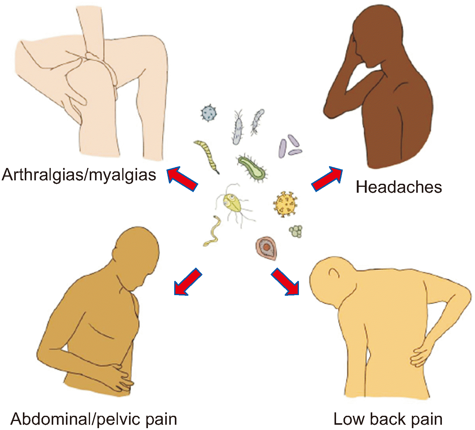
kjp-36-3-281-f1.tif
Fig. 2
Artistic rendition illustrating representative acute and chronic pain conditions that may result from infectious processes. Antibacterial, antiviral, and anti-parasitic agents have been found to confer analgesia in each of the following conditions via direct effects on pathogen load (e.g., spinal pain, dyspepsia) and/or by off-target effects (e.g., mitigating autoimmune and sensitization processes). Drawing by Seffrah Jin Cohen.
kjp-36-3-281-f2.tif
Table 1
Summary of the evidence for antimicrobial agents with possible off-target analgesic effects
Antimicrobial agent Mechanism(s) of action Preclinical evidence for antinociception Clinical evidence for a therapeutic effect
Cephalosporins [24,27] Upregulation of glial glutamate transporter-1, preventing glutamate neurotoxicity and potentially reducing pro-inflammatory cytokine concentrations. Evidence for neuroprotection in neurodegenerative diseases (e.g., amyotrophic lateral sclerosis) and the prevention or treatment of neuropathic pain. Ceftriaxone possibly mitigates post-surgical pain. Only specific cephalosporins, or specific generations of cephalosporins, might confer analgesic effects, but this requires confirmation.
Ribavirin [34,61] Possibly competitive inhibition of inosine monophosphate dehydrogenase, increasing the frequency of deleterious viral mutations. Partial reversal by naloxone suggests opioidergic effects. Evidence for antinociception in models of acute inflammatory pain. Effect partially reversed by naloxone and enhanced by propranolol, baclofen, ibuprofen, and others. Scant evidence for benefit compared to standard analgesics for viral-associated (e.g., chikungunya) joint pain.
Hydroxychloroquine, Chloroquine [40,50,61,65] Antirheumatic effects may result from interference with "antigen processing" in macrophages and other cells, and inhibition of autophagy. Evidence for complex regional pain syndrome and numerous inflammatory disorders and neoplastic diseases. No evidence for benefit compared to placebo or standard analgesics for viral-associated joint pain. Less efficacious than other disease-modifying agents for rheumatoid arthritis but may provide value as add-on therapy. Low-level evidence for other inflammatory diseases (e.g., lupus, dermatomyositis). Anecdotal evidence for complex regional pain syndrome.
Rapamycin [73,91] Inhibits mammalian target of rapamycin complex 1 (mTOR) and inhibits synaptic plasticity. Evidence for neuropathic pain, opioid-induced hyperalgesia, central sensitization, affective components of pain, inflammatory myopathies, mitochondrial disorders, and cancer-associated pain. Evidence for anti-tumor effects and cancer-associated pain. Anecdotal evidence in genetic heterotopic ossification and inflammatory myopathies.
Minocycline [99,104104,107,120,125,128] Inhibits central and peripheral glial cell activity, attenuates release of inflammatory cytokines, and binds to NR2B subunit of N-methyl-D-aspartate receptors. Evidence for neuropathic (e.g., painful diabetic neuropathy) and nociceptive (e.g., visceral) pain conditions and cancer-associated bone pain. May reduce affective components of pain, such as depression, anxiety, and fear. No benefit compared to placebo or tricyclic antidepressants for lumbar radicular pain. Potential benefit for peripheral neuropathic conditions in small prospective studies.
Dapsone [142,143] Inhibition of neutrophil activity and release of inflammatory cytokines. Evidence for inflammatory disorders. May cause neuropathy and hemolysis with prolonged use. Superior to placebo for rheumatoid arthritis, comparable to chloroquines. Anecdotal evidence for bullous systemic lupus erythematosus and several inflammatory dermatoses.
Piscidin-1 [173,174] Glial cell inhibition, suppression of cyclooxygenase-2 and inducible nitric oxide synthase. Evidence for neuropathic pain and tumor apoptosis. Clinical evidence is lacking.
TOOLS
Similar articles