

 PDF
PDF Citation
 Print
Print


INTRODUCTION
Knee pain and knee osteoarthritis are highly prevalent diseases [1–3]. Aging and obesity are related to the prevalence of knee osteoarthritis [1,3,4], and periarticular block of the knee joint is gaining importance for diseases in the knee joint, as it could be used as a modality to treat these patients.
Innervation of the knee joint is complicated yet has been a topic of interest to clinicians for decades [5–8], as precise identification of the peripheral nerves of the knee joint could be utilized for the nerve block itself and other pain interventions. The indications of periarticular nerve block have been widely applied in various settings, including in perioperative analgesia, chronic pain control, and emergency medicine [9–12].
The knee joint is innervated by the femoral, sciatic, and obturator nerves. These three nerves divide into smaller branches. The anterior aspect of the knee joint is often explained as being innervated by 11 to 15 periarticular branches [13,14]. These periarticular branches can be divided into four quadrants: the superolateral, superomedial, inferomedial, and inferolateral. In the superior lateral quadrant, branches include the nerve to the vastus intermedius, as well as the nerves to the vastus lateralis, superior lateral genicular nerve (SLGN), lateral retinacular nerve, lateral articular nerve, and common peroneal nerve (CPN). The superioromedial quadrant is innervated by the nerve to the intermedius, the nerves to the vastus medius, superior medial genicular nerve (SMGN), and medial retinacular nerve. The inferior medial quadrant is innervated by the anterior division of the obturator nerve, the infrapatellar branch of the saphenous nerve, and the inferior medial genicular nerve (IMGN). The inferior lateral quadrant is innervated by the inferior lateral genicular nerve (ILGN) and recurrent peroneal nerve (RPN) [14]. Of these complex innervations, the three genicular nerves, the SLGN, SMGN, and IMGN, are commonly targeted. The saphenous nerve is another commonly targeted nerve of the knee joint [15]. In addition, the sciatic, tibial, common peroneal, or femoral nerve block could be applied for controlling knee joint pain, although these can pose some risks for motor involvement [16,17]. The ILGN has been frequently omitted from these genicular blocks to avoid injury to the CPN and foot drop [18].
There are few anatomical studies identifying the route of the ILGN [5,6,19]. Of these few studies, a comprehensive cadaveric study by Tran et al. [6] states the relative relationship of the ILGN to the lateral collateral ligament and CPN. In that study, the ILGN was described as a long articular branch coursing deep to the lateral collateral ligament, turning anteriorly just inferior to the lateral femoral condyle and joining the inferior genicular vessels. Therefore, in this study, the aim was to investigate the route of the ILGN in relation to the CPN and explore the possibilities of techniques for blocking the ILGN.
Go to : 
MATERIALS AND METHODS
This trial was held at the Catholic Institute for Applied Anatomy, Seoul, Korea. This trial was approved by the institutional hospital review board of the College of Medicine, the Catholic University of Korea (IRB no. MC21EIDI0078). After a pilot trial with three cadavers, the main trial was conducted with another five fresh frozen cadavers. Cadavers with a history of knee surgery or trauma were excluded from the trial. The pilot trial was conducted to confirm the feasibility of the block technique and the adequacy of the injectate material.
Both knees of the cadavers were included in the trial. Angio-catheters were inserted into both sides of the common iliac artery. Fifty milliliters of red-colored latex solution was injected to distinguish the vascular structure during dissection. Cadavers were kept at 4°C for two weeks, and an ultrasound-guided block was conducted. A high frequency ultrasound device (HS50; Samsung Healthcare) was used over a linear range from 3–14 MHz, given the depth of the targeted area. After ten days of blocking, dissection was performed, and the presence and staining of the ILGN with dye were evaluated.
1. Ultrasound-guided block
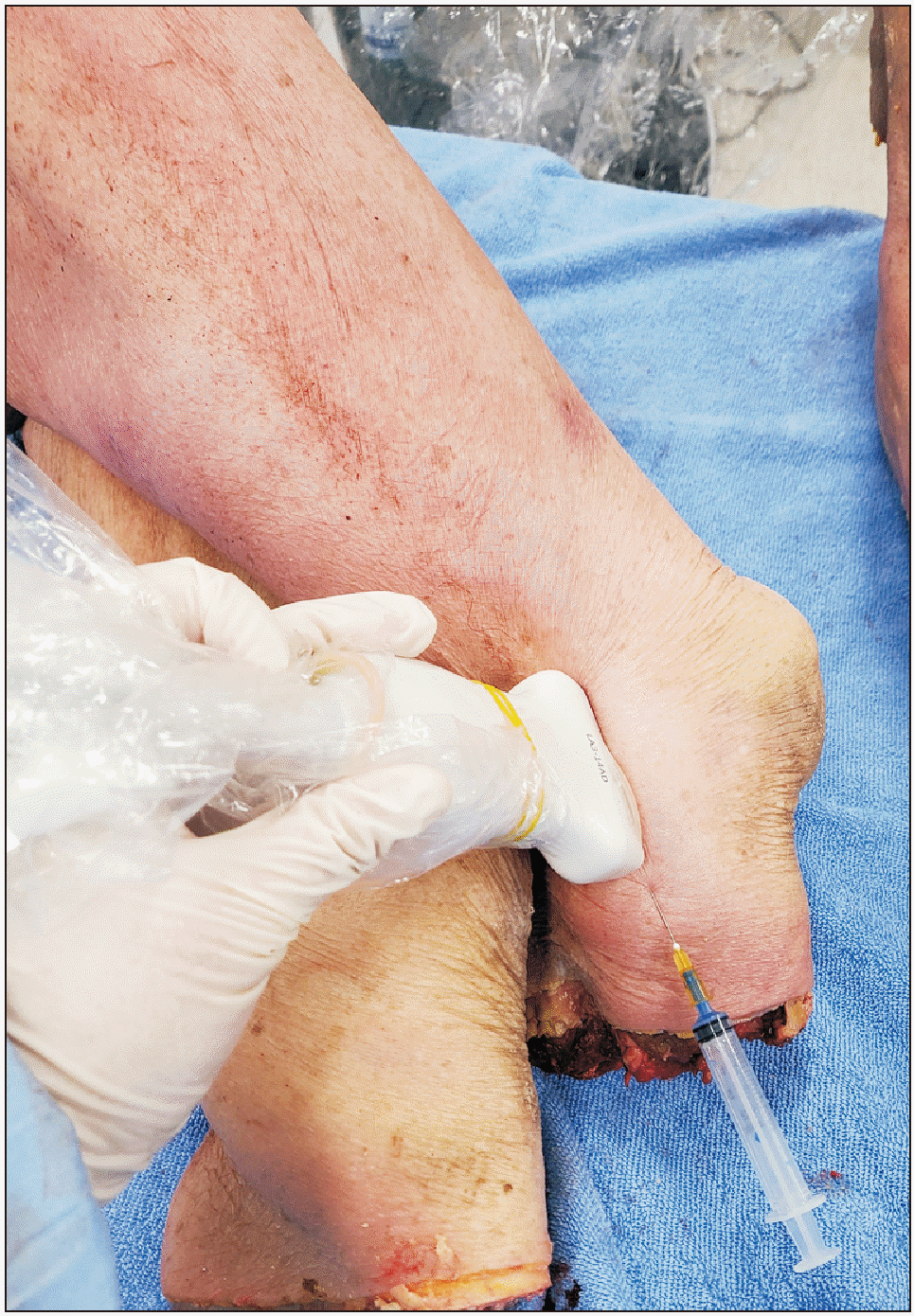
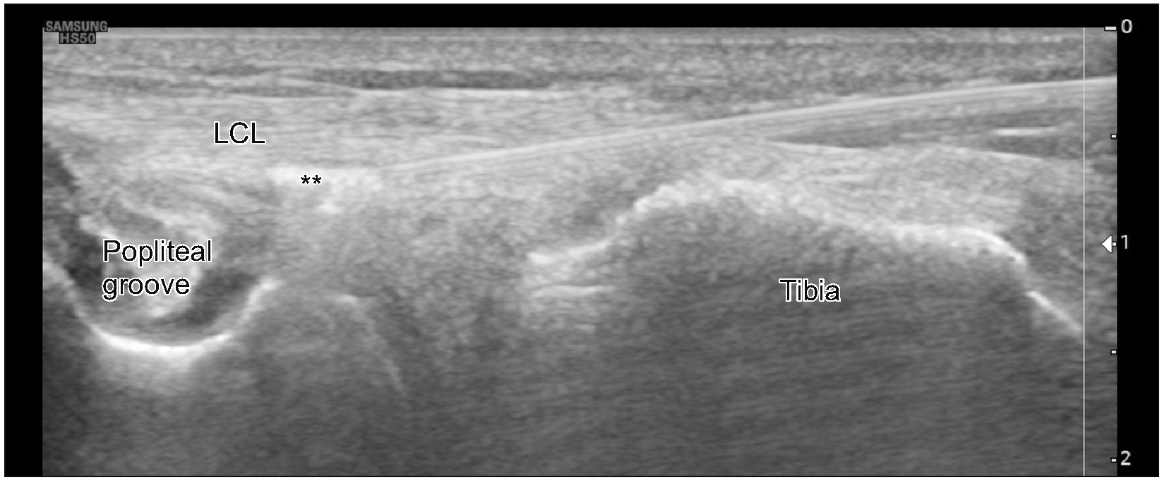
The specimen was kept in the figure-four position. The probe was placed in the longitudinal direction. The iliotibial band at the knee level was identified, then the distal tip of the ultrasound probe was rotated posteriorly to find the groove for the popliteus, and the proximal tip was moved posteriorly to locate the lateral collateral ligament of the knee. The probe was then moved distally to confirm the lateral collateral ligament’s insertion into the fibula head. To avoid bony contact with the fibular head, the probe was then moved in a cephalad direction. Then, the needle was inserted from the caudal to the cephalad direction (Figs. 1, 2). Two milliliters of solution was injected below the lateral collateral ligament with a 25-gauge, 5-cm needle (Softjec; Hwajin Medical). The injection material was prepared as a 140:40:20 volume ratio of distilled water to latex solution to colored water-soluble ink. The area of puncture was marked on the skin by a skewer.
2. Cadaveric anatomic dissection
The cadavers were placed in the lateral position, and then the skin around the injection site was widely dissected. After the tissue flap was raised and reflected, the CPN was identified coursing from the popliteal region. The iliotibial tract was pushed aside from its insertion, and branches of the CPN were carefully dissected near the biceps femoris muscle. If the articular branch emerged just above the femoral epicondyle coursing superiorly, this structure was regarded as the SLGN originating from the CPN [6]. Branches of the CPN coursing inferior to the lateral epicondyle were regarded as the ILGN. The whole dissection procedure was performed by an anatomist with more than 17 years of experience (S.H.K.).
3. Statistical analysis
For continuous demographic data such as the age, height, weight and body mass index (BMI) of the cadavers, mean and standard deviation were used for description. For categorical data such as the gender of the population or frequency of staining on ILGN and SLGN, the proportion was described with numbers and percentages. SAS Version 9.4 (SAS Institute) was used for calculation.
Go to :
RESULTS
Ten knees from five cadavers were inspected, and the ILGN was found in eight specimens (80.0%). A pilot study was performed with three cadavers, but the results from these cadavers were excluded from the main trial. The injected material for the pilot trial was colored yellow, and precise differentiation of the dye from the nerves and tissue was difficult; therefore, the authors decided to change the color of the dye from yellow to blue and proceeded to perform the main study.
Of the five cadavers enrolled, the mean age was 87.3 ± 7.9 years, and the BMI was 17.6 ± 3.6; 4 were male, and one was female (Table 1).
Of the eight knees with an ILGN, the branch was dyed in 5 specimens (62.5%) (Fig. 3) with the authors’ method. The SLGN was presented in two knees in this experiment (20.0%) (Fig. 4A) Of these two specimens, only one was stained with the authors’ method (50.0%) (Fig. 4B, Table 2).
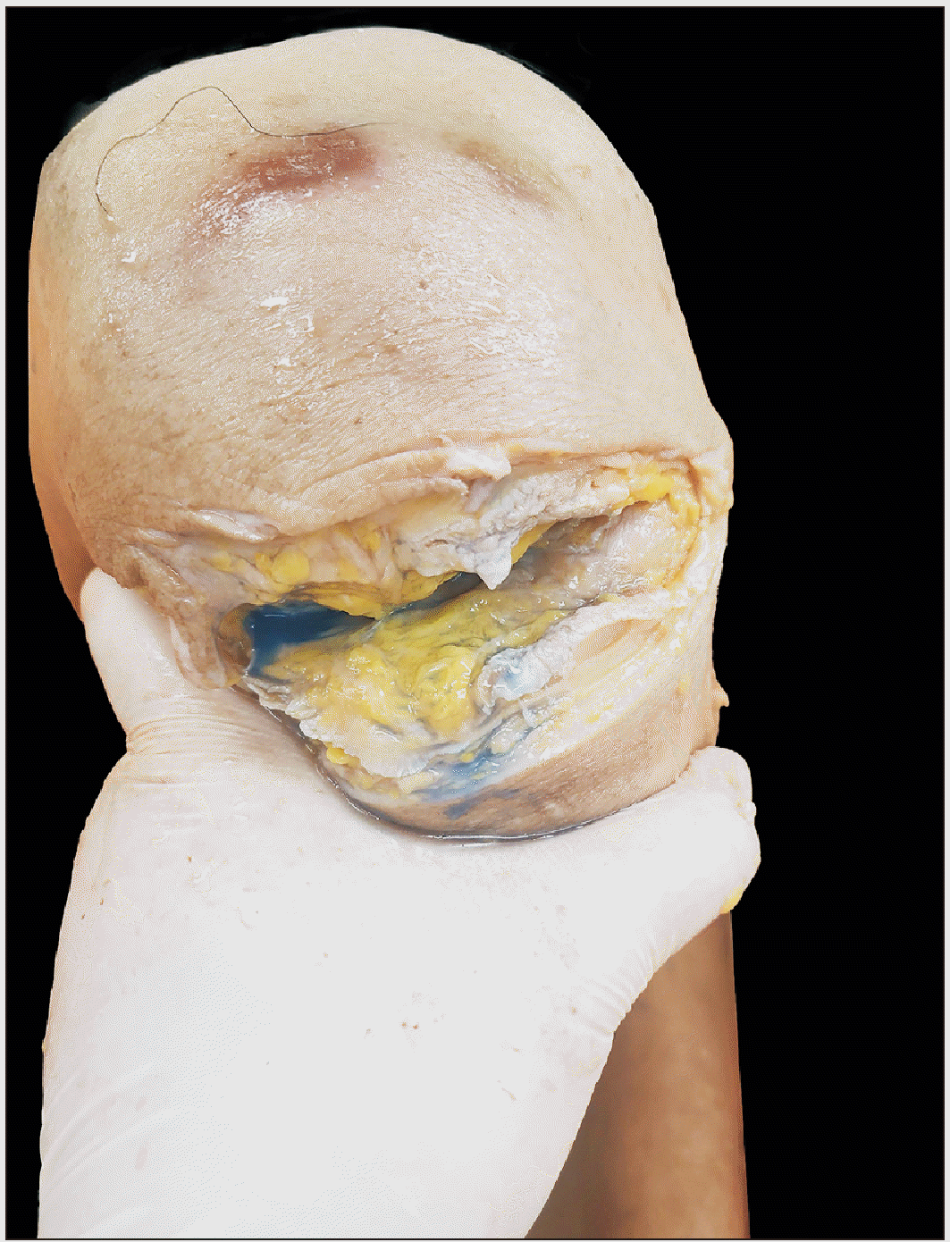
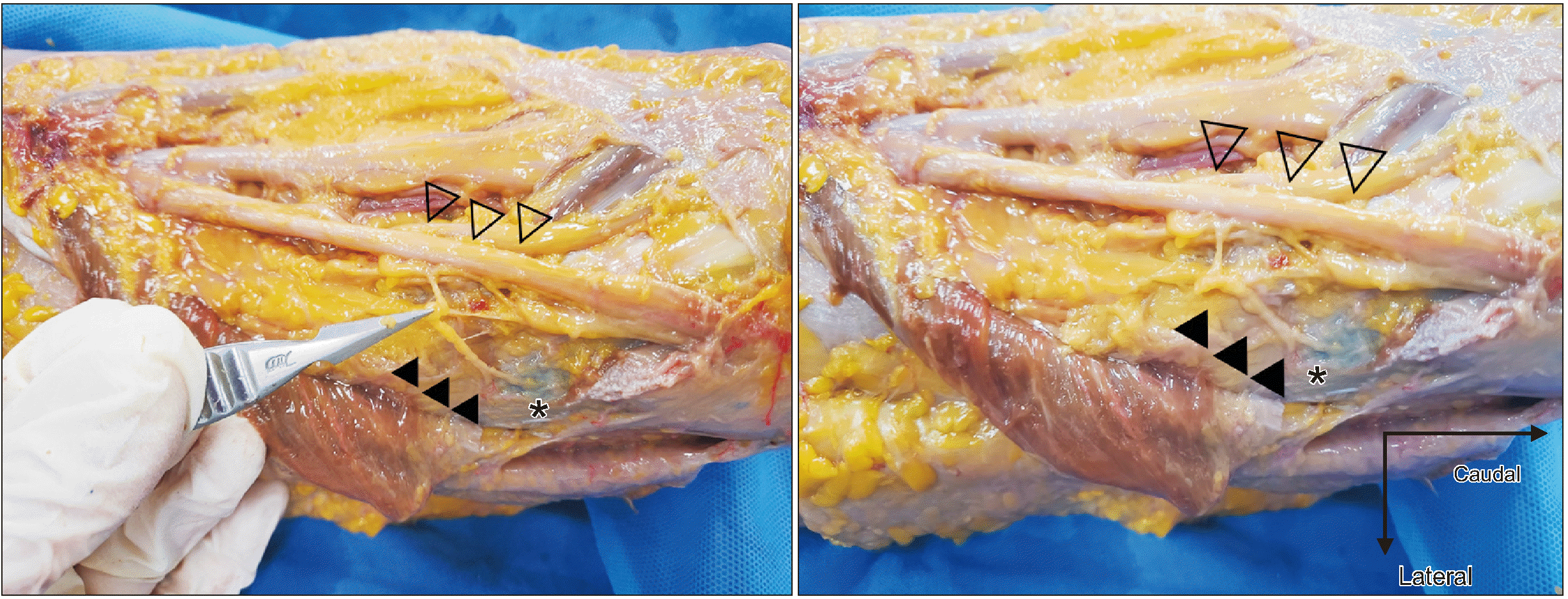 | Fig. 3Inferior lateral genicular nerve (ILGN) branching to the lower part of the lateral epicondyle of femur. Asterisks indicate the lateral epicondyle. The hollow arrowhead indicates the main branch of the common peroneal nerve, and the black arrowhead indicates the ILGN. Blue dye has reached the ILGN in this specimen.
|
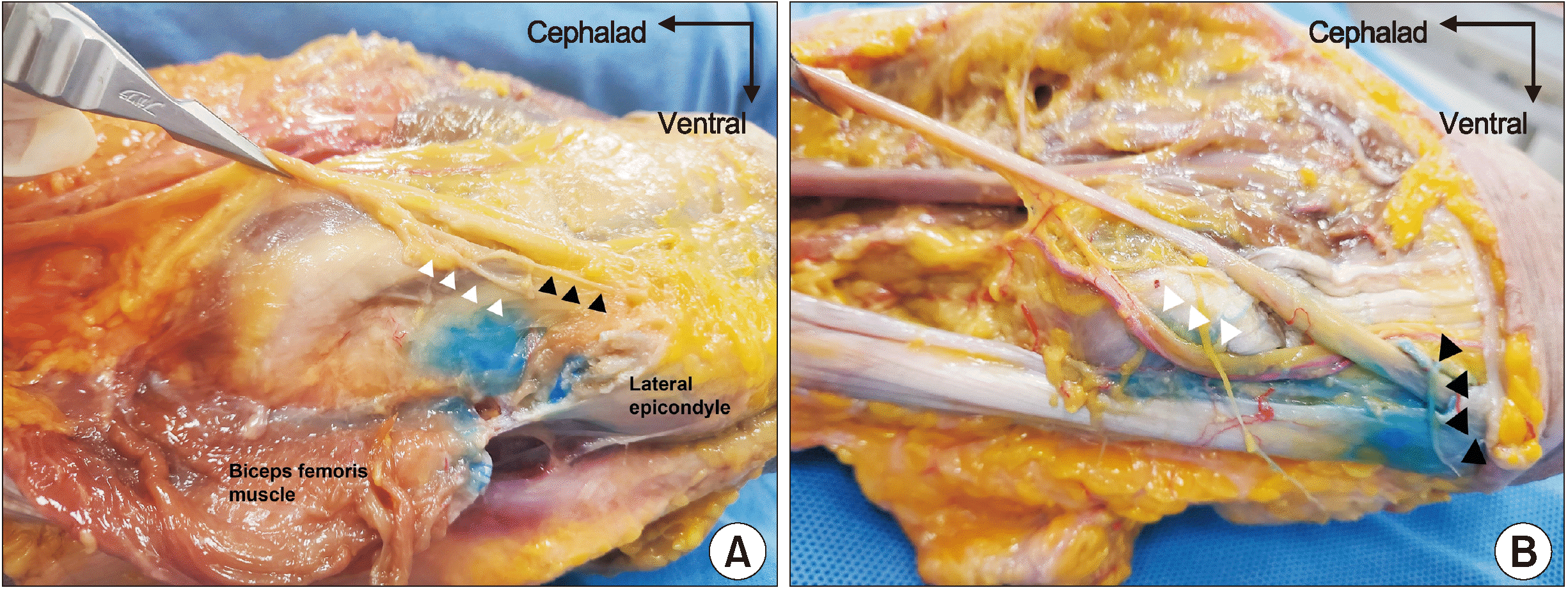 | Fig. 4Cadaveric dissection of inferior lateral genicular nerve (ILGN) and superior lateral genicular nerve (SLGN). (A) Presence of the SLGN and ILGN in the authors’ specimen. Blue dye is not reaching either nerve in this specimen. The white arrowhead indicates the SLGN, and the black arrowhead indicates the ILGN. (B) In this specimen, blue dye is infiltrating the ILGN while sparing the SLGN. Forceps is pinching the common peroneal nerve for better visualization. The white arrowhead indicates the SLGN, and the black arrowhead indicates the ILGN.
|
Intraarticular spread of dye material appeared in 1 of 10 specimens (Fig. 5), and the CPN was not dyed in any of the specimens.
Go to :
DISCUSSION
This cadaveric study confirmed the presence of the ILGN from the CPN and that it courses below the lateral collateral ligament. This research is meaningful in that it investigated the route of the ILGN, which had not yet been studied well, and the possibility of blocking it.
According to the anatomic study of Tran et al. [14], three knee articular branches originate from the CPN, the SLGN, ILGN, and recurrent fibular nerve (RFN). They reported that if the SLGN originates from the CPN, it does so just superior to the lateral condyle as a short branch that joins the superior lateral genicular vessels. The ILGN was reported to course in a more caudal direction, inferior to the lateral femoral condyle, and the RFN was reported to arise more inferiorly and course around the neck of the fibula.
The course of the ILGN differs among studies. A comprehensive study of knee joint innervation by Gardner [5] described that fine filaments of the CPN could be traced to the edge of the lateral tibial condyle. Kennedy et al. [20] used the term ‘lateral articular nerve’ to describe the fiber that emerges from the joint line superior to the fibular head and innervates the inferior knee capsule and collateral ligament. Terms describing the genicular nerve do not exist in this literature, but this might be describing the same structure the authors describe as the ‘ILGN’ [20]. Similarly, a cadaveric study by Horner and Dellon [21] described the ‘inferior lateral articular nerve’, which originates from the CPN coursing deep to the biceps femoris terminating near the fibular head under the iliotibial band. In this study, the SLGN is described to originate from the sciatic nerve.
Some authors regard the recurrent branch of the CPN as the inferolateral branch [22]; hence, the inferior lateral nerve should not be targeted for radiofrequency procedures because this could compromise the main branch of the CPN around the neck of the fibula [23]. However, according to the present findings, the ILGN follows a different route from the RFN, and any nerve blocks and pain-related interventions of the ILGN and RFN should be approached differently.
One mini-review reported ultrasound-guided visualization of the ILGN under the lateral collateral ligament. The authors found that the ILGN coursed with the artery and could be traced with doppler ultrasound [24]. However, in the present study, the pulsating artery could not be visualized on fresh frozen cadavers, and more importantly, vascular structures were often not accompanied by the ILGN in this study. Therefore, the authors would not consider the inferior lateral genicular artery as a landmark for the ILGN.
In summary, two main articular branches of the knee originating from the CPN have been described as the ‘ILGN’, which is often named the ‘inferior lateral articular nerve’ or the ‘RFN’. One exception was the ‘lateral retinacular nerve’ described in a single cadaveric study [25], but the authors could not find other studies regarding this structure arising from the CPN. Additionally, two other anatomical studies defined this structure as originating from the sciatic nerve or its branches [8,21].
In the present study, the authors wanted to verify the rate of occurrence of the ILGN and identify a feasible method of blocking this structure. The volume used for the experiment was decided upon in previous clinical studies on genicular nerve block [11,12,26,27]. Based on these studies, the authors used a 2 mL volume for the block of the genicular nerve. The occurrence of the ILGN was verified in the majority (80.0%) of specimens, while a successful nerve block was achieved in only 62.5%. The authors attributed the failure of the block to two reasons. The space under the lateral collateral ligament is densely occupied by ligaments such as the lateral collateral ligament, as well as tendons and bony structures. The water-soluble dye was restricted in terms of its spread and injection, and it could not sufficiently stain the thin terminal ILGN branch. Intraarticular staining occurred in one specimen, but the ILGN was not despite its presence. If the needle accidentally pierces the synovial membrane, the dye might accidentally flow toward the synovial space.
The SLGN is often reported as branches of the sciatic nerve [7,21], and a minor variant is described as originating from the CPN [6]. In the present study, the SLGN occurred in only 20% of the specimens. Therefore, from the present findings, the SLGN often originates from the sciatic nerve and rarely from the CPN.
A limitation of this study is the relatively small number of specimens. Additionally, this is a cadaveric study, and the distribution of dye material could differ from that in the living human body. Due to technical difficulties, we did not investigate variants of the ILGN branching directly from the sciatic nerve as in some other studies. A longitudinal branch descending toward the femorotibial space was reported [7]. In conclusion, the ILGN is a branch of the CPN that courses under the lateral collateral ligament of the knee and was found in 80.0% of specimens in this study. Additionally, the success rate of block with a 2 mL injection was 62.5%, and there was no suspicion that motor weakness would occur. However, the success rate was rather low. If authors used larger volume for the block, the success rate could be higher. Further comparative study could be needed to validate the effect and side effects of this block. Further clinical studies on successful methods and doses for blocking the ILGN are needed.
Go to :
 XML Download