Journal List > Korean J Pain > v.36(3) > 1516083089

Lee, Yang, Won, and Kim: Coccydynia: anatomic origin and considerations regarding the effectiveness of injections for pain management

Abstract

Coccydynia is a debilitating pain disorder. However, its pathophysiology is not well understood. When approaching coccydynia, the exact underlying cause of pain must be identified to develop an appropriate treatment plan. The specific approach to coccydynia can vary depending on an individual's condition and the underlying cause. Thorough evaluation by a pain physician is essential to determine the most appropriate course of treatment. The purpose of this review is to examine the various causes contributing to coccygeal pain and specifically focus on the exact anatomical neurostructures, such as the anococcygeal nerve, perforating cutaneous nerve, and ganglion impar. We also reviewed the relevant clinical outcomes and suggested recommendations for each anatomical structure.

INTRODUCTION

Chronic pain is a complex phenomenon, and a combination of definitions are used to define it [1]. Pain arising from various spinal structures constitutes the majority of chronic pain disorders. The lifetime prevalence of spinal pain is reportedly more than 50% [2]. Lumbosacral pain is one of the most common reasons for visiting health care providers. It is a chronic pain condition that has the highest prevalence not only in Korea but also globally [3,4]. The importance of pain medicine for spinal pain has been refined and continues to evolve. With advancements, there has been a continuous increase in the number of structures beyond the spine.
Coccydynia, also known as coccygodynia, is a debilitating pain in the coccyx but its pathophysiology, including accurate prevalence, is still not well understood. When approaching coccydynia, the exact cause of the pain needs to be identified based on a deeper understanding of the accurate anatomy of the coccygeal region to develop an appropriate treatment plan. It can arise from various sources including musculoskeletal issues, nerve impingement, inflammation, and referred pain. It is important to note that the specific approach to coccydynia can vary depending on the individual's condition and underlying cause. Thorough evaluation by a pain physician is essential to determine the most appropriate course of treatment.
In this article, we aim to discuss the various causes that contribute to coccyx pain and specifically focus on the anatomical neurostructures. We conducted a comprehensive review of different injections for coccydynia patients and verified their supporting evidence. Moreover, we endeavored to offer recommendations and suggestions for their practical application in a clinical setting.

MAIN BODY

1. Basic anatomies

1) Nerves

(1) The perforating cutaneous nerve

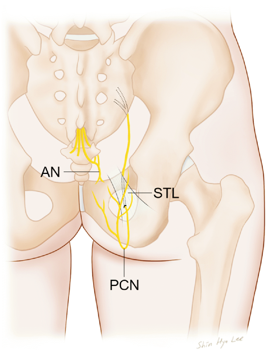
The perforating cutaneous nerve usually originates from the posterior aspects of the second and third sacral ventral spinal rami. It pierces the sacrotuberous ligament, curves around the inferior border of the gluteus maximus, and supplies the skin over the inferomedial aspect of this muscle (Fig. 1). The nerve may arise from the pudendal nerve or, if absent, may be replaced by a branch from the posterior femoral cutaneous nerve or from the third and fourth or fourth and fifth sacral spinal ventral rami [5,6].

(2) Anococcygeal nerve

The coccygeal nerve is not usually well described in anatomical textbooks. In human development, it appears in up to five pairs [7], and typically regresses, except for the first pair, although a second coccygeal nerve may occasionally persist [8]. This nerve forms the coccygeal plexus as follows. The fifth sacral ventral ramus emerges from the sacral hiatus, curves around the lateral margin of the sacrum below its cornu, and pierces the coccygeus muscle to reach its pelvic surface, where it is joined by a descending branch of the fourth sacral ventral ramus. The small trunk descends on the pelvic surface of the coccygeus muscle to join the minute coccygeal ventral ramus emerging from the sacral hiatus and curves around the lateral coccygeal margin to pierce the coccygeus and reach the pelvis. The small trunk is the coccygeal plexus. Anococcygeal nerves arise from the coccygeal plexus (Fig. 2), which pierces the sacrotuberous ligament to form fine filaments to supply the adjacent skin [5,6].

(3) Ganglion impar

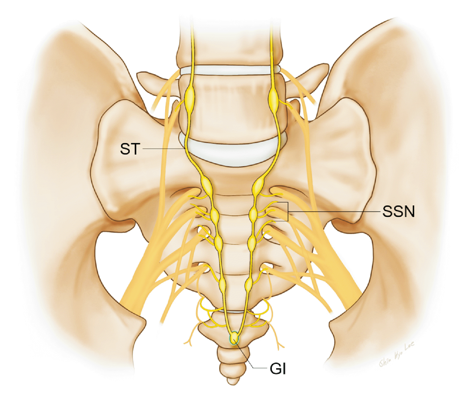
The pelvic sympathetic trunk lies in the extraperitoneal tissue anterior to the sacrum, and medial or anterior to the anterior sacral foramina and has four or five interconnected ganglia. Above, it continues into the lumbar sympathetic trunk, and below, the two trunks converge to unite in the small ganglion anterior to the coccyx (Fig. 3). This small ganglion is called the ganglion impar [5,6], which has diverse locations and sizes between the sacrococcygeal joint and coccygeal tip [9]. The gray rami communicantes pass from the ganglia to the sacral and coccygeal spinal nerves; however, the white rami communicantes are absent. The medial branches of distribution connect across the midline; twigs from the first two ganglia join the inferior hypogastric plexus or hypogastric nerve; others form a plexus on the median sacral artery [6]. The ganglion impar plays a significant role in the development of pelvic and coccyx pain because it transmits sympathetic efferent signals to nociceptive afferent signals from various areas, including the perineum, distal rectum, distal vagina, distal urethra, and anus. Involvement of the ganglion impar highlights its importance in the generation and perception of pain in these regions [10].

2) Bone and joints

The coccyx is the final segment of the spine and is located at the end of the tail. The term "coccyx" originates from a Greek word that refers to the beak of a cuckoo bird, as it resembles the shape of the coccyx when observed from a side view [11]. The coccyx is a triangular bone formed by the fusion of 3–5 segments. The largest segment of the coccyx is articulated with the lowest segment of the sacrum.

(1) Sacrococygeal joint

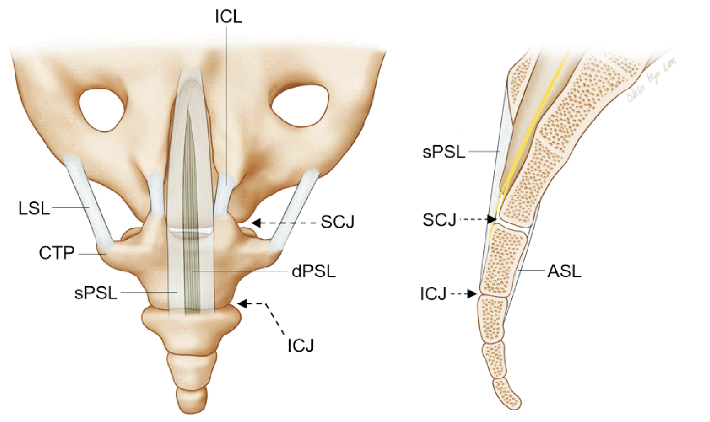
The sacrococygeal joint is a symphysis between the sacral apex and coccygeal base, united by a fibrocartilaginous disc, remnants of hyaline cartilage, and the anterior, posterior, and lateral ligaments (Fig. 4). The anterior sacrococcygeal ligament comprises irregular fibers descending on the pelvic surfaces of both the sacrum and coccyx, which are attached to the anterior longitudinal ligament. The superficial posterior sacrococcygeal ligament is flat and passes from the sacral hiatus margin to the dorsal coccygeal surface, roofing the lower sacral canal. The deep posterior sacrococcygeal ligament extends from the back of the fifth sacral vertebral body to the dorsum of the coccyx. This ligament corresponds to the posterior longitudinal ligament. The lateral sacrococcygeal ligament is on each side, similar to the intertransverse ligament. This ligament connects the coccygeal transverse process to the inferolateral sacral angle, completing the foramen for the fifth sacral spinal nerve. The intercornual ligament connects the sacral and coccygeal cornus on each side [12].

(2) Intercoccygeal joints

These are symphyses with thin discs of fibrocartilage between the coccygeal segments in young individuals. Segments are also connected by the extension of the anterior and posterior sacrococcygeal ligaments (Fig. 4). All segments unite relatively early in adult males but later on in females. Occasionally, the joint between the first and second segments is synovial, and the apex of the terminal segment is connected to the overlying skin by white fibrous tissue [12]. A rudimentary intervertebral disc may be present between the first and second segments [13]. In a study examining the gross anatomy of the intercoccygeal joints, Maigne et al. [13] observed significant variation in the structure of these joints. The joints ranged from intact discs that resembled lumbar intervertebral discs to intermediate structures with cystic or fibrotic changes and synovial joints. In certain instances, the intercoccygeal joints have been found to be fused together [13]. Certain types of coccygeal morphologies have been reported to be associated with an increased susceptibility to coccydynia.

2. Etiology and clinical features

The term "coccydynia" was introduced by Simpson in 1859 to describe pain originating from the coccyx [14]. Furthermore, Foye [15] has referred to coccyx pain as the lowest site of lower back pain (LBP). Coccydynia, which lacks a reported overall incidence, is commonly observed anecdotally. According to the limited available reports, the coccyx has been identified as a potential source of pain in approximately 1%–2.7% of individuals seeking medical attention for back pain among all non-traumatic complaints related to the spine [16,17]. Coccydynia often arises from direct vertical trauma, repetitive microtrauma, or childbirth. However, it is crucial to exclude several underlying causes, including infections such as soft tissue abscesses or osteomyelitis, as well as malignancies such as chordoma, which have a high fatality rate [18]. Direct vertical trauma to the coccyx can yield various outcomes, ranging from contusion to fracture-dislocation. When the coccygeal ligaments are compromised, either through trauma or non-traumatic factors, dynamic coccygeal instability can occur. This instability is characterized by excessive movement of the coccyx during weight bearing or sitting, resulting in coccygeal pain. Abnormal coccyx mobility can manifest as hypermobility with lax ligaments or as hypomobility, indicating rigidity. In addition, the coccyx can experience subluxation, instability, or even complete dislocation [19].
Specific coccyx shapes have a high propensity for coccydynia. Abnormalities in the coccygeal morphology or position, including lateral deviation due to a scoliotic deformity, can increase the likelihood of coccygeal pain. Furthermore, excessive flexion or extension of the coccyx can make individuals more susceptible to pain [20]. A mobile sacrococcygeal joint, a more ventrally curved coccyx, and the presence of posterior spicule formation have also been associated with the development of coccydynia as anatomical risk factors. In men, intercoccygeal joint subluxation has also been reported as a risk factor for experiencing coccydynia [21]. Discomfort can arise from a distal coccyx bone spur when pressure is applied to the skin beneath the spur while sitting. Additionally, a distal coccyx bone spur may cause pain when the skin is pinched beneath the spur while sitting [22].
Coccydynia is associated with several risk factors, including female sex and obesity. Body mass index plays a role as it can influence sitting posture and the amount of pressure exerted on the coccyx. Coccydynia is more prevalent in females than in males, with a five-fold increased risk. Rapid weight loss has also been identified as a risk factor owing to the diminished cushioning effect of adipose tissue in the buttock region. Other reported risk factors include osteoarthritis, osteomyelitis, and participation in contact sports [23,24].
The general symptoms of coccydynia without any condition are tenderness and pain, which may manifest as a localized ache in the lower sacral area, coccyx, or adjacent muscles and soft tissues. Traumatic coccydynia is characterized by a history of trauma preceding the onset of pain, usually with an acute presentation. Conversely, idiopathic coccydynia tends to have a more gradual and insidious onset without any identifiable precipitating factors. In cases of coccydynia caused by other factors, a meticulous and comprehensive patient history often offers clues regarding potential underlying causes [24]. Patients usually point to the coccyx as the site of pain. Severity is dependent on various predisposing factors, including the duration of time spent sitting, especially when in a partially reclined or backward-leaning position. Additionally, standing up from a seated position may induce a temporary but intense escalation of coccygeal pain. Other factors that can exacerbate this condition include prolonged standing, sexual intercourse, and defecation. Women have reported an exponential increase in pain during the premenstrual period. Dyspareunia and piriformis syndrome are infrequently associated with coccydynia [25,26].
Coccydynia is predominantly diagnosed through a thorough clinical examination supported by static standard radiographs, computed tomography (CT) scans, and routine blood tests. These diagnostic measures are primarily aimed at excluding any notable abnormalities, except in cases involving tumors or infections that may exhibit specific abnormalities [27]. Actually, the evaluation process of symptoms is even more crucial. The established referred pain pattern originating from the lumbar region is often confused with pain originating from the coccyx, making it seemingly insignificant but crucial in clinical settings [28]. The correlation between pre-existing back pain or coccydynia and lumbar disc pathology has been extensively explored, offering comprehensive information on this clinical presentation. Various studies have reported a range of 15% to 31% in the prevalence of concurrent or pre-existing low back pain among patients [20,29]. Some studies have also found higher frequencies. Perkins et al. [30] noted that 77% of patients had accompanying lumbar spinal disorders. Similarly, Balain et al. [31] documented an incidence of 71% for concomitant lumbar spinal disorders. The prevalence of such conditions highlights the potential for coccydynia to be misinterpreted as spinal pain in clinical settings.

3. Management of pain using interventional techniques

Various treatment approaches have been suggested for managing coccydynia. However, there is a lack of extensive clinical studies that have been analyzed to thoroughly assess the efficacy of these different treatments. Conservative measures remain the cornerstone of coccydynia treatment and are successful in most patients [32]. Non-steroidal anti-inflammatory drugs, sitting aids, manual manipulation, and local anesthetics with steroid injections are widely recognized effective treatment methods [23]. Surgical interventions, including coccygectomy, are considered the final option and are pursued only when all other available treatment options have proven ineffective. Coccygectomy involves the surgical removal of the coccyx, typically at the proximal end near the sacrococcygeal junction [30]. This review discusses interventional techniques, focusing on injection therapies associated with neuroanatomical structures. Injection therapies are utilized as both diagnostic tools and therapeutic interventions despite the relative scarcity of supporting literature, particularly for chronic coccydynia. Although there is no definitive consensus on the most appropriate injection site for coccydynia, imaging guidance is generally recommended when administering injections in the vicinity of the coccyx [26]. Fluoroscopy and ultrasound are the preferred imaging techniques used by most practitioners to accurately guide injections. In some cases, CT is recommended [33]. Care must be taken when using sterile techniques to avoid infection, and universal precautions should be taken to avoid risks to the operator.

1) Caudal epidural steroid injection

Caudal epidural steroid injections are frequently used to treat lower sacral radicular pain, including coccydynia [34]. Despite its frequent utilization in clinical practice and suggestions as a potential approach for patients unresponsive to conservative treatment [35], the availability of high-quality studies on coccydynia is limited. Sencan et al. [36] compared the treatment outcomes of ganglion impar block (GIB) and caudal epidural steroid injection in patients with chronic coccydynia unresponsive to conservative treatment. GIB may provide a more significant pain reduction in the short term (3 months). However, the long-term efficacy of the treatment in both groups remains unclear, and it is uncertain whether repeated injections are necessary. Another important point regarding caudal epidural steroid injection is the lack of consensus regarding the ideal volume for coccydynia.

2) Anococcygeal nerve block

There are only a few available literature reports on techniques for anococcygeal nerve block. Yamada et al. [37] described neurolysis techniques in seven patients with intractable perineal pain following surgery for rectal cancer. The final position of the tip of the needle was near the frontal face of the coccygeal bone in the lateral view under fluoroscopic guidance. Although the results were favorable, they were limited to a small number of patients with cancer-related pain. Alimehmeti et al. [38] also reported cases of coccydynia related to anococcygeal nerve blocks. The cases dealt with in that study were not for cancer pain, but it was reported that most patients (11 of 13) had a history of trauma or surgery; among them, nine underwent surgery. The specific technique or dosage of the nerve block was not mentioned in this report. No further literature regarding the use of this nerve block technique for the management of both cancer-related and non-cancer-related pain could be found, which could make it a challenge to definitively classify the anococcygeal nerve block as a precise block technique. Thus, the precise technique and medical evidence are still considered to be lacking for chronic coccydynia.

3) GIB

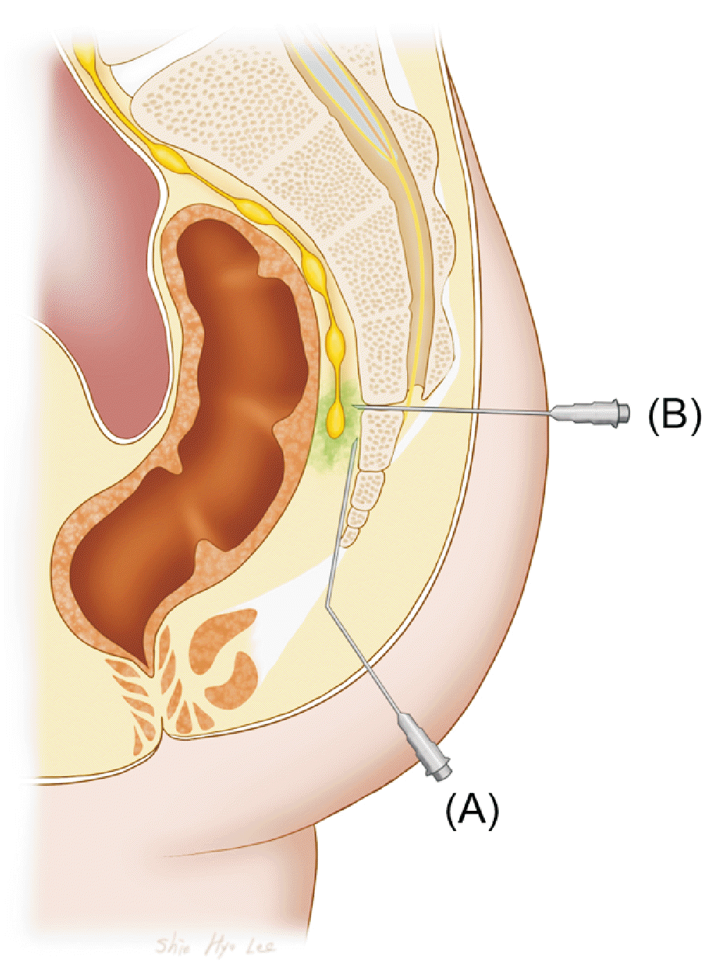
GIB has a relatively higher reported clinical efficacy than other techniques. GIB has proven to be valuable in both evaluating and managing pain originating from sympathetic involvement or maintenance of the perineum and coccyx [39]. In recent years, this technique has been used in the treatment of pain secondary to malignancy as well as in the management of chronic nonmalignant pain. Utilizing a local anesthetic, GIB has been utilized as a diagnostic tool for assessing pelvic and rectal pain, primarily before neurolytic blockade. This enables the execution of a differential neural blockade by employing anatomical principles. It also serves as a therapeutic intervention for pain relief in these areas [40]. Most studies describe the use of GIB in the management of malignant pain [41]. Multiple reports have detailed the application of GIB in various conditions including interstitial cystitis and radiation enteritis. According to the evaluation conducted by Day, evidence supporting the use of GIB was graded as 1C, indicating low-quality evidence. Despite the limited evidence, a strong recommendation was made based on specific indications, including vulvar cancer pain, chronic perineal pain, and sacral postherpetic neuralgia [42]. It is important to note that pain associated with certain conditions, such as postherpetic neuralgia, can have a sympathetically mediated component even if the somatic nerves are primarily affected [34,40,43]. It can be performed with the assistance of imaging guidance by utilizing various techniques for the needle approach [44] (Fig. 5).

4) Sacrococcygeal joint injection

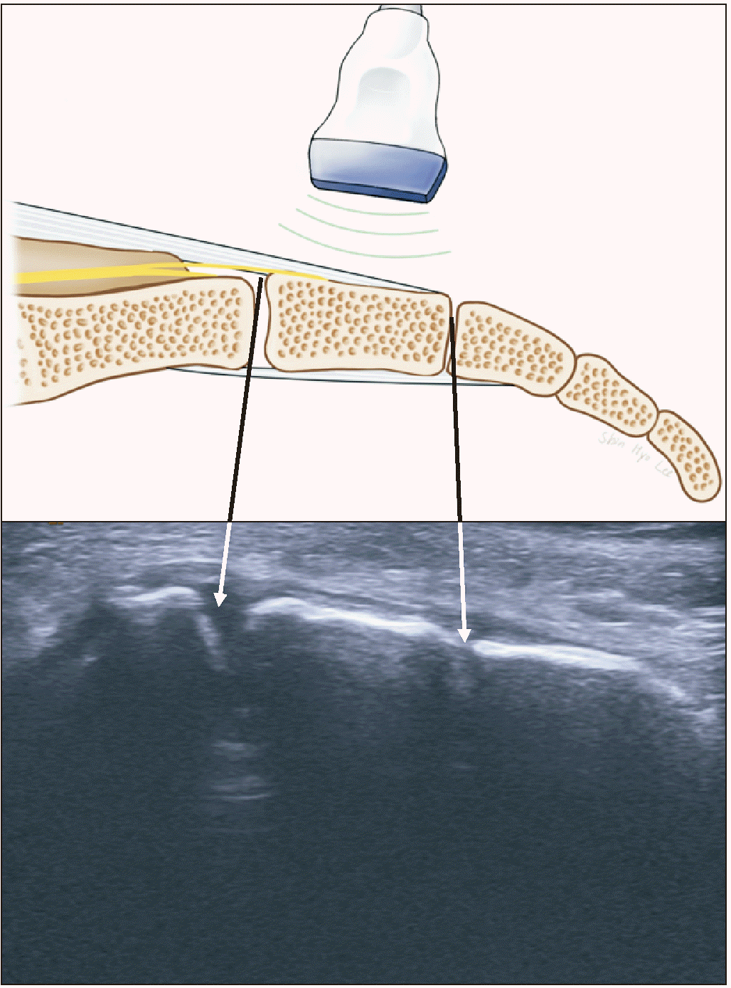
Most of the reported literature appears to be associated with injections administered in the sacrococcygeal and intercoccygeal joints. However, treatment involving the infiltration of local anesthetics and steroids around the coccyx is presumed to be administered into the coccygeal soft tissue or in the surrounding area, rather than intra-articularly. According to the findings of Mitra et al. [45], patients were more likely to experience significant pain relief from steroid injections when the pain persisted for less than 6 months. Wray et al. [46] reported a success rate of 60% in a prospective study on the treatment of coccygodynia using ultrasound-guided injections as in Fig. 6. This study did not provide explicit information on the methodology used to identify infiltration sites. It is considered that the patients in this study received blind coccygeal soft tissue injections but not joint injections.

CONCLUSIONS

The coccyx is commonly considered as a vestigial structure, leading to a relative lack of comprehensive knowledge regarding its anatomy. Paradoxically, it is associated with pain and other disorders, highlighting the significant gaps in our understanding. These gaps include the detailed anatomy of the coccygeal plexus and its distribution as nerves, function of the coccygeal body, and anatomy and potential significance of the sacrococcygeal zygapophyseal joints in relation to coccydynia. LBP is the most common form of spinal pain in terms of chronicity and severity, and the lumbar facet joints have been implicated in chronic LBP in up to 45% of patients. In addition to degenerative changes in the sacrococcygeal intervertebral and intercoccygeal joints of the coccyx, it is crucial to consider these supplementary factors in addition to the participation of the sacrococcygeal zygapophyseal joint, which is directly adjacent to the lumbar facet joint.
Coccydynia, which is frequently a self-limiting, mild condition, sometimes necessitates more aggressive treatments for certain patients. In such cases, the underlying cause of coccydynia may be complex or multifactorial. Injection procedures have been known to be effective in relieving pain and treating specific medical conditions. For this reason, although there is a lack of strong evidence-based literature supporting the effectiveness of interventional procedures, many pain physicians recommend injection procedures for patients experiencing chronic or unresponsive coccydynia. However, there is no unanimous agreement on the most optimal injection site for these procedures yet. Thus, it is essential to make a knowledgeable choice regarding the decision to perform a nerve block by consulting with a qualified healthcare professional. Especially, patient-specific factors, including individual circumstances, risks, and potential benefits, should be carefully evaluated.
Further investigation of this structure will contribute to a comprehensive understanding of its involvement in modulating sensory and autonomic pathways in the sacrococcygeal region. This knowledge may pave the way for the development of novel and more efficient therapeutic interventions for patients with coccydynia.

Notes

DATA AVAILABILITY

Data sharing is not applicable to this article as no datasets were generated or analyzed in this study.

CONFLICT OF INTEREST

No potential conflict of interest relevant to this article was reported.

AUTHOR CONTRIBUTIONS

Shin-Hyo Lee: Writing/manuscript preparation; Miyoung Yang: Writing/manuscript preparation; Hyung-Sun Won: Writing/manuscript preparation; Yeon-Dong Kim: Writing/manuscript preparation.

REFERENCES

1. Breivik H, Collett B, Ventafridda V, Cohen R, Gallacher D. 2006; Survey of chronic pain in Europe: prevalence, impact on daily life, and treatment. Eur J Pain. 10:287–333. DOI: 10.1016/j.ejpain.2005.06.009. PMID: 16095934.
crossref
2. Manchikanti L, Singh V, Datta S, Cohen SP, Hirsch JA. American Society of Interventional Pain Physicians. 2009; Comprehensive review of epidemiology, scope, and impact of spinal pain. Pain Physician. 12:E35–70. DOI: 10.36076/ppj.2009/12/E35. PMID: 19668291.
3. Carey TS, Garrett J, Jackman A, McLaughlin C, Fryer J, Smucker DR. 1995; The outcomes and costs of care for acute low back pain among patients seen by primary care practitioners, chiropractors, and orthopedic surgeons. The North Carolina Back Pain Project. N Engl J Med. 333:913–7. DOI: 10.1056/NEJM199510053331406. PMID: 7666878.
crossref
4. Kim YD, Moon HS. 2015; Review of medical dispute cases in the pain management in Korea: a medical malpractice liability insurance database study. Korean J Pain. 28:254–64. Erratum in: Korean J Pain 2016; 29: 62. DOI: 10.3344/kjp.2015.28.4.254. PMID: 26495080. PMCID: PMC4610939.
crossref
5. Romanes GJ. 1981. Cunningham's textbook of anatomy. 12th ed. Oxford University Press;p. 797–806.
6. Berry M, Standring SM, Bannister LH. Williams PL, editor. 1995. Nervous system. Gray's anatomy. 38th ed. Churchill Livingstone;p. 901–1397. DOI: 10.37019/e-anatomy/49576.
7. O'Rahilly R, Müller F, Meyer DB. 1990; The human vertebral column at the end of the embryonic period proper. 4. The sacrococcygeal region. J Anat. 168:95–111. PMID: 2182589. PMCID: PMC1256893.
8. Pearson AA, Sauter RW, Buckley TF. 1966; Further observations on the cutaneous branches of the dorsal primary rami of the spinal nerves. Am J Anat. 118:891–903. DOI: 10.1002/aja.1001180313. PMID: 4163167.
crossref
9. Oh CS, Chung IH, Ji HJ, Yoon DM. 2004; Clinical implications of topographic anatomy on the ganglion impar. Anesthesiology. 101:249–50. DOI: 10.1097/00000542-200407000-00039. PMID: 15220800.
crossref
10. Woon JT, Stringer MD. 2012; Clinical anatomy of the coccyx: a systematic review. Clin Anat. 25:158–67. DOI: 10.1002/ca.21216. PMID: 21739475.
crossref
11. Sugar O. 1995; Coccyx. The bone named for a bird. Spine (Phila Pa 1976). 20:379–83. DOI: 10.1097/00007632-199502000-00024. PMID: 7732478.
12. Soames RW. Williams PL, editor. 1995. Skeletal system. Gray's anatomy. 38th ed. Churchill Livingstone;p. 425–711. DOI: 10.1308/rcsann.2006.88.4.425a.
13. Maigne JY, Guedj S, Straus C. 1994; Idiopathic coccygodynia. Lateral roentgenograms in the sitting position and coccygeal discography. Spine (Phila Pa 1976). 19:930–4. DOI: 10.1097/00007632-199404150-00011. PMID: 8009351.
crossref
14. Simpson JY. 1859; Clinical lectures on the diseases of women. Lecture XVII: on coccydynia and diseases and deformities of the coccyx. Med Times Gaz. 40:1–7.
15. Foye PM. 2010; Stigma against patients with coccyx pain. Pain Med. 11:1872. DOI: 10.1111/j.1526-4637.2010.00999.x. PMID: 21134124.
crossref
16. Ghormley RK. 1958; An etiologic study of back pain. Radiology. 70:649–53. DOI: 10.1148/70.5.649. PMID: 13554831.
crossref
17. Pennekamp PH, Kraft CN, Stütz A, Wallny T, Schmitt O, Diedrich O. 2005; Coccygectomy for coccygodynia: does pathogenesis matter? J Trauma. 59:1414–9. DOI: 10.1097/01.ta.0000195878.50928.3c. PMID: 16394915.
crossref
18. Foye PM. 2017; Coccydynia: tailbone pain. Phys Med Rehabil Clin N Am. 28:539–49. DOI: 10.1016/j.pmr.2017.03.006. PMID: 28676363.
19. Przybylski P, Pankowicz M, Boćkowska A, Czekajska-Chehab E, Staśkiewicz G, Korzec M, et al. 2013; Evaluation of coccygeal bone variability, intercoccygeal and lumbo-sacral angles in asymptomatic patients in multislice computed tomography. Anat Sci Int. 88:204–11. DOI: 10.1007/s12565-013-0181-2. PMID: 23700101.
crossref
20. Postacchini F, Massobrio M. 1983; Idiopathic coccygodynia. Analysis of fifty-one operative cases and a radiographic study of the normal coccyx. J Bone Joint Surg Am. 65:1116–24. DOI: 10.2106/00004623-198365080-00011. PMID: 6226668.
crossref
21. Woon JT, Maigne JY, Perumal V, Stringer MD. 2013; Magnetic resonance imaging morphology and morphometry of the coccyx in coccydynia. Spine (Phila Pa 1976). 38:E1437–45. DOI: 10.1097/BRS.0b013e3182a45e07. PMID: 23917643.
crossref
22. Doursounian L, Maigne JY, Jacquot F. 2015; Coccygectomy for coccygeal spicule: a study of 33 cases. Eur Spine J. 24:1102–8. DOI: 10.1007/s00586-014-3753-5. PMID: 25559295.
crossref
23. Nathan ST, Fisher BE, Roberts CS. 2010; Coccydynia: a review of pathoanatomy, aetiology, treatment and outcome. J Bone Joint Surg Br. 92:1622–7. DOI: 10.1302/0301-620X.92B12.25486. PMID: 21119164.
24. Patijn J, Janssen M, Hayek S, Mekhail N, Van Zundert J, van Kleef M. 2010; 14. Coccygodynia. Pain Pract. 10:554–9. DOI: 10.1111/j.1533-2500.2010.00404.x. PMID: 20825565.
25. Thiele GH. 1963; Coccygodynia: cause and treatment. Dis Colon Rectum. 6:422–36. DOI: 10.1007/BF02633479. PMID: 14082980.
26. Lirette LS, Chaiban G, Tolba R, Eissa H. 2014; Coccydynia: an overview of the anatomy, etiology, and treatment of coccyx pain. Ochsner J. 14:84–7. PMID: 24688338. PMCID: PMC3963058.
27. De Andres J, Chaves S. 2003; Coccygodynia: a proposal for an algorithm for treatment. J Pain. 4:257–66. DOI: 10.1016/S1526-5900(03)00620-5. PMID: 14622695.
crossref
28. Won HS, Yang M, Kim YD. 2020; Facet joint injections for management of low back pain: a clinically focused review. Anesth Pain Med (Seoul). 15:8–18. DOI: 10.17085/apm.2020.15.1.8. PMID: 33329784. PMCID: PMC7713865. PMID: 63f25c93e64244039e30bd124e2632a0.
crossref
29. Bayne O, Bateman JE, Cameron HU. 1984; The influence of etiology on the results of coccygectomy. Clin Orthop Relat Res. 190:266–72. DOI: 10.1097/00003086-198411000-00047.
crossref
30. Perkins R, Schofferman J, Reynolds J. 2003; Coccygectomy for severe refractory sacrococcygeal joint pain. J Spinal Disord Tech. 16:100–3. DOI: 10.1097/00024720-200302000-00016. PMID: 12571492.
crossref
31. Balain B, Eisenstein SM, Alo GO, Darby AJ, Cassar-Pullicino VN, Roberts SE, et al. 2006; Coccygectomy for coccydynia: case series and review of literature. Spine (Phila Pa 1976). 31:E414–20. DOI: 10.1097/01.brs.0000219867.07683.7a. PMID: 16741442.
crossref
32. Trollegaard AM, Aarby NS, Hellberg S. 2010; Coccygectomy: an effective treatment option for chronic coccydynia: retrospective results in 41 consecutive patients. J Bone Joint Surg Br. 92:242–5. DOI: 10.1302/0301-620X.92B2.23030. PMID: 20130316.
33. Datir A, Connell D. 2010; CT-guided injection for ganglion impar blockade: a radiological approach to the management of coccydynia. Clin Radiol. 65:21–5. DOI: 10.1016/j.crad.2009.08.007. PMID: 20103417.
crossref
34. Foye PM, Buttaci CJ, Stitik TP, Yonclas PP. 2006; Successful injection for coccyx pain. Am J Phys Med Rehabil. 85:783–4. DOI: 10.1097/01.phm.0000233174.86070.63. PMID: 16924191.
crossref
35. Govardhani Y, RamMohan G, Abhijith S, Savithri B. 2021; A comparative retrospective study of the efficacy of caudal epidural with manipulation versus ganglion impar block with manipulation in patients with coccydynia. Indian J Pain. 35:42–5. DOI: 10.4103/ijpn.ijpn_152_20. PMID: 78f2d87aab1945a68dcc81410440728f.
crossref
36. Sencan S, Yolcu G, Bilim S, Kenis-Coskun O, Gunduz OH. 2022; Comparison of treatment outcomes in chronic coccygodynia patients treated with ganglion impar blockade versus caudal epidural steroid injection: a prospective randomized comparison study. Korean J Pain. 35:106–13. DOI: 10.3344/kjp.2022.35.1.106. PMID: 34966017. PMCID: PMC8728552.
crossref
37. Yamada K, Ishihara Y, Saito T. 1994; Relief of intractable perineal pain by coccygeal nerve block in anterior sacrococcygeal ligament after surgery for rectal cancer. J Anesth. 8:52–4. DOI: 10.1007/BF02482755. PMID: 28921200.
crossref
38. Alimehmeti RH, Schuenke MD, Dellon AL. 2022; Anococcygeal nerve and sitting pain: differential diagnosis and treatment results. Ann Plast Surg. 88:79–83. DOI: 10.1097/SAP.0000000000002920. PMID: 34670963.
39. Lema MJ. 2001; Invasive analgesia techniques for advanced cancer pain. Surg Oncol Clin N Am. 10:127–36. DOI: 10.1016/S1055-3207(18)30089-9. PMID: 11406455.
crossref
40. Toshniwal GR, Dureja GP, Prashanth SM. 2007; Transsacrococcygeal approach to ganglion impar block for management of chronic perineal pain: a prospective observational study. Pain Physician. 10:661–6. DOI: 10.36076/ppj.2007/10/661. PMID: 17876362.
41. Gunduz OH, Kenis-Coskun O. 2017; Ganglion blocks as a treatment of pain: current perspectives. J Pain Res. 10:2815–26. DOI: 10.2147/JPR.S134775. PMID: 29276402. PMCID: PMC5734237.
crossref
42. Day M. 2008; Sympathetic blocks: the evidence. Pain Pract. 8:98–109. DOI: 10.1111/j.1533-2500.2008.00177.x. PMID: 18366465.
crossref
43. Munir MA, Zhang J, Ahmad M. 2004; A modified needle-inside-needle technique for the ganglion impar block. Can J Anaesth. 51:915–7. DOI: 10.1007/BF03018890. PMID: 15525617.
crossref
44. Ho KY, Nagi PA, Gray L, Huh BK. 2006; An alternative approach to ganglion impar neurolysis under computed tomography guidance for recurrent vulva cancer. Anesthesiology. 105:861–2. DOI: 10.1097/00000542-200610000-00048. PMID: 17006101.
crossref
45. Mitra R, Cheung L, Perry P. 2007; Efficacy of fluoroscopically guided steroid injections in the management of coccydynia. Pain Physician. 10:775–8. DOI: 10.36076/ppj.2007/10/775. PMID: 17987101.
46. Wray CC, Easom S, Hoskinson J. 1991; Coccydynia. Aetiology and treatment. J Bone Joint Surg Br. 73:335–8. DOI: 10.1302/0301-620X.73B2.2005168. PMID: 2005168.
crossref

Fig. 1
Cutaneous nerves distributing the skin of coccygeal region. AN: anococcygeal nerve, PCN: the perforating cutaneous nerve, STL: sacrotuberous ligament.
kjp-36-3-272-f1.tif
Fig. 2
Schematic drawing of the anococcygeal nerves and adjacent structures. AN: anococcygeal nerve, GI: ganglion impar, CP: coccygeal plexus.
kjp-36-3-272-f2.tif
Fig. 3
Intrapelvic view of ganglion impar (GI). SSN: sacral splanchnic nerve, ST: sympathetic trunk.
kjp-36-3-272-f3.tif
Fig. 4
Sacrococcygeal joint (SCJ) and adjacent ligaments. ASL: anterior sacrococcygeal ligament, dPSL: deep posterior sacrococcygeal ligament, sPSL: superficial posterior sacrococcygeal ligament, ICL: intercornual ligament, LSL: lateral sacrococcygeal ligament, CTP: coccygeal transverse process, ICJ: intercoccygeal joint.
kjp-36-3-272-f4.tif
Fig. 5
Schematic drawing showing commonly used methods for ganglion impar blockade. Directions of needles indicate lateral (A) and trans-coccygeal approaches (B), respectively.
kjp-36-3-272-f5.tif
Fig. 6
Ultrasound guided joint injection for patients with coccydynia. Arrows indicate each injection site on ultrasound image and coccyx.
kjp-36-3-272-f6.tif
TOOLS
Similar articles