

 PDF
PDF Citation
 Print
Print


Introduction
The lumbricals are a group of deep muscles in the hand and foot that on contraction can flex the metacarpophalangeal or metatarsophalangeal joints, and extend the interphalangeal joints [1, 2]. They contribute to the functionality of the hand and the foot. In the foot, the lateral three lumbrical muscles are attached proximally to the adjacent sides of the tendons of the flexor digitorum longus (FDL) and the first lumbrical is attached only to the medial aspect of the first tendon of FDL [1]. They pass anteriorly, on the medial sides of the metatarsophalangeal joints of each of the lateral four toes, and incline superiorly to be attached to the base of the proximal phalanx and the dorsal digital expansions of the lateral four toes [1, 3].
The lumbricals contain numerous muscle spindles, which are proprioceptors [1, 4]. This proprioceptive information is very important for the coordinated movement between the plantar flexors and extensors, and the control of joint movements while standing, walking, and running [1].
Apart from flexing the metatarsophalangeal joints and extending the interphalangeal joints of the lateral four toes [4], the lumbricals also maintain the extension of the digits at the interphalangeal joints, while the FDL tendons are flexing the toes [5]. Thus, they prevent toes from buckling under while walking and running [5]. They have been reported to help in maintaining the medial longitudinal arch [6-8]. Further, along with the interossei, they play a role in the stabilization of the forefoot during the stance phase, and rearfoot during the pre-swing phase of gait [6, 9]. The absence or degeneration of lumbricals can result in the extension of the metatarsophalangeal joint and flexion of interphalangeal joints. This can result in clawed toes [6, 10]. In tibial nerve injuries and Charcoat- Marie-Tooth disease, which is a hereditary motor-sensory neuropathy, clawing of toes is commonly seen [1]. However, there are very few reports of morphological variations and degeneration of the foot lumbricals. Here, we report our findings of degenerated lumbricals in otherwise normal feet of cadavers.
Go to : 
Materials and Methods
This was an observational study during routine dissection of cadavers for the teaching of anatomy to undergraduate and postgraduate students in the Department of Anatomy. We studied the lumbricals in 20 male and 8 female cadavers that were 60–80 years of age at the time of death (Table 1) and had been preserved in 10% formalin solution. During the dissection of the sole as per the instructions in the manual dissection, the lumbrical muscles were carefully dissected out, observed, and isolated [3]. They were then traced to their tendon and we also noted their attachments, course, and the morphological appearance of the lumbricals. Samples were taken from apparently degenerated lumbrical muscles for histopathological examination. Both normal lumbrical muscle and degenerated lumbrical muscle samples were processed for paraffin-embedding, sectioning, and staining [11]. To observe microscopic changes, staining of the sections was done by hematoxylin and eosin, and to confirm degenerating changes Masson’s trichrome (MT) staining technique was used [11].
Table 1
Demographic data of cadavers
![]()
Go to :
Results
Of the 224 lumbricals studied, we found four visibly degenerated lumbricals in two male cadavers. In the first cadaver (male, aged 65 years at the time of death), the 2nd and 4th lumbricals in the left foot and 2nd in the right foot (Fig. 1A, B) were degenerated in appearance- pale and fibrous instead of brown. In the second cadaver (male, aged 68 years at the time of death), the right fourth lumbrical was degenerated (Fig. 1C, D).
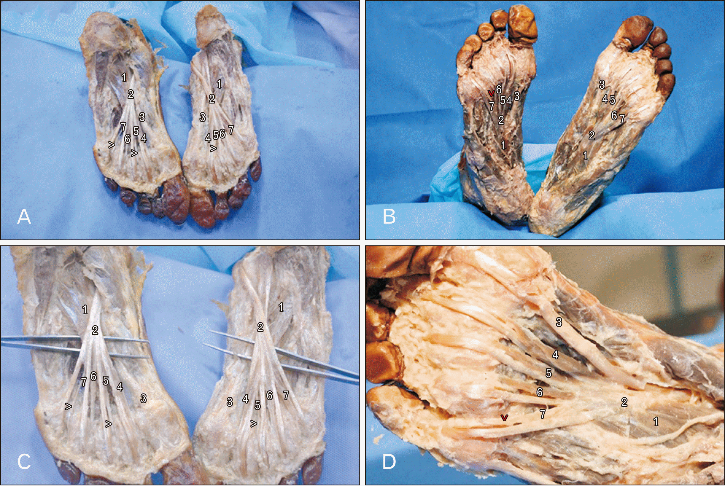 | Fig. 1Photographs of lumbricals of the foot showing: (A) degenerated 2nd and 4th lumbricals in the left and 2nd lumbrical in the right foot; (B) enlarged view of (A) showing degenerated 2nd and 4th lumbricals in the left and 2nd lumbrical in the right foot; (C) the degenerated fourth lumbrical of the right foot and normal lumbricals in the left foot; and (D) enlarged view of the right foot of (C) showing degenerated fourth lumbrical. 1, flexor accessorius; 2, tendon of flexor digitorum longus; 3, tendon of flexor hallucis longus; 4, first lumbrical; 5, second lumbrical; 6, third lumbrical; 7, fourth lumbrical; >, degenerated lumbrical.
|
On light microscopic examination of hematoxylin and eosin stained sections of the normal lumbrical muscle, we observed striated muscle tissue that was arranged in a series of fascicles and the individual muscle fibres had numerous peripheral nuclei (Fig. 2A, B). In the MT stained sections, the muscle fibres appeared to be bright red, the endomysium and perimysium appeared to be green and the peripheral nuclei in the muscle fibres were dark blue in colour (Fig. 3A, B). On examining the degenerated muscle tissue, we observed that it was made of bundles of collagen fibres that were aligned parallel to the skeletal muscle fibres (Fig. 2C, D). On MT-stained sections, the fibrous bundles appeared green (Fig. 3C, D); whereas, some of the fibres that were seen among them stained red.
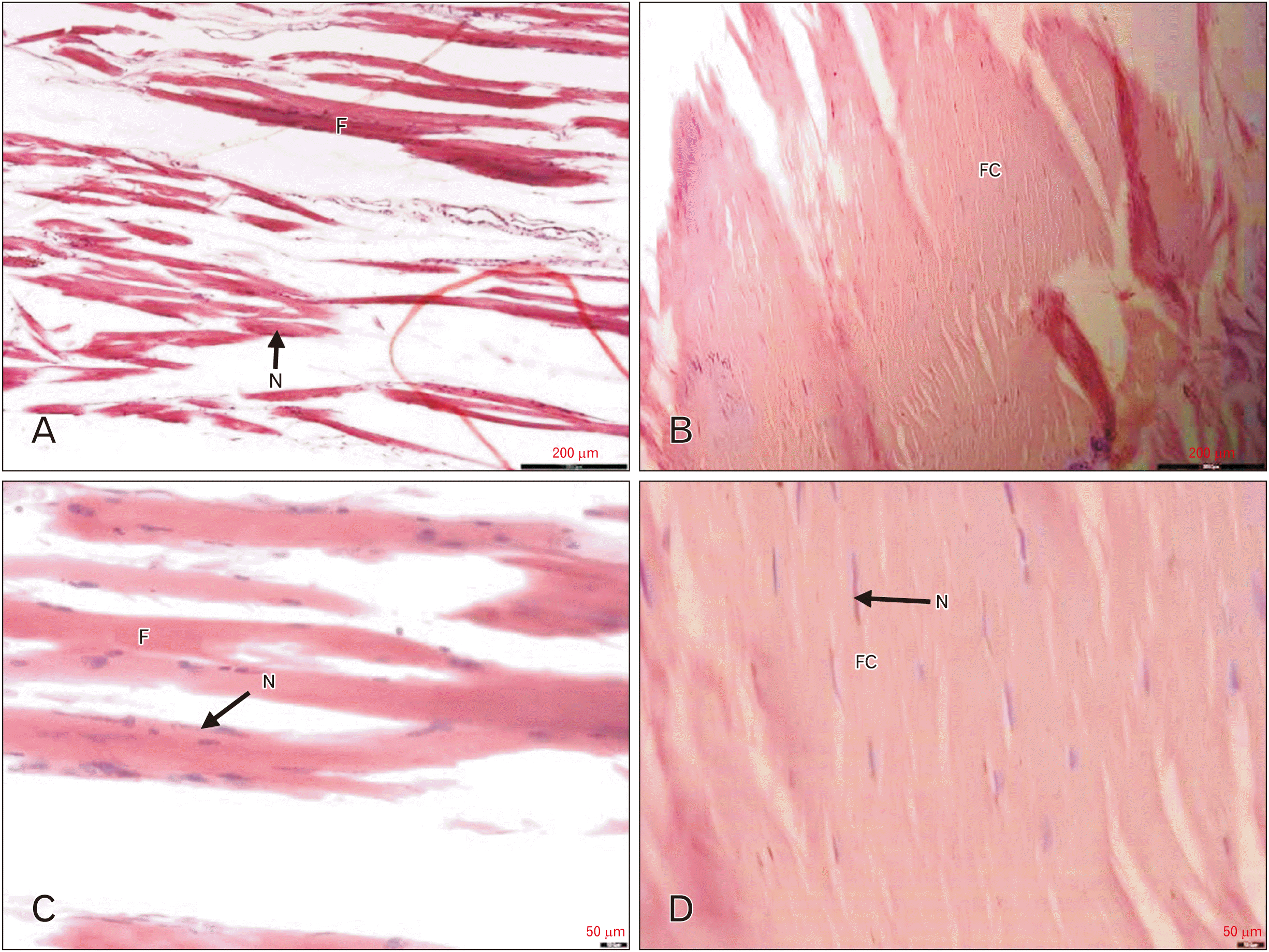 | Fig. 2The photomicrographs of longitudinally sectioned lumbrical muscles, stained with hematoxylin and eosin, showing: (A) normal lumbrical muscle at low power (10×); (B) normal lumbrical muscle at high power (40×); (C) degeneration of lumbrical muscle at low power (10×); and (D) degeneration of lumbrical muscle at high power (40×). Scale bars=200 µm (A, C); scale bars=50 µm (B, D). F, muscle fascicle; N, nucleus; FC, fascicle of collagen fibers.
|
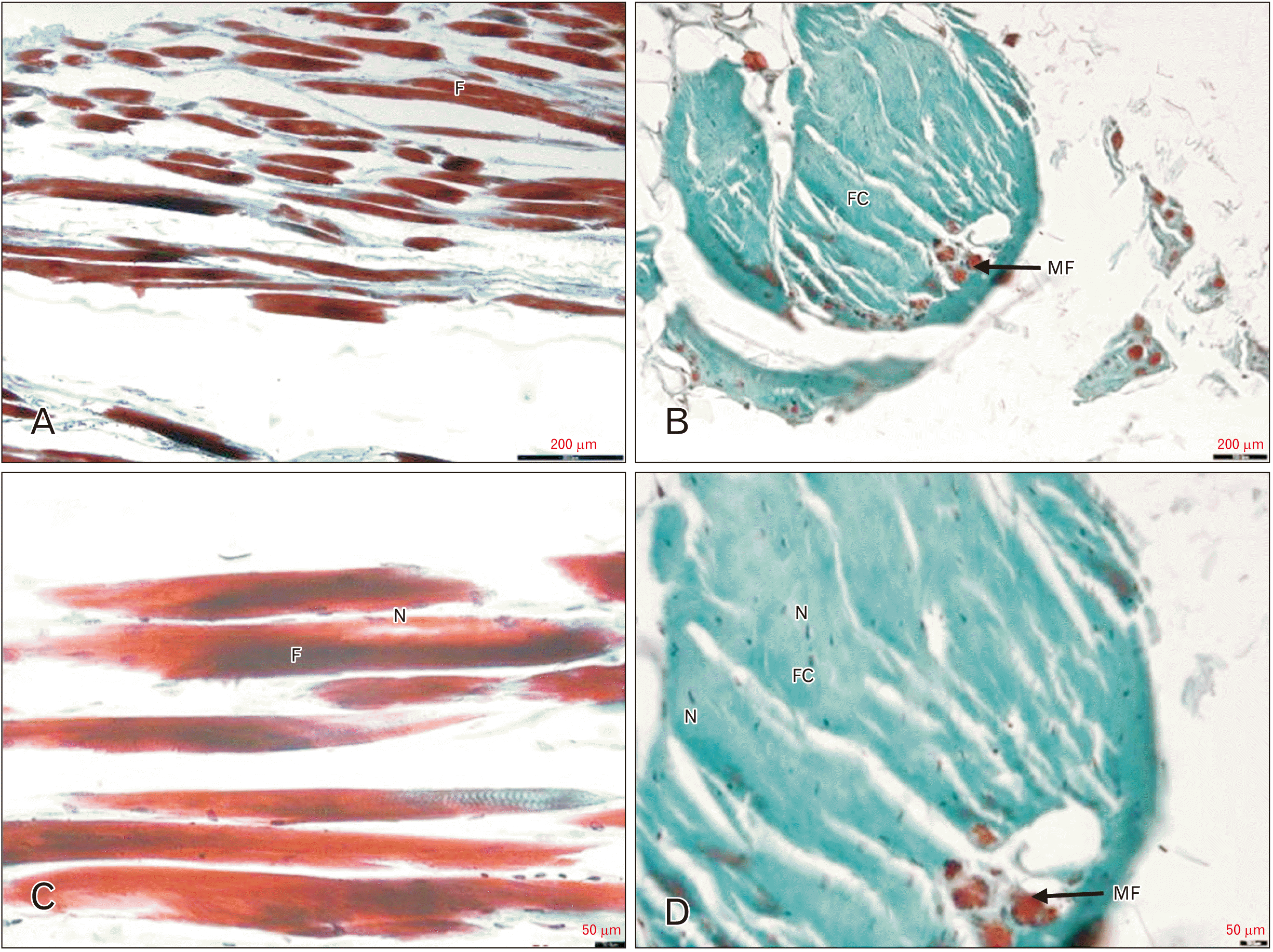 | Fig. 3The photomicrographs of longitudinally sectioned lumbrical muscles, stained with Masson’s trichrome technique, showing: (A) normal lumbrical muscle at low power (10×); (B) normal lumbrical muscle at high power (40×); (C) degeneration of lumbrical muscle at low power (10×); and (D) degeneration of lumbrical muscle at high power (40×). Scale bars=200 µm (A, C), scale bars=50 µm (B, D). F, muscle fascicle ; N, nucleus; FC, fascicle of collagen fibres; MF, muscle fibres.
|
Go to :
Discussion
In the present study, we found four apparently degenerated lumbricals in two male cadavers aged 65 years and 68 years, respectively that on microscopy revealed replacement of striated muscle fibres with collagenous fibrous tissue.
Skeletal muscle atrophy occurs due to various reasons like disuse, aging, starvation, and cachexia in several diseases [12]. It is evident by the replacement of the striated muscle fibres by collagen fibres. As we saw in our study, collagen fibres on staining with the Masson’s trichrome method take up the colour of the counterstain- neutral green or methylene blue [11]. The degeneration of lumbrical muscles may be due to compression of the branches of the lateral plantar nerve (LPN) that supply the lumbricals. There also could be space-occupying lesion along the course of the LPN- including ganglia, bone tumours, aneurysm of arteries, and joint abnormalities, or even tenosynovitis, which may cause compression of LPN [1, 13]. The branches of LPN may also be injured due to stress in a hypermobile pronating foot [14].
There are a few reports on the morphological variation of foot lumbricals. Chaney et al. [10], dissected 257 limbs, and all four lumbricals were present in only 54.5% of limbs. There were no lumbricals in 3.3% of the feet studied; one lumbrical only in 3.5%, two lumbricals in 12.1% and three in 27.2% [10]. In most of the studies, the third and fourth lumbricals were most often absent in the foot [15]. However, these authors have not reported any neural, bony or joint related anomalies in the feet that they had studied.
In conclusion, degeneration of the lumbricals is a rare phenomenon. However, the detection of a degenerated lumbrical should instigate the surgeon to look for causes of compression of the branches of the LPN that supply the lumbricals in the foot.
Go to :
 XML Download