Abstract
Upper limb muscle variations can be encountered on imaging or at surgery. We report an unusual muscle and band found during routine dissection of the arm in a cadaver. This case is described and salient literature reviewed. A band was found that traveled from the insertion of the pectoralis major tendon distally and obliquely toward the medial intermuscular septum and medical epicondyle. Fibers of the brachialis were found to interdigitate into the band. A tunnel was formed that carried the median nerve and brachial vessels. Evidence of median nerve compression was observed. We considered this an example of a pectorobrachioepicondylaris muscle. However, some can lead to clinical presentations. Although the significance of the case reported herein is not certain, signs of median nerve compression were identified. We believe that the term pectorobrachioepicondylaris bests describes the muscle reported herein and that our case represents a previously unreported variant of this muscle.
Accessory muscular slips crossing the floor of the axilla between the latissimus dorsi and the pectoralis major have been described. These muscle variants can take their origin from the costal cartilages, the ribs or the borders of the pectoralis major muscle [1]. They have been variably referred to as chondroepitrochlearis, axillary arches and costocoracoideus. A dorsoepitrochlearis slip from the latissimus dorsi muscle has also been reported [2]. The insertion of these slips is variable but the chondroepitrochlearis and dorsoepitrochlearis muscles most often insert into the fascia of the arm, the medial intermuscular septum or the medial epicondyle of the humerus [2-4].
Herein, we report an unusual muscular variant of the anterior arm found during routine cadaveric dissection. The details of this case are discussed along with germane reports from the literature.
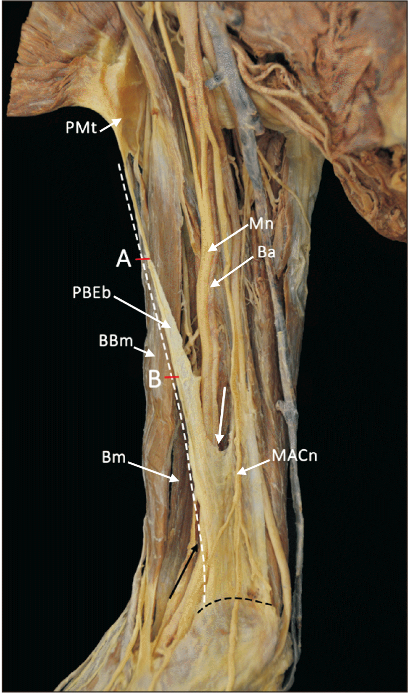
During a routine dissection of an embalmed right upper limb (69-year-old at death male cadaver), a connective tissue band (with some muscle fibers) in the anteromedial aspect of the arm was observed. It originated proximally from the inferior border of the pectoralis major muscle near its insertion into the humerus and extended distally to the medial epicondyle of the humerus and the medial intermuscular septum. It ran superficial to the biceps brachii muscle and deep to the medial cutaneous nerves for the arm and forearm (Fig. 1).
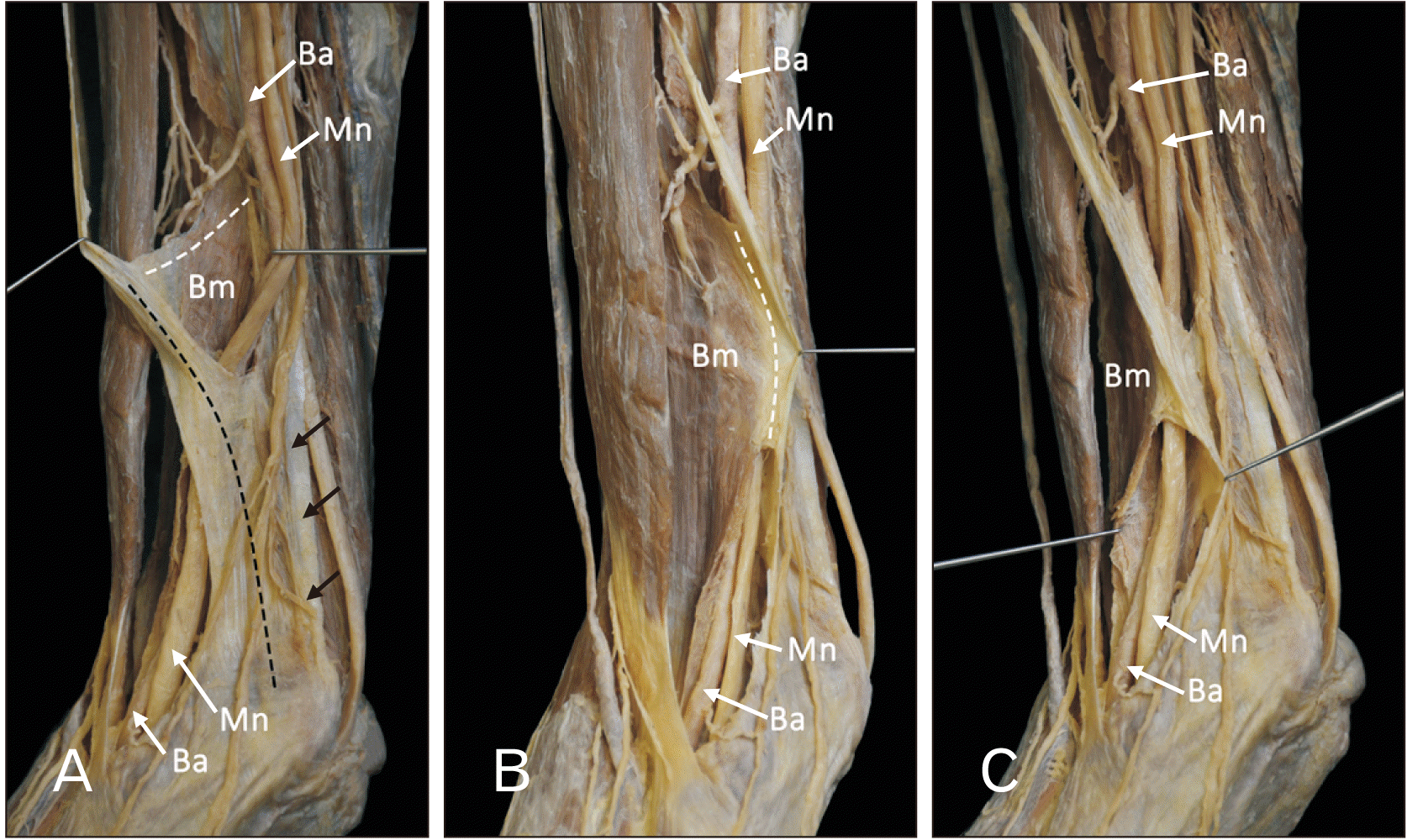
The total length of this band was 23.5 cm. The width at its origin from the pectoralis major tendon was 8 mm. It progressively decreased in size distally becoming thinnest 5 cm distal to its origin. From this point, in the proximal third of the arm, the band became progressively wider (Fig. 1). At 14 cm from its origin, the band enlarged in size and divided into two layers, anterolateral and posteromedial, which delimited a canal for the passage of the median nerve and the brachial artery in the anterior compartment of the arm (Fig. 2).
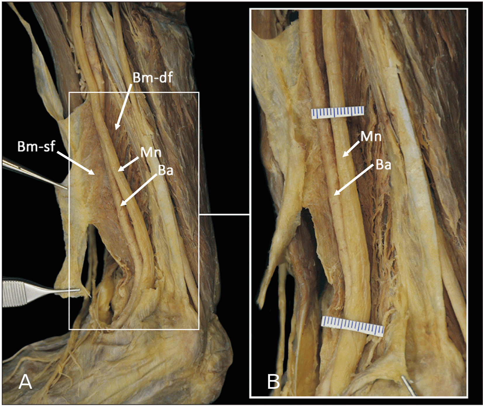
The anterolateral fibers fused with the anterior surface of the brachialis muscle, which contributed a thin layer of muscle fibers into the band. These muscular fibers were in continuity with a muscular extension of the brachialis muscle. The two layers formed by the division of the band, and the medial border of the brachialis muscle, which was clearly divided into superficial and deep fibers, created a musculotendinous canal (4 cm in length) for the median nerve and the brachial artery along the distal third of the arm (Figs. 2, 3). The median nerve and the brachial artery exited the canal 4.5 cm proximal to the medial epicondyle to then enter the cubital fossa. Although no direct nerve innervation was seen traveling to the musculoaponeurotic band, the brachialis muscle, of which this was attached, was innervated by the musculocutaneous nerve. The morphology of the median nerve at the exit of the canal was flattened, showing an increase of 55% of its diameter in relation with the diameter of the nerve at the entrance of the musculoaponeurotic canal (Fig. 3).
The posteromedial fibers (with the absence of any muscle) traveled distally to join the intermuscular septum and the most posterior brachialis muscle fibers. At this point of the proximal opening of the canal, the musculoaponeurotic fibers had a width of 12 mm, becoming progressively wider and more consistent in size distally at approximately 4 cm in width. It finally ended in the fascia over the common flexor tendon (Fig. 2). The band tapered in width in the distal third of the arm with an approximate width of 2 cm. The anterolateral part of the band was continuous with the superficial fibers of the brachialis muscle, whereas the posteromedial fibers of the band expanded and ended in the medial epicondyle and the fascia over the common flexor tendon.
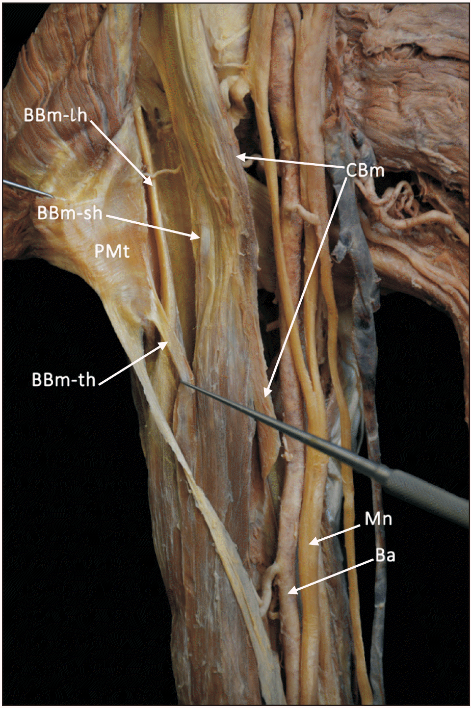
A third head of the biceps brachii was present. This small head connected the short head of the biceps brachii with the pectoralis major tendon. Lastly, the distal fibers of the corocobrachialis muscle (which showed a flattened morphology) were elongated farther than normal and thus, could be classified as a corocobrachialis longus [5]. This latter muscle was 18 cm long and its insertion into the humerus was 4 cm beyond the upper most fibers of origin of the brachialis muscle (Fig. 4).
No additional anatomical variations were noted in this specimen. The authors state that every effort was made to follow all local and international ethical guidelines and laws that pertain to the use of human cadaveric donors in anatomical research [6].
Pectoralis major and minor develop from the thoracohumeral portion of the panniculus carnosus during the 5th week of development [7]. This atavistic muscle and specifically, its thoracic part, travel from the anterior thorax and axilla to descend distally and insert into the humerus [8]. Although such a muscle usually regresses in man, several variant muscles in this region are thought to arise from its persistence [1, 3]. For example, the chondroepitrochlearis, which usually arises from the costochondral junctions of the fifth to seventh ribs and inserts on the medial intermuscular septum or the medial epicondyle of the humerus, is considered a derivative [9]. Other terms used to describe this and similar muscles include the costoepitrochlearis, chondrohumeralis, chondrofascialis [3, 4], thoracoepicondylaris [10], chondrobrachialis, costohumeralis [2], and chondroepicondylaris [9]. Some authors have also used the term chondroepitrochlearis for muscles having only an origin from the pectoralis major muscle’s tendon with no diret attachment to the thorax. For these muscles, some authors have suggested using the term pectoroepicondylaris to better reflect this anatomy [11].
Wood [1] described a muscle arising from the anterior rectus sheath and inserting separately from the rest of the pectoralis major into the deep surface of the upper fibers of its tendon. Macalister [3] described two cases of a brachiofascialis muscle with an origin from the coracoid process and an insertion into the brachial fascia. For muscles such as the chondroepitrochlearis muscle, Wood [1] reminded us that a similar muscle exists in bats, monkeys, and is similar to the brachioabdominal muscle found in the batrachian reptiles.
The muscle fibers in our case were only from a superficial layer of the brachialis muscle, which has been reported to rarely have two layers [4]. Our case was also found to have a third head of the biceps brachii. This variant found in about 10% of the population has also been found concomitantly in cases having an axillary arch muscle [12]. A longer than normal coracobrachialis muscle i.e., coracobrachialis longus was also identified ipsilaterally [5]. To our knowledge, no similar cases as the one presented herein have been reported with these additional muscular variations. However, El-Naggar and Al-Saggaf [13] reported a case of a coracobrachialis muscle, which they thought might represent a variant of the coracobrachialis longus muscle. In their case, the median nerve and brachial artery traveled through a tunnel created by the muscle. Lastly, as documented by Calori [14], anomalous muscular bands in the anterior arm can travel between the biceps brachii and medial intermuscular septum and thus compress the median nerve and brachial artery.
Wood [1] described a case of a band of fibers (2.5 cm in width), detached from the lower border of the pectoralis major that traveled down the arm, crossing the brachial vessels and median nerve obliquely, to end in the medial intermuscular septum. Of all the previous cases, his case is most similar to the case presented here although there was no mention of attachments into the brachialis muscle or the formation of a musculoaponeurotic tunnel.
Based on the available literature, we believe that the term pectorobrachioepicondylaris bests describes the muscle reported herein as no other cases match its description exactly. Such a case is of archival value and could help explain rare cases of median nerve compression in the arm.
Acknowledgements
The authors sincerely thank those who donated their bodies to science so that anatomical research could be performed. Results from such research can potentially increase mankind’s overall knowledge that can then improve patient care. Therefore, these donors and their families deserve our highest gratitude [15].
Notes
Author Contributions
Conceptualization: AC (Ana Carrera), RST. Data acquisition: AC (Arada Chaiyamoon), AC (Aida Cateura), FR. Data analysis or interpretation: JI, MAR. Drafting of the manuscript: AC (Ana Carrera), RST. Critical revision of the manuscript: JRS. Approval of the final version of the manuscript: all authors.
References
1. Wood J. 1867; IV. Variations in human myology observed during the Winter session of 1866-67 at King's college, London. Proc R Soc Lond. 15:518–46. DOI: 10.1098/rspl.1866.0119.

2. Loukas M, Tubbs RS. 2009; Musculus dorsoepitrochlearis. Clin Anat. 22:782. DOI: 10.1002/ca.20826. PMID: 19637307.

3. Macalister A. 1867; Further notes on muscular anomalies in human anatomy, and their bearing upon homotypical myology. Proc R Irish Acad. 10:121–64.
4. Tubbs RS, Shoja MM, Loukas M. 2016. Bergman's comprehensive encyclopedia of human anatomic variation. John Wiley & Sons;DOI: 10.1002/9781118430309.
5. Georgiev GP, Landzhov B, Tubbs RS. 2017; A novel type of coracobrachialis muscle variation and a proposed new classification. Cureus. 9:e1466. DOI: 10.7759/cureus.1466.

6. Iwanaga J, Singh V, Takeda S, Ogeng'o J, Kim HJ, Moryś J, Ravi KS, Ribatti D, Trainor PA, Sañudo JR, Apaydin N, Sharma A, Smith HF, Walocha JA, Hegazy AMS, Duparc F, Paulsen F, Del Sol M, Adds P, Louryan S, Fazan VPS, Boddeti RK, Tubbs RS. 2022; Standardized statement for the ethical use of human cadaveric tissues in anatomy research papers: recommendations from anatomical journal editors-in-chief. Clin Anat. 35:526–8. DOI: 10.1002/ca.23849. PMID: 35218594.
7. Afroze MKH, Sangeeta M, Tiwari S. 2020; A case report of additional slip of pectoralis major- pectorotubero fascialis muscle. J Med Sci Health. 6:18–20. DOI: 10.46347/JMSH.2020.v06i01.004.
8. Hartman CG, Straus WL. 1971. The anatomy of the rhesus monkey, Macaca mulatta. Hafner;DOI: 10.1038/134047a0.
9. Palagama SP, Tedman RA, Barton MJ, Forwood MR. 2016; Bilateral chondroepitrochlearis muscle: case report, phylogenetic analysis, and clinical significance. Anat Res Int. 2016:5402081. DOI: 10.1155/2016/5402081. PMID: 27242928. PMCID: PMC4875967.

10. Loukas M, Louis RG Jr, Kwiatkowska M. 2005; Chondroepitrochlearis muscle, a case report and a suggested revision of the current nomenclature. Surg Radiol Anat. 27:354–6. DOI: 10.1007/s00276-005-0337-4. PMID: 15912263.

11. Kotian SR, Bhat KMR. 2013; Pectoro-epicondylaris: a rare extension of the pectoralis major muscle. Rev Argent Anat Clin. 5:29–32. DOI: 10.31051/1852.8023.v5.n1.14049. PMID: 5c2d606f1b1f4d548d21cffdc78310c0.
12. Horrocks P, Hale White W, Lane WA. 1884; An account of the abnormalities observed in the dissecting room during the winter session 1882-1883. Guys Hosp Rep. 42:39–48.
13. El-Naggar MM, Al-Saggaf S. 2004; Variant of the coracobrachialis muscle with a tunnel for the median nerve and brachial artery. Clin Anat. 17:139–43. DOI: 10.1002/ca.10213. PMID: 14974102.

14. Calori L. 1868; On important anomalies of bones, vessels, nerves and muscles observed in last two years doing human anatomy. Mem Acad Sci Bol. 8:417–82. Italian.
15. Iwanaga J, Singh V, Ohtsuka A, Hwang Y, Kim HJ, Moryś J, Ravi KS, Ribatti D, Trainor PA, Sañudo JR, Apaydin N, Şengül G, Albertine KH, Walocha JA, Loukas M, Duparc F, Paulsen F, Del Sol M, Adds P, Hegazy A, Tubbs RS. 2021; Acknowledging the use of human cadaveric tissues in research papers: recommendations from anatomical journal editors. Clin Anat. 34:2–4. DOI: 10.1002/ca.23671. PMID: 32808702.

Fig. 1
Anteromedial view of the arm showing the PBEb running from the inferior border of the pectoralis major muscle to the medial epicondyle of the humerus and the medial intermuscular septum. White dashed line, total length 23.5 cm; black dashed line, attachment in the fascia of epitrochlear muscles; (A) 5 cm form origin. Point of smaller width of the band; (B) 14 cm from origin. Division of the band in anterolateral and posteromedial layers; white arrow, proximal opening of the canal; Black arrow, distal opening of the canal; PBEb, pectorobrachioepicondylaris band; PMt, pectoralis major tendon; BBm, biceps brachii muscle; Bm, brachialis muscle; MACn, medial antebrachial cutaneous nerve.

Fig. 2
Anteromedial view of the distal third of the arm. (A) Formation of the musculotendinous arch for the median nerve and the brachial artery by the division of the band into anterolateral and posteromedial layers. (B) Layer of muscular fibers from the brachialis muscle fused with the anterolateral fibers of the band. (C) Distal opening of the canal between the medial border of the brachialis muscle and the posteromedial fibers of the band. White dashed line, anterolateral layer; black dashed line, posteromedial layer; black arrows, medial intermuscular septum; Bm, brachialis muscle; Mn, median nerve; Ba, brachial artery.

Fig. 3
(A) Demonstration of the opened musculotendinous canal by sectioning and reflecting the posteromedial fibers of the band. Superficial and deep fibers of the brachialis muscle can be observed. (B) Enlarged image showing evidence of possible median nerve compression. Bm-sf, brachialis muscle-superficial fibers; Bm-df, brachialis muscle-superficial fibers; Mn, Median nerve; Ba, Brachial artery.

Fig. 4
Anterior view of the proximal third of the arm showing the accessory head of biceps brachii muscle and the coracobrachialis muscle. PMt, pectoralis major tendon; BBm-sh, biceps brachii muscle-long head; BBm-lh, biceps brachii muscle -short head; BBm-th, biceps brachii muscle-third head; CBm, coracobrachialis muscle; Mn, median nerve; Ba, brachial artery.


 PDF
PDF Citation
 Print
Print


 XML Download