

 PDF
PDF Citation
 Print
Print


Introduction
The liver is a large, highly vascularized organ in the right upper quadrant of the abdomen. It typically receives arterial support from branches of the celiac trunk (CT). The CT originates from the abdominal aorta and forms three major vessels: common hepatic artery (CH), left gastric artery (LGA), and splenic artery. After giving off the gastroduodenal artery, the CH continues toward the liver as the proper hepatic artery (PHA) before bifurcating into left and right hepatic arteries as it approximates the hepatic hilum. The right hepatic artery (RHA) supplies the right and caudate lobes– segments I, V-VIII– while the left hepatic artery (LHA) supplies the left and quadrate lobes– segments II-IV [1]. This aforementioned pattern of blood supply occurs in approximately 52%–80% of subjects. However, variations in this pattern are common, and nearly half of the population has some variation in arterial support of their liver. Numerous studies have reported aberrant vessels originating from the CT, LGA, and superior mesenteric artery. Aberrant LHA has been documented in 4%–18% of cases with 3% occurring as a replaced vessel originating from the LGA (rLHA). Aberrant right hepatic arteries occur in approximately 8.4%–18% of individuals, and 3.7% exist as a replaced RHA (rRHA) from the superior mesenteric artery. Both rLHA and rRHA occur in 0.8% of subjects [2, 3]. The gallbladder may also be affected by variations in hepatic vasculature because the RHA supplies the cystic artery in 70%–80% of cases. Variations in the cystic artery are relatively common and may be found in 25%–50% of individuals. The cystic artery has been documented originating from the superior mesenteric artery (SMA) in 0.3%–1.4% of cases [2]. Overall, documented variations suggest hepatic arteries can arise from multiple sources. As such, knowledge of these vascular variations is necessary to control and avoid unnecessary bleeding during surgical procedures involving the liver.
Go to : 
Case Report
During routine dissection of the CT and SMA in a 58-year-old male anatomical donor who died of metastatic lung cancer, an unusual pattern of hepatic arteries was identified (Fig. 1). The common hepatic artery (CH) branched into the gastroduodenal artery and the PHA (Fig. 1, red arrow b). The PHA did not divide into right and left hepatic arteries as typically seen (Fig. 1B, red arrow b), but rather continued as the middle hepatic artery. Due to this abnormal branching pattern, further dissection was completed to determine the origin of the left and right hepatic arteries. For consistency we adapt the terminology in Bergman (2016). Specifically, an aberrant hepatic artery can be classified as either replaced or accessory. Replaced arteries are a substitute for the normal hepatic artery while accessory hepatic arteries are in addition to normal hepatic arteries [2]. Accordingly, the vessels discovered during this dissection are classified as replaced hepatic arteries. As shown in Fig. 1, two replaced hepatic arteries were identified - one originating from the LGA (Fig. 1, red arrow c) and one from the SMA (Fig. 1, red arrow a). The hepatic portal vein and tributaries were removed for clarity. A small segment of the portal vein can be seen in Fig. 1, magenta asterisk. In this subject, the LGA branches from the CT and divides into a rLHA (Fig. 1; red arrow c) before supplying the lesser curvature of the stomach. The SMA also provides a rRHA (Fig. 1, red arrow a) that travels laterally toward the common bile duct. This replaced branch was situated posterior to the typical structures of the portal triad within the hepatoduodenal ligament, forming a portal quadrad. In this subject, the SMA also provided the cystic artery (Fig. 1B, yellow wire). Because of the rightward course of the hepatic branch from the SMA and the pattern of branching from this vessel, the cystic artery arose close to the inferior surface of the liver and outside Calot’s triangle. There were no additional anomalies or pathologies present in the abdomen of this donor.
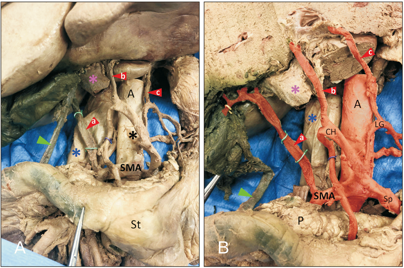 | Fig. 1(A) is the foregut from the subject, removed from the body cavity. The celiac trunk (asterisk) and SMA branch from the aorta (A) in typical fashion. The left gastric artery issued a branch directed towards the inferior aspect of the left lobe of the liver (red arrowhead c). The common hepatic artery (CH) split to form the proper hepatic artery, which continues as the middle hepatic artery (red arrowhead b), and the gastroduodenal artery (purple wire). The SMA formed a larger branch directed superiorly towards the porta hepatis (red arrowhead a). The common bile duct is indicated by the green arrowhead. The inferior vena cava is indicated by the blue asterisk and the portal vein is indicated by the magenta asterisk. (B) The liver has been dissected to provide a better view of the arteries entering the inferior aspect. The aorta and its branches are colored red. The branch from the SMA to the liver also provides the cystic artery (yellow wire). SMA, superior mesenteric artery; St, stomach; P, pancreas; Sp, splenic artery; LG, left gastric.
|
Further dissection of the liver was undertaken to investigate the segments of liver supplied by each of the three hepatic arteries. Based on the dissection of the liver, we devised the arterial map of the liver shown in Fig. 2. Specifically, the rRHA from the SMA supplied the right lobe of the liver–segments V-VII. The middle hepatic artery from PHA supplied liver segment IV and the caudate lobe (segment I). The rLHA from the LGA supplied segments II and III.
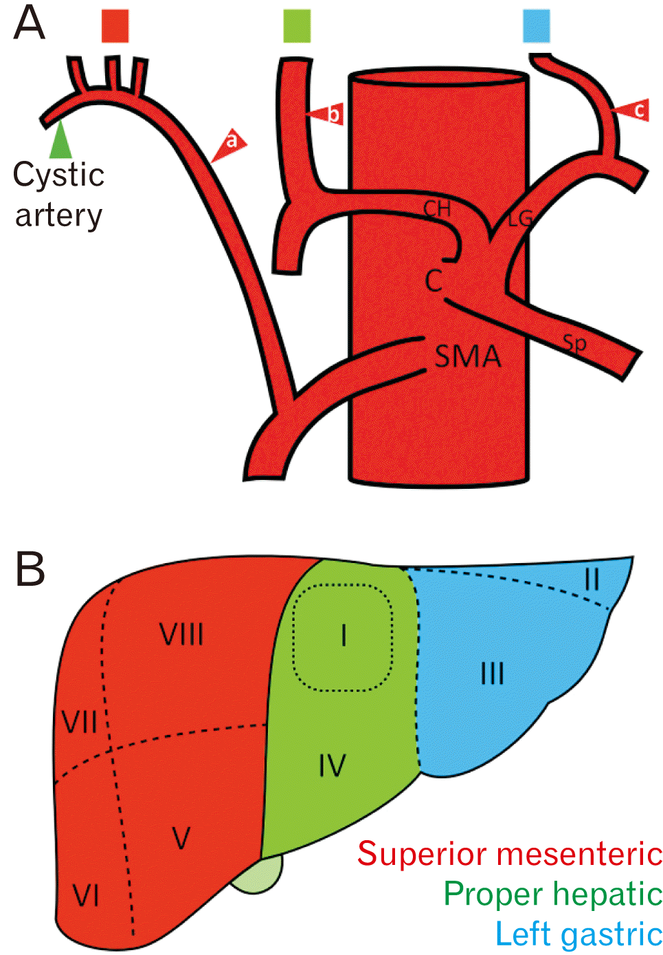 | Fig. 2(A) shows a schematic of the arterial pattern to the liver in this subject. The red arrowheads are the same as in Fig. 1. Red arrowheads indicate the three arteries supplying the liver- these vessels originate from the SMA (a), proper hepatic artery (b), and LG (c). The hepatic branch from the SMA provided the cystic artery (green arrowhead). The colored blocks above the hepatic arteries correspond to the liver regions shown in (B). (B) is a schematic representation of the liver from the subject of this case study. Based on the arrangement of arteries along the inferior aspect of the liver, we propose this arterial map: the right lobe of the liver was supplied by the replaced right hepatic artery from the SMA, while the remaining liver was supplied by the middle hepatic artery from the celiac trunk, with segment II and III supplied by the replaced left hepatic artery from the left gastric artery. SMA, superior mesenteric artery; CH, common hepatic artery; LG, left gastric artery; Sp, splenic artery; C, celiac trunk.
|
Go to :
Discussion
In the present case study, we describe an unusual pattern of hepatic blood supply. Three extrahepatic arteries arose from the superior mesenteric artery, proper hepatic artery, and LGA rather than the typical pattern of two extrahepatic arteries arising solely from the proper hepatic artery. The variation observed in this case study is a combination of Michels types II and III, and is classified as Michels type IV variation–both the right and left hepatic arteries are replaced with vessels originating from the SMA and LGA [2]. The proper hepatic artery continues as the middle hepatic artery. This variation is rare and was found in 0.8% of individuals [3]. As observed in Fig. 1, the three extrahepatic arteries were similar in diameter suggesting substantial contributions from each, as indicated in Fig. 2.
Variation in hepatic blood supply is common and may be traced to embryologic development. The liver begins developing during week 4 of gestation. It grows into the septum transversum, and by the end of week 6, the liver occupies most of the abdominal cavity. During the embryonic period the liver divides into three parts, each with its own primitive blood supply. A left lateral lobe is supplied by the left embryonic artery from the LGA. A central lobe is supplied by the middle embryonic artery from the CH. A right lateral lobe is supplied by the right embryonic artery originating from the omphalomesenteric artery, and it ascends posterior to the portal vein. The arteries are initially connected by a ventral longitudinal anastomosis, and if this fails to obliterate, a common celiomesenteric trunk may be observed. As the central lobe enlarges and overtakes the lateral lobes, the middle embryonic artery develops into the proper hepatic artery–this splits into left and right branches and becomes the predominant blood supply to the liver. The left and right embryonic arteries typically regress. Aberrant hepatic arteries may represent the persistence of embryologic hepatic arteries, or an incomplete obliteration of the ventral longitudinal anastomosis [4]. In the current case study, the replaced left and right hepatic arteries appear in the location of the temporary embryonic vessels and may reflect a lack of regression of these arteries.
An understanding of hepatic arterial variation is important to prevent iatrogenic injury and surgical complications. Aberrant arteries may require a change to surgical procedures [3]; therefore, preoperative imaging and knowledge of common variations are crucial [5]. During liver procedures, all hepatic arteries must be preserved as each serves particular liver segments. There are surgical costs and benefits associated with hepatic arterial variations. In the current donor, the presence of three separate hepatic arteries may offer flexibility in resection of liver segments. However, it may require complex intraoperative reconstruction, increasing operative time and the likelihood of complications [6]. A rLHA from the LGA can be damaged during procedures involving the lower esophagus or stomach, leading to hepatic necrosis. However, the aberrant vessel may provide collateral support in cases of porta hepatis obstruction [7] and is likely to be unaffected in bile duct cancer [8]. In the current donor, the middle hepatic artery supplies liver segment IV, which is often included in planes of resection. Understanding blood supply to this segment affects donor outcomes, liver regeneration, and the likelihood of liver ischemia [9, 10]. A rRHA from the SMA can be injured or accidentally ligated in cholecystectomy [11, 12] and pancreaticoduodenectomy [13], compromising blood supply to the liver and bile duct. This increases risk of liver necrosis and breakdown of the hepatobiliary anastomosis, leading to biliary fistula. Occasionally, a rRHA travels through the head of the pancreas, increasing the likelihood of iatrogenic damage during resection [14]. However, an rRHA may improve the prognosis for patients with cholangiocarcinoma as the vessel is farther away from the lesion and tends to be spared [1]. A vessel branching from the SMA is also longer and reduces the likelihood of hepatic thrombosis following grafts in living liver donors [15]. Overall, knowledge of hepatic artery variants is key to surgical planning and preventing intra- and post-operative complications.
In conclusion, this case study describes a rare variation of hepatic arterial supply discovered during routine dissection of the abdominal aorta and associated viscera. Embryonic development of the CT and SMA may contribute to a wide variety of aberrant hepatic arteries. Due to the relatively common occurrence, hepatic vasculature must be well understood to ensure successful outcomes during surgical procedures involving the liver.
Go to :
 XML Download