This article has been
cited by other articles in ScienceCentral.
Abstract
We present the first case of buckled thyroid cartilage identified in a human cadaver. This rare anatomical variant, in patients, often produces dysphonia and is a potential source for diagnostic confusion. In the cadaveric case described, the laryngeal prominence is deviated to the left without deviation of the internal structures of the larynx, such as vocal folds and vocalis muscles. The medical history of the patient is not known. Finally, a review of current literature on the buckled thyroid cartilage is presented. Such a case represents a rare opportunity to visualize this deformity via anatomical dissection.
Go to :

Keywords: Thyroid cartilage, Anatomy, Cadaver, Larynx
Introduction
The larynx is a complex anatomical structure of the throat involved in vital functions such as swallowing, respiration, and phonation. It is formed by three unpaired cartilages, the thyroid, cricoid, and epiglottis, as well as four paired cartilages, the arytenoid, cuneiform, triticeal and corniculate cartilages. The thyroid cartilage is the largest structure of the larynx and serves to protect the internal laryngeal structures. The thyroid cartilage can be palpated and inspected on physical examination by locating the laryngeal prominence, colloquially known as the Adam’s apple. Developmentally, the laryngeal cartilages are developed from the fusion of the fourth and sixth pharyngeal arch cartilages.
Any variation in the anatomy can disturb normal function of an organ and cause symptoms [
1]. Etiologies of anatomic variation include benign lesions, malignancy, trauma, autoimmune disease, and congenital anatomical variants. The study of anatomical variation of the larynx has yielded improved diagnostic and surgical accuracy. For example, Friedrich and Lichtenegger performed a cadaveric study of 50 larynges and established standard proportions of the male and female larynx, providing a flexible benchmark for the detection of abnormalities and surgical correction [
2].
Chang et al. [
3] performed a retrospective case series of 11 patients with a buckled thyroid cartilage between the years 1996 and 2016, establishing this rare anatomic variant in the literature. Additional reports of this finding will improve inclusion of this anatomical variant in the wide differential for laryngeal symptoms, such as dysphonia. Here, we present, to our knowledge, the first case of a buckled thyroid cartilage in a human cadaver.
Go to :

Case Report
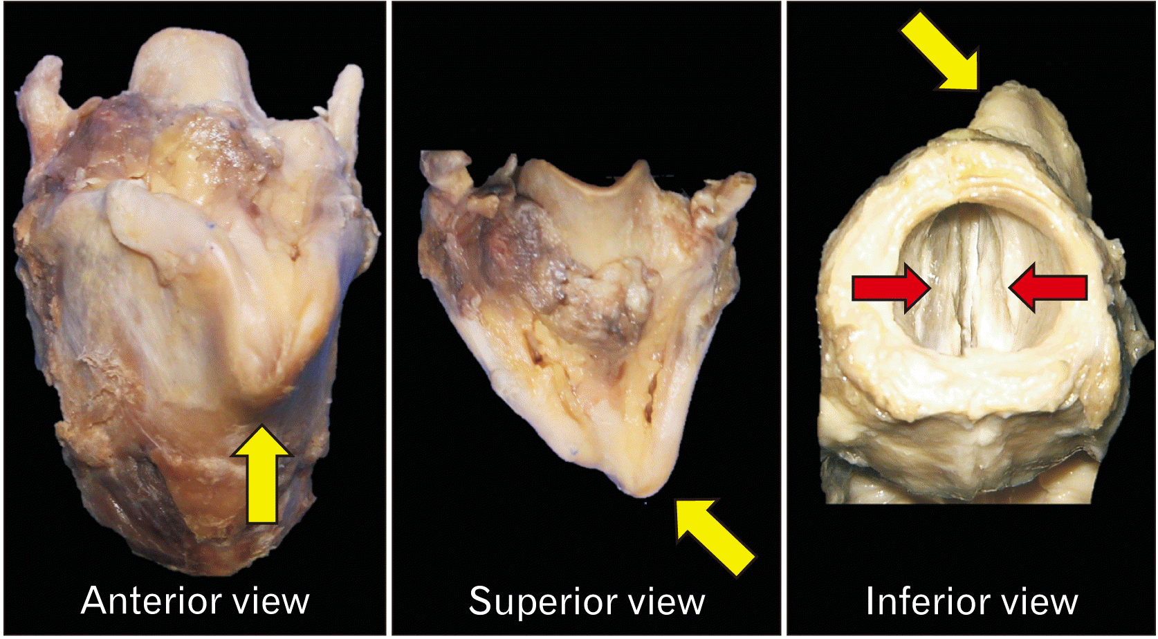
This routine dissection was of a male human cadaver whose age at the time of death was 64-years-old. There was no known medical history or obvious signs of previous surgery or trauma to the regions dissected. In the anterior and superior views, the laryngeal protuberance was deviated to the left (
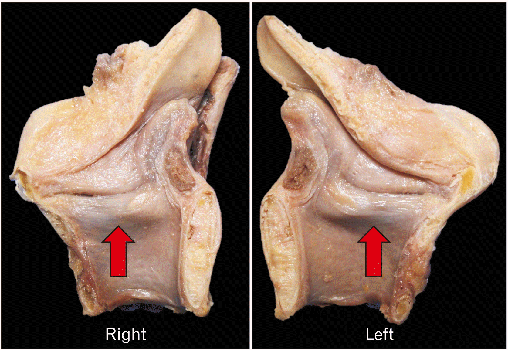
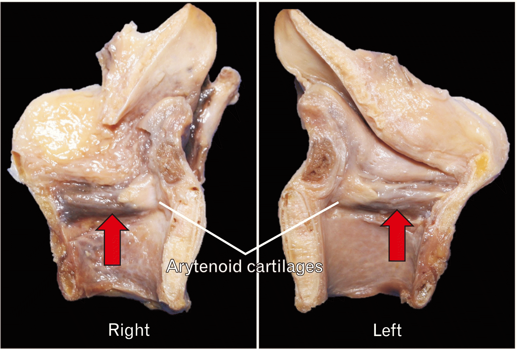
Fig. 1). Then the larynx was sagittally sectioned to see if there was any deviation of the vocal cords (
Figs. 2,
3). There was no sign of deviation of internal structures of the larynx, such as vocal folds and cords.
 | Fig. 1Anterior, superior, and inferior views of the buckled thyroid cartilage. Note the laryngeal protuberance (arrows) is deviated to the left. Also, note that the vocal folds (red arrows, vocalis muscles) are symmetrically positioned (following the sagittal cut and removal of the mucosa over the vocal folds). 
|
 | Fig. 2Medial view of the sagittally sectioned buckled thyroid cartilage. Note the absence of deviation of the vocal folds (red arrows). 
|
 | Fig. 3Medial view of the sagittally sectioned buckled thyroid cartilage following the removal of the mucosa over the vocal folds (red arrows). 
|
The authors state that every effort was made to follow all local and international ethical guidelines and laws that pertain to the use of human cadaveric donors in anatomical research [
4].
Go to :

Discussion
We present here, to our knowledge, the first case report of a buckled thyroid cartilage in a cadaver. In the absence of pathological or trauma etiologies in our case, such a variation might have been due to an unknown embryological derailment.
Chang et al. [
3] found the most common symptom associated with this anatomical variant was dysphonia. They hypothesize that the mechanism of hoarseness is dampening of vibration of the true vocal fold secondary to medial displacement of the false vocal folds due to the buckled thyroid cartilage [
3]. Urban et al. [
5] similarly hypothesized that dysphonia can be caused by true vocal fold paresis secondary to thyroid cartilage anatomical variations. These authors presented six individuals with hemilaryngeal microsomia. The common variable in these conditions was asymmetry of the thyroid cartilage angles. While Honjo et al. [
6] proposed deformity of the thyroid cartilage to be related to the normal aging process, Chang et al. [
3] presented a case of trauma in a 23-year-old, and Urban et al. [
5] presented six cases of congenital anomalies. Although the asymmetrical external view was found in the present case, the vocal folds and cords seemed symmetrical internally. Thus, this variation might not have affected either the patient’s phonation or respiration.
In conclusion, this cadaveric case study contributes to the body of literature on anatomical variations of the larynx, and specifically, to the buckled thyroid cartilage variant. Additional cadaveric studies will improve our understanding of the prevalence and etiology of this variant. Physicians should consider anatomical variation of the larynx in their differential of dysphonia.
Go to :


 PDF
PDF Citation
 Print
Print


 XML Download