Journal List > Anat Cell Biol > v.56(2) > 1516083081

Keereewan, Monum, Prasitwattanaseree, and Mahakkanukrauh: Stature estimation using the sacrum in a Thai population

Abstract

Stature is an essential component of biological profile analysis since it determines an individual’s physical identity. Long bone dimensions are generally used to estimate the stature of skeletal remains; however, non-long bones such as the sternum, cranium, and sacrum may be necessary for some forensic situations. This study aimed to generate a regression equation for stature estimation of the skeletal remains in the Thai population. Ten measurements of the sacrum were measured from 200 dry sacra. The results revealed that the maximum anterior breadth (MAB) provided the most accurate stature prediction model among males (correlation coefficient [r]=0.53), standard error of estimation (SEE=5.94 cm), and females (r=0.48, SEE=6.34 cm). For the multiple regression model, the best multiple regression models were stature equals 41.2+0.374 (right auricular surface height [RASH])+1.072 (anterior-posterior outer diameter of S1 vertebra corpus [APOD])+0.256 (dorsal height [DH])+0.417 (transverse inner diameter of S1 vertebra corpus [TranID])+0.2 (MAB) with a SEE of 6.42 cm for combined sex. For males, stature equals 63.639+0.478 (MAB)+0.299 (DH)+0.508 (APOD) with a SEE of 5.35, and stature equals 75.181+0.362 (MAB)+0.441 (RASH)+0.132 (maximum anterior height [MAH]) with a SEE of 5.88 cm for females. This study suggests that regression equations derived from the sacrum can be used to estimate the stature of the Thai population, especially when a long bone is unavailable.

Introduction

Identification of skeletal remains is an essential process for postmortem examinations. A lack of definitive personal identification hampers the legal process concerning the skeletal remains. Matching DNA, fingerprints, or dental profiles is the key criterion for identifying catastrophe victims [1]. However, these methods need the antemortem data of the unknown deceased before matching with the possible missing person. Therefore, the biological profile of the skeletal remains, which includes ancestry, age, sex, and stature, needs to be analyzed to reduce the number of potential missing people.
Stature is an essential parameter of biological profile analysis.This parameter may offer valuable evidence of personal identification when the unknown deceased or missing person is tall or short in their population. Currently, the primary methods for estimating the stature of skeletal remains are anatomical and mathematical [2, 3]. The anatomical method is the most precise since it calculates stature by adding the dimensions of the skull, vertebrae, femur, tibia, talus, and soft tissues. On the other hand, the mathematical method calculates stature by the dimension of one or more bone lengths using regression analysis. The regression model derived from long bones was considered the most accurate for the mathematical method [2, 4]. Although the anatomical method has a greater accuracy rate than the mathematical method, due to taphonomy and animal scavenging, entire skeleton remains are sometimes not accessible in actual forensic scenarios. Therefore, in forensic circumstances, the mathematical technique could be preferable.
The shortcomings of the stature estimation model using the mathematical method are the specifications of population models because of the different genetics, nutrition, environment, and other factors in each population [5, 6]. Therefore, a stature estimation model generated from one population may not be appropriate for application to other populations. Previously, Mahakkanukrauh et al. [7] reported the stature estimation model using the long bone lengths for the Thai population. The correlation coefficient (r) between bone length and height of males ranged from 0.66 to 0.77, and the lowest standard error of estimation (SEE) was 4.59 cm, and for female samples, the lowest SEE was 5.21 cm, and the r-value between bone length and height ranged from 0.55 to 0.76.
However, in natural catastrophes, aviation accidents, and terrorist strikes, long bones might be shattered or unavailable for analysis [8]. Consequently, other bones are required for stature estimation. Several studies have reported the correlation between stature and the non-long bones of the human body, such as the calcaneus and talus [9], skull [10, 11], and lumbar vertebrae [12] in the Thai population and the metacarpal [13], clavicle [14], sternum [15], skull [16], and vertebral column [17] in other populations. The results indicated that these bones could be used for estimating the stature of the skeletal remains. The sacrum is a robust bone usually well-preserved at the scene and has been previously investigated for estimating stature in many populations [3, 8, 18-22]. However, the accuracy rate of the stature estimation model varied significantly amongst populations.
No previous reports of stature estimation using sacral dimension were identified in the Thai population. This information might be required in case the long bone of the skeletal remains was not recovered or not well-preserved enough to calculate stature. Therefore, this study aimed to generate a stature estimation model using the sacral dimension in the Thai population.

Materials and Methods

A total of 240 dry sacra were divided into 200 sacra (male 100, female 100) for the training sample and 40 sacra for the test sample (male 20, female 20). These were randomly chosen from the Osteology Research and Training Center, Faculty of Medicine, Chiang Mai University, Thailand. The sacra were obtained from donated bodies of Thai people who lived 200 to 300 kilometers away from Chiang Mai and died from 2007 to 2019. The mean age of the male sample was 63.0 years with a standard deviation of 15.9 years (range, 28–94 years), and the mean age of the female sample was 64.7 years with a standard deviation of 16.2 years (range, 26–91 years). Sacra that demonstrated trauma, anomalies, and congenital or acquired deformities that affected the measurements were excluded from our study. The mean age of the male test sample was 59.8 years with a standard deviation of 10.94 years (range, 43–73 years), and the mean age of the female test sample was 71.8 years with a standard deviation of 11.54 years (range, 55–94 years).
The stature of the cadaver was measured from the vertex of the head to the heel of the foot, with the cadaver in a supine position before skeletal processing. Then, all of the sacra were macerated and allowed to dry. The range of cadaver stature of the male training sample was between 150 and 190 cm (mean 166.5±6.99 cm), and for the female training sample, the range was between 140 and 172 cm (mean 154.0±7.19 cm). The male test sample’s cadaver stature range was between 150 and 172 cm (mean 165.5±5.71 cm), and the female test sample was between 145 and 165 cm (mean 153.1±5.08 cm) (Table 1).

Measurement

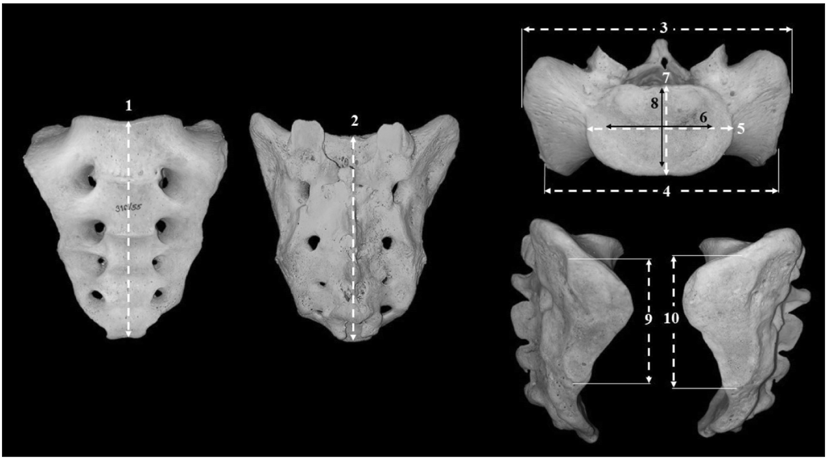
The sacral dimensions were measured in millimeters using digital vernier calipers by one of us (WK), and 30 sacra were randomly chosen from the training sample for repeating the measurements by WK and other forensic science graduate students to test the intra- and inter-observer agreements. The details of the measurements are shown in Table 2 and Fig. 1.

Statistical analysis

Data were analyzed using the IBM SPSS Statistics for Windows version 22.0 (IBM Corp., Armonk, NY, USA). The intra- and inter-observer agreements were assessed by the technical error of measurement (TEM), r-value, and the relative TEM (rTEM). The normality was assumed based on the Kolmogorov–Smirnov test at P>0.05. The descriptive statistic was performed to obtain maximum, minimum, mean, and standard deviation. An independent sample t-test for normally distributed variables and the Mann–Whitney test for not normally distributed variables were used to assess the sexual dimorphism of each measurement. The correlations between stature and the measured variables were assessed using Pearson correlation coefficients. The regression equations for stature estimation were generated from the significantly correlated variables. The presented equations also contain an adjustment to account for estimated live stature, which was done by deducting 2.0 cm. from the constant. Deducting 2 cm. from the cadaver’s stature is typically considered approximate to the living stature [7, 8]. The multiple regression equations were applied to 40 test samples (20 males and 20 females). The mean and median absolute errors were calculated to obtain the difference between predicted stature and corrected stature; the accuracy percentage within the first SEE was also calculated to evaluate the regression equations. A P-value of less than 0.05 was considered statistically significant.

Results

First, we consider the intra- and inter-observer agreements of the variables. The ranges of TEM for intra- and inter-observer were 0.20–0.42 and 0.31–0.62, respectively. The rTEM values of all measurements were less than 1.5 for intra-observation error and less than 2 for inter-observation error. These were within the acceptable range [25]. In addition, all variables showed a high value of r (0.98–0.99). An r-value of more than 0.75 was considered precise in anthropometry [26].
Descriptive statistics for the measures in both sexes are presented in Table 3. Kolmogorov–Smirnov test revealed that normality was assumed at a P-value>0.05 for all variables except the anterior-posterior outer diameter of S1 vertebra corpus (APOD) and anterior-posterior inner diameter of S1 vertebra corpus (APID) for males and the transverse outer diameter of S1 vertebra corpus (TranOD) for females. The independent sample t-test and the Mann–Whitney test showed that all variables had significant differences between males and females with P-values<0.05, except for the maximum anterior breadth (MAB). In addition, the mean values of the variables of males were significantly larger than those of females, except for the anterosuperior breadth (ASB), which was the inverse in females.
The correlations between stature and sacral measurements were statistically significant, except ASB for the combined sex sample, the transverse inner diameter of the S1 vertebra corpus (TranID), and APID for males and APID for females. The highest correlation coefficient was obtained between the RASH and stature in the combined sex sample (r=0.61). The highest correlation coefficients for males and females were obtained between the MAB and stature (r=0.53 for males and 0.48 for females, respectively).
The significant stature correlated variable for generating the regression equation is listed in Table 4. The correlation coefficient and the SEE of the simple regression model in the combined sex sample were higher than those of the sex-specific samples. The SEE of the model in the combined sex sample ranged from 7.54 cm to 8.93 cm, whereas the SEE of the sex-specific simple regression model ranged from 5.94 cm to 6.88 cm for males and 6.34 cm to 6.97 cm for females. The lowest SEE was obtained from MAB, followed by dorsal height (DH) and maximum anterior height (MAH) for males and DH and RASH for females.
The stepwise regression equation is presented in Table 5. The result of this study indicated that the RASH, APOD, DH, TranID, and MAB were selected to generate an equation to estimate stature for the combined sex sample, which produced the highest correlation coefficient (r=0.74). The MAB, DH, and APOD were selected for the male equation with a correlation coefficient of 0.56, and the MAB, RASH, and MAH were selected for the female equation with a correlation coefficient of 0.59.
The multiple regression equations were tested on test samples (n=40) (Table 6). The female-specific equation demonstrated the lowest mean absolute error and the highest accuracy percentage within the first SEE (3.98 cm and 80 percent). However, the absolute error range of the male-specific equation was lower than those of the female and combined sex equations, and the percentage of accuracy within the first SEE of the male equation was equal to that of the combined sex equation at 65 percent.

Discussion

Our study obtained equations for estimating the stature of the sacrum in the Thai population. The stature estimation equations of our study evaluated the stature with a SEE of 5.35 cm for males, 5.88 cm for females, and 6.42 cm for the combined sex sample.
The results of our study indicated that all sacral dimensions of males were larger than those of females, except for the ASB. This result is similar to that of Pininski and Brits [3] and Zhan et al. [8]. These results confirmed that the male sacrum is longer and narrower; on the other hand, the female sacrum is shorter and broader. It was believed that the morphology of the pelvis is adjusted for parturition in females [24].
The multiple regression equation increased the correlation coefficient and reduced the SEE, which indicates higher accuracy than the simple regression equation, following Pelin et al. [19]. Compared with previous studies (Table 7), the accuracy of the stature estimation model of our study was better than those for Black South Africans, Malaysians, and White South African populations. For males, the accuracy of stature estimation in this study was higher than those for Black and White South Africans and American populations but lower than those for the Western Chinese population. The model’s accuracy for female samples in this study was higher than those of White South African and Black American populations but lower than those of Black South Africans and Western Chinese populations [3, 8, 18, 22].
The divergence of the results might be due to several factors that affected the bone dimension and the stature of each population, such as nutrition, genetics, demographics, and population [5, 6]. Moreover, the different sample types, such as dry skeleton, computed tomography images, and the age and height distribution of the samples might also affect the accuracy of the results. Although our study did not investigate the effect of age on the sacral dimension, Karakas et al. [21] and Lazarevski [27] reported that age affected sacral height in females, which decreases with age due to parturition. Additionally, bowing and an inferior-posterior displacement of the sacrum increased with age. Giroux and Wescott [18] reported that stature decreases around 30 years of age. However, it is still being determined how age-related decreases in stature affect estimation based on sacral height.
For testing of the multiple regression in Table 6, the lower mean absolute error (MAE) and the higher percentage of accuracy in the first SEE of the female test samples might be caused by the more significant variability in the sacrum dimension with the stature of the male test samples than those of the female test samples. However, the highest range of MAE found in the combined and female test samples might be from the extreme disproportion of sacral dimension with stature in one female test sample.
The SEE of our simple linear regression ranged from 7.54 to 8.93 cm for the combined sex sample, 5.94 to 6.88 cm for males, and 6.34 to 6.97 cm for females. The RASH provided the lowest SEE and the highest r and R2 in the combined sex sample of our study (SEE=7.54 cm, r=0.61, R2=0.37). This result was similar to that of Soon et al. [22]. The auricular surface height indicated the most useful stature estimation in the Malaysian population with a SEE of 7.97 cm and an r-value of 0.53. The MAB of this study provided the most accurate result for the male and female samples (SEE=5.94 cm and 6.34 cm). This result is similar to Zhan’s study, but the SEE of our study was lower than those for the Chinese and African populations [3,8]. Other variables were considered the most accurate in other populations. For example, the total height of the sacral and the first four coccygeal vertebrae together (∑SC) indicated the highest stature estimation indicator for Anatolian Caucasian males [19]. In 2008, Giroux and Wescott showed that the SEE of sacral height for Black males and females was 6.96 and 7.21 cm in the American population. In addition, Torimitsu et al. [20] reported that the SEE of the posterior sacral length for males was 5.83 cm, and the SEE of the posterior sacrococcygeal length for females was 6.68 cm in the Japanese population. These results confirmed that the regression equations depend on each study’s population and variables (Table 8).
The correlation coefficient, the R2, and the SEE of our models were not as high as the stature estimation using the long bone and non-long bone dimensions such as calcaneus and talus [9], vertebral column [8], sternum [29], skull [10, 11], and lumbar vertebral [12] of a previous study in the Thai population. However, the accuracy of stature estimation using the sacrum was superior to that using skull and lumbar vertebrae images in the Thai population (Table 9). The highest correlation coefficients of the stature estimation model using the calcaneus and talus in males and females were 0.71 and 0.66, and the lowest SEE was 5.70 and 5.68 cm [9]. The highest correlation coefficient of stature estimation using the vertebral column was 0.79, and the lowest SEE was 5.80 cm [28], whereas the stature estimation using the skull and mandible dimensions was 0.72, and the lowest SEE was 7.05 cm [12]. The highest R2 of stature estimation using the lumbar vertebral (L3) was 0.33, and the lowest SEE was 7.70 cm. Therefore, the regressions of this study indicate that sacral measurement may be helpful in stature estimation in cases where better predictors such as long bones, calcaneus, talus, and vertebral column are unavailable.
One limitation of our study was the range of cadaver stature in males and females of our samples (150 to 190 cm and 140 to 172 cm, respectively). Stature estimation in different populations or height ranges should be used with care. For this reason, future study is suggested in different populations.
In conclusion, this study suggests that variables of sacral measurement correlate to stature. The multiple regression equations can estimate stature in the Thai population for generating biological profiles in a forensic context when long bones are unavailable.

Acknowledgements

The authors sincerely appreciate and respect the individuals and families who donated bodies and skeletons to the Faculty of Medicine for medical research. Moreover, the authors are grateful for the support from the Forensic Osteology Research Center, Faculty of Medicine, Chiang Mai University, and partial support from Chiang Mai University. This study was also supported by the Faculty of Medicine, Chiang Mai University.

Notes

Author Contributions

Conceptualization: WK, TM. Data acquisition: WK. Data analysis or interpretation: WK, TM, SP. Drafting of the manuscript: WK, TM. Critical revision of the manuscript: TM, SP, PM. Approval of the final version of the manuscript: all authors.

Conflicts of Interest

No potential conflict of interest relevant to this article was reported.

References

1. INTERPOL. 2018. INTERPOL Disaster Victim Identification guide 2018 [Internet]. INTERPOL;Lyon: Available from: https://www.interpol.int/How-we-work/Forensics/Disaster-Victim-Identification-DVI. cited 2022 May 25.
2. Lundy JK. 1985; The mathematical versus anatomical methods of stature estimate from long bones. Am J Forensic Med Pathol. 6:73–6. DOI: 10.1097/00000433-198503000-00013. PMID: 3984986.
crossref
3. Pininski M, Brits D. 2014; Estimating stature in South African populations using various measures of the sacrum. Forensic Sci Int. 234:182.e1–7. DOI: 10.1016/j.forsciint.2013.08.030. PMID: 24094668.
crossref
4. Dayal MR, Steyn M, Kuykendall KL. 2008; Stature estimation from bones of South African whites. S Afr J Sci. 104:124–8.
5. Perkins JM, Subramanian SV, Davey Smith G, Özaltin E. 2016; Adult height, nutrition, and population health. Nutr Rev. 74:149–65. DOI: 10.1093/nutrit/nuv105. PMID: 26928678. PMCID: PMC4892290.
crossref
6. Jelenkovic A, Sund R, Hur YM, Yokoyama Y, Hjelmborg JV, Möller S, Honda C, Magnusson PK, Pedersen NL, Ooki S, Aaltonen S, Stazi MA, Fagnani C, D'Ippolito C, Freitas DL, Maia JA, Ji F, Ning F, Pang Z, Rebato E, Busjahn A, Kandler C, Saudino KJ, Jang KL, Cozen W, Hwang AE, Mack TM, Gao W, Yu C, Li L, Corley RP, Huibregtse BM, Derom CA, Vlietinck RF, Loos RJ, Heikkilä K, Wardle J, Llewellyn CH, Fisher A, McAdams TA, Eley TC, Gregory AM, He M, Ding X, Bjerregaard-Andersen M, Beck-Nielsen H, Sodemann M, Tarnoki AD, Tarnoki DL, Knafo-Noam A, Mankuta D, Abramson L, Burt SA, Klump KL, Silberg JL, Eaves LJ, Maes HH, Krueger RF, McGue M, Pahlen S, Gatz M, Butler DA, Bartels M, van Beijsterveldt TC, Craig JM, Saffery R, Dubois L, Boivin M, Brendgen M, Dionne G, Vitaro F, Martin NG, Medland SE, Montgomery GW, Swan GE, Krasnow R, Tynelius P, Lichtenstein P, Haworth CM, Plomin R, Bayasgalan G, Narandalai D, Harden KP, Tucker-Drob EM, Spector T, Mangino M, Lachance G, Baker LA, Tuvblad C, Duncan GE, Buchwald D, Willemsen G, Skytthe A, Kyvik KO, Christensen K, Öncel SY, Aliev F, Rasmussen F, Goldberg JH, Sørensen TI, Boomsma DI, Kaprio J, Silventoinen K. 2016; Genetic and environmental influences on height from infancy to early adulthood: an individual-based pooled analysis of 45 twin cohorts. Sci Rep. 6:28496. DOI: 10.1038/srep28496. PMID: 27333805. PMCID: PMC4917845.
crossref
7. Mahakkanukrauh P, Khanpetch P, Prasitwattanseree S, Vichairat K, Troy Case D. 2011; Stature estimation from long bone lengths in a Thai population. Forensic Sci Int. 210:279.e1–7. DOI: 10.1016/j.forsciint.2011.04.025. PMID: 21616616.
crossref
8. Zhan MJ, Fan F, Qiu LR, Peng Z, Zhang K, Deng ZH. 2018; Estimation of stature and sex from sacrum and coccyx measurements by multidetector computed tomography in Chinese. Leg Med (Tokyo). 34:21–6. DOI: 10.1016/j.legalmed.2018.07.003. PMID: 30098582.
crossref
9. Scott S, Peckmann TR, Patriquin ML, Varas CG, Meek S. 2019; The estimation of stature from the tarsals: enhancing the disaster victim identification process in Thailand using the calcaneus and the talus. Forensic Anthropol. 2:332. DOI: 10.5744/fa.2019.1026.
crossref
10. Iamsila M. 2011. Estimation of stature from skull measurements in Thai population [thesis]. Silpakorn University;Bangkok:
11. Inchai C. 2016. Stature estimation from skull using craniometric method in a Thai population [thesis]. Chiang Mai University;Chiang Mai:
12. Suwanlikhid N, Prasitwattanaseree S, La Tegola L, Palee P, Sinthubau A, Guglielmi G, Mahakkanukrauh P. 2020; Sex and stature estimation from adult lumbar vertebrae in a Thai population based on image analysis. Int J Morphol. 38:1651–6. DOI: 10.4067/S0717-95022020000601651.
crossref
13. Zaher JF, El-Ameen NFM, Seedhom AE. 2011; Stature estimation using anthropometric measurements from computed tomography of metacarpal bones among Egyptian population. Egypt J Forensic Sci. 1:103–8. DOI: 10.1016/j.ejfs.2011.03.002.
crossref
14. Torimitsu S, Makino Y, Saitoh H, Sakuma A, Ishii N, Yajima D, Inokuchi G, Motomura A, Chiba F, Yamaguchi R, Hashimoto M, Hoshioka Y, Iwase H. 2017; Stature estimation in a contemporary Japanese population based on clavicular measurements using multidetector computed tomography. Forensic Sci Int. 275:316.e1–6. DOI: 10.1016/j.forsciint.2017.02.037. PMID: 28343812.
crossref
15. Torimitsu S, Makino Y, Saitoh H, Sakuma A, Ishii N, Inokuchi G, Motomura A, Chiba F, Hoshioka Y, Iwase H. 2015; Estimation of sex in Japanese cadavers based on sternal measurements using multidetector computed tomography. Leg Med (Tokyo). 17:226–31. DOI: 10.1016/j.legalmed.2015.01.003. PMID: 25684712.
crossref
16. Chiba M, Terazawa K. 1998; Estimation of stature from somatometry of skull. Forensic Sci Int. 97:87–92. DOI: 10.1016/S0379-0738(98)00145-5. PMID: 9871988.
crossref
17. Nagesh KR, Pradeep Kumar G. 2006; Estimation of stature from vertebral column length in South Indians. Leg Med (Tokyo). 8:269–72. DOI: 10.1016/j.legalmed.2006.05.007. PMID: 16963305.
crossref
18. Giroux CL, Wescott DJ. 2008; Stature estimation based on dimensions of the bony pelvis and proximal femur. J Forensic Sci. 53:65–8. DOI: 10.1111/j.1556-4029.2007.00598.x. PMID: 18279242.
crossref
19. Pelin C, Duyar I, Kayahan EM, Zağyapan R, Ağildere AM, Erar A. 2005; Body height estimation based on dimensions of sacral and coccygeal vertebrae. J Forensic Sci. 50:294–7. DOI: 10.1520/JFS2004010. PMID: 15813539.
crossref
20. Torimitsu S, Makino Y, Saitoh H, Ishii N, Hayakawa M, Yajima D, Inokuchi G, Motomura A, Chiba F, Iwase H. 2014; Stature estimation in Japanese cadavers using the sacral and coccygeal length measured with multidetector computed tomography. Leg Med (Tokyo). 16:14–9. Erratum. DOI: 10.1016/j.legalmed.2014.10.009. PMID: 24262652.
crossref
21. Karakas HM, Celbis O, Harma A, Alicioglu B. 2011; Total body height estimation using sacrum height in Anatolian Caucasians: multidetector computed tomography-based virtual anthropometry. Skeletal Radiol. 40:623–30. DOI: 10.1007/s00256-010-0937-x. PMID: 20495800.
crossref
22. Soon LP, Noor MHM, Abdullah N, Hadi H. 2020; Stature estimation of the Malaysian population based on sacrum CT scans. Egypt J Forensic Sci. 10:18. DOI: 10.1186/s41935-020-00192-5. PMID: 4d5c7496e43447e182979efc28fa3ce7.
crossref
23. White TD, Black MT, Folkens PA. White TD, Black MT, Folkens PA, editors. 2011. Chapter 11 - pelvis: sacrum, coccyx, and os coxae. Human Osteology. 3rd ed. Academic Press;San Diego: p. 219–40. DOI: 10.1016/B978-0-12-374134-9.50011-8.
24. Naksuwan N, Parasompong N, Praihirunkit P, Aobaom S, Khamphikham P. 2021; Sacral morphometrics for sex estimation of dead cases in Central Thailand. Leg Med (Tokyo). 48:101824. DOI: 10.1016/j.legalmed.2020.101824. PMID: 33310090.
crossref
25. Perini TA, de Oliveira GL, dos Santos Ornellas J, de Oliveira FP. 2005; Technical error of measurement in anthropometry. Rev Bras Med Esporte. 11:86–90. DOI: 10.1590/S1517-86922005000100009.
26. Weinberg SM, Scott NM, Neiswanger K, Marazita ML. 2005; Intraobserver error associated with measurements of the hand. Am J Hum Biol. 17:368–71. DOI: 10.1002/ajhb.20129. PMID: 15849702.
crossref
27. Lazarevski MB. 2004; Pelvic bone system changes and pathogenesis of genital prolapse - a radiopelvimetric study. Gynaecol Perinatol. 13:1–12. DOI: 10.3109/9780203500125-9.
28. Sinthubua A, Inchai C, Ruengdit S, Singsuwan P, Mahakkanukrauh P. 2019; Stature estimation from vertebral column in a Thai population. Int Med J. 26:213–5.
29. Jeamamornrat V, Monum T, Keereewan W, Mahakkanukrauh P. 2022; Stature estimation using the sternum in a Thai population. Anat Cell Biol. 55:170–8. DOI: 10.5115/acb.22.045. PMID: 35773219. PMCID: PMC9256492.
crossref

Fig. 1
Ten sacrum measurements. 1, maximum anterior height; 2, dorsal height; 3, maximum anterior breadth; 4, anterosuperior breadth; 5, transverse outer diameter of S1 vertebra corpus; 6, transverse inner diameter of S1 vertebra corpus; 7, anterior-posterior outer diameter of S1 vertebra corpus; 8, anterior-posterior inner diameter of S1 vertebra corpus; 9, right auricular surface height; 10, left auricular surface height.
acb-56-2-259-f1.tif
Table 1
Demographics of the samples
Sample Sex Number Age (yr) Stature (cm)
Mean Range SD Mean Range SD
Training Male 100 63.04 28–94 15.98 166.54 150–190 6.99
Female 100 64.69 26–91 16.25 153.97 140–172 7.19
Test Male 20 59.75 43–73 10.94 165.5 150–172 5.71
Female 20 71.85 55–94 11.54 153.1 145–165 5.08
Table 2
Description of sacral measurements
No. Measurement Abbreviation Description
1 Maximum anterior height MAH The distance from the ventral midline point of the sacral promontory to the midline of the inferoventral midline point of the last sacral vertebral body [23].
2 Dorsal height DH The distance from the superodorsal midline point of the S-1 body to the inferodorsal midline point of the S-5 body [23].
3 Maximum anterior breadth MAB The greatest breadth of the first sacral vertebra (including the alae) [23].
4 Anterosuperior breadth ASB The transverse distance between the most superoventral
points of the auricular margins [23].
5 Transverse outer diameter of S1 vertebra corpus TranOD The maximum transverse outer diameter of S1 vertebra corpus [24].
6 Transverse inner diameter of S1 vertebra corpus TranID The maximum transverse inner diameter of S1 vertebra corpus [present study].
7 Anterior-posterior outer diameter of S1 vertebra corpus APOD The maximum anterior-posterior outer diameter of S1 vertebra corpus [present study].
8 Anterior-posterior inner diameter of S1 vertebra corpus APID The maximum anterior-posterior inner diameter of S1 vertebra corpus [24].
9 Right auricular surface height RASH The maximum craniocaudal dimension of the right auricular surface [24].
10 Left auricular surface height LASH The maximum craniocaudal dimension of the left auricular surface [24].

MAH, maximum anterior height; DH, dorsal height; MAB, maximum anterior breadth; ASB, anterosuperior breadth; TranOD, transverse outer diameter of S1 vertebra corpus; TranID, transverse inner diameter of S1 vertebra corpus; APOD, anterior-posterior outer diameter of S1 vertebra corpus; APID, anterior-posterior inner diameter of S1 vertebra corpus; RASH, right auricular surface height; LASH, left auricular surface height.

Table 3
Descriptive statistics of sacral measurements (in mm)
Variable Male (n=100) Female (n=100) P-value
Range Mean SD Range Mean SD
MAH 77.88–129.76 102.96 9.21 72.22–123.43 96.81 10.18 <0.001
DH 87.65–131.75 107.98 8.26 81.41–120.71 101.51 8.14 <0.001
MAB 97.36–123.01 109.19 5.34 93.72–125.29 109.80 6.29 0.460
ASB 82.86–104.15 95.03 5.02 79.88–113.47 96.90 6.54 0.024
TranOD 34.84–53.03 45.64 3.25 35.61–52.92 42.14 3.27 <0.001
TranID 26.47–43.63 34.13 3.24 22.12–39.79 30.60 3.34 <0.001
APOD 26.97–43.65 32.49 2.45 25.96–35.29 29.46 1.89 <0.001
APID 17.87–34.43 24.52 2.35 16.34–27.48 21.84 2.01 <0.001
RASH 51.45–70.10 60.53 4.41 42.70–65.66 55.13 4.51 <0.001
LASH 50.35–69.35 60.57 4.54 43.45–65.59 55.51 4.56 <0.001

MAH, maximum anterior height; DH, dorsal height; MAB, maximum anterior breadth; ASB, anterosuperior breadth; TranOD, transverse outer diameter of S1 vertebra corpus; TranID, transverse inner diameter of S1 vertebra corpus; APOD, anterior-posterior outer diameter of S1 vertebra corpus; APID, anterior-posterior inner diameter of S1 vertebra corpus; RASH, right auricular surface height; LASH, left auricular surface height.

Table 4
Simple linear regression for the estimation of stature
Sex Variable (mm) Number Regression formulae SEE (cm) r R2 P-value
Overall MAH 200 S=111.602+0.467 (MAH) 8.22 0.50 0.25 <0.001
DH 200 S=93.275+0.62 (DH) 7.76 0.57 0.33 <0.001
MAB 200 S=97.829+0.552 (MAB) 8.93 0.34 0.11 <0.001
TranOD 200 S=103.125+1.256 (TranOD) 8.28 0.49 0.24 <0.001
TranID 200 S=119.101+1.21 (TranID) 8.35 0.48 0.22 <0.001
APOD 200 S=95.681+2.02 (APOD) 7.82 0.57 0.32 <0.001
APID 200 S=116.498+1.802 (APID) 8.29 0.49 0.23 <0.001
RASH 200 S=94.318+1.106 (RASH) 7.54 0.61 0.37 <0.001
LASH 200 S=99.453+1.013 (LASH) 7.89 0.56 0.31 <0.001
Male MAH 100 S=129.827+0.337 (MAH) 6.30 0.44 0.19 <0.001
DH 100 S=119.083+0.421 (DH) 6.09 0.50 0.24 <0.001
MAB 100 S=88.317+0.698 (MAB) 5.94 0.53 0.28 <0.001
ASB 100 S=126.442+0.401 (ASB) 6.73 0.29 0.07 0.004
TranOD 100 S=144.458+0.44 (TranOD) 6.88 0.20 0.03 0.041
APOD 100 S=132.616+0.983 (APOD) 6.60 0.35 0.11 <0.001
APID 100 S=146.65+0.73 (APID) 6.81 0.25 0.06 0.014
RASH 100 S=129.011+0.587 (RASH) 6.53 0.37 0.13 <0.001
LASH 100 S=129.445+0.579 (LASH) 6.51 0.38 0.13 <0.001
Female MAH 100 S=124.593+0.283 (MAH) 6.62 0.40 0.15 <0.001
DH 100 S=110.994+0.404 (DH) 6.42 0.46 0.20 <0.001
MAB 100 S=91.902+0.547 (MAB) 6.34 0.48 0.22 <0.001
ASB 100 S=117.792+0.353 (ASB) 6.84 0.32 0.09 0.001
TranOD 100 S=122.043+0.71 (TranOD) 6.83 0.32 0.10 0.001
TranID 100 S=128.096+0.78 (TranID) 6.73 0.36 0.12 <0.001
APOD 100 S=122.46+1.002 (APOD) 6.97 0.26 0.06 0.008
RASH 100 S=112.484+0.716 (RASH) 6.45 0.45 0.19 <0.001
LASH 100 S=122.638+0.528 (LASH) 6.80 0.34 0.10 0.001

SEE, standard error of estimation; r, correlation coefficient; R2, R-squared, coefficient of determination; MAH, maximum anterior height; DH, dorsal height; MAB, maximum anterior breadth; ASB, anterosuperior breadth; TranOD, transverse outer diameter of S1 vertebra corpus; TranID, transverse inner diameter of S1 vertebra corpus; APOD, anterior-posterior outer diameter of S1 vertebra corpus; APID, anterior-posterior inner diameter of S1 vertebra corpus; RASH, right auricular surface height; LASH, left auricular surface height. P-value of linearity testing.

Table 5
Multiple regression for the estimation of stature
Sex Regression formulae SEE (cm) r R2 P-value
Overall S=41.2+0.374 (RASH)+1.072 (APOD)+0.256 (DH)+0.417 (TranID)+0.2 (MAB) 6.42 0.74 0.54 <0.001
Male S=63.639+0.478 (MAB)+0.299 (DH)+0.508 (APOD) 5.35 0.66 0.41 <0.001
Female S=75.181+0.362 (MAB)+0.441 (RASH)+0.132 (MAH) 5.88 0.59 0.33 <0.001

SEE, standard error of estimation; r, correlation coefficient; R2, R-squared, coefficient of determination; RASH, right auricular surface height; APOD, anterior-posterior outer diameter of S1 vertebra corpus; DH, dorsal height; TranID, transverse inner diameter of S1 vertebra corpus; MAB, maximum anterior breadth; MAH, maximum anterior height.

Table 6
Mean absolute error after application of regression to the test set
Sex Number Mean absolute error (cm) Range (cm) SD Percent of accuracy within the first SEE
Overall 40 5.60 0.16–21.10 4.56 65
Male 20 4.63 0.28–9.74 2.82 65
Female 20 3.98 0.38–14.08 3.19 80

SEE, standard error of estimation.

Table 7
Comparison of multiple regression for the estimation of stature reported by previous studies and present study
Sex Author Method Population SEE (cm) r R2
Combined This study Dry bone Thai 6.42 0.74 0.54
Pininski and Brits [3] Dry bone Black South Africans 6.48 0.67 0.45
Soon et al. [22] CT Malaysian 7.11 0.63
Pininski and Brits [3] Dry bone White South Africans 8.08 0.49 0.24
Male Zhan et al. [8] MDCT Chinese 4.89 0.66
This study Dry bone Thai 5.35 0.66 0.41
Pelin et al. [19] MRI Caucasian 5.67 0.68
Pininski and Brits [3] Dry bone Black South Africans 6.05 0.55 0.31
Giroux and Wescott [18] Dry bone Black American 7.39 0.73
Pininski and Brits [3] Dry bone White South Africans 8.08 0.42 0.18
Giroux and Wescott [18] Dry bone White American 8.16 0.53
Female Zhan et al. [8] MDCT Chinese 4.47 0.66
Pininski and Brits [3] Dry bone Black South Africans 5.75 0.51 0.33
This study Dry bone Thai 5.88 0.59 0.33
Pininski and Brits [3] Dry bone White South Africans 6.19 0.48 0.23
Giroux and Wescott [18] Dry bone Black American 7.24 0.77
Giroux and Wescott [18] Dry bone White American 8.43 0.44

SEE, standard error of estimation; r, correlation coefficient; R2, R-squared, coefficient of determination; CT, computed tomography; MDCT, multidetector computed tomography.

Table 8
Comparision of linear regression for the estimation of stature reported by previous studies and the present study
Sex Author Method Population Number formulae SEE (cm) r R2
Combined Pininski and Brits [3] Dry bone Black South Africans 108 S=103.646+11.701 (S1+S2) 6.52 0.66 0.44
Torimitsu et al. [20] MDCT Japanese 216 S=81.69+0.61 (PSCL) 7.15 0.72 0.51
This study Dry bone Thai 200 S=94.318+1.106 (RASH) 7.54 0.61 0.37
Soon et al. [22] CT Malaysian 305 S=114.619+8.480 (LASH) 7.81 0.56
Pininski and Brits [3] Dry bone White South Africans 102 S=138.947+15.869 (S4) 8.28 0.43 0.19
Male Zhan et al. [8] MDCT Chinese 190 S=97.997+5.714 (MTDB) 5.47 0.52
Torimitsu et al. [20] MDCT Japanese 110 S=143.67+0.43 (PSL) 5.83 0.51 0.26
This study Dry bone Thai 100 S=88.317+0.698 (MAB) 5.94 0.53 0.28
Pininski and Brits [3] Dry bone Black South Africans 50 S=113.003+19.013 (S1) 6.31 0.48 0.23
Pelin et al. [19] MRI Caucasian 42 S=131.3+2.74 (∑SC) 6.40 0.43
Giroux and Wescott [18] Dry bone Black American 57 S=143.773+3.117 (SH) 6.96 0.46
Giroux and Wescott [18] Dry bone White American 92 S=149.812+2.461 (SH) 7.17 0.39
Pininski and Brits [3] Dry bone White South Africans 51 S=149.517+12.374 (S4) 8.11 0.39 0.15
Female Zhan et al. [8] MDCT Chinese 160 S=94.427+4.967 (MTDB) 5.06 0.49
Pininski and Brits [3] Dry bone Black South Africans 58 S=116.21+8.855 (S1+S2) 5.74 0.56 0.32
Pininski and Brits [3] Dry bone White South Africans 51 S=137.468+14.401 (S4) 6.20 0.46 0.21
This study Dry bone Thai 100 S=91.902+0.547 (MAB) 6.34 0.48 0.22
Torimitsu et al. [20] MDCT Japanese 106 S=85.29+ 0.56 (PSCL) 6.68 0.66 0.43
Giroux and Wescott [18] Dry bone Black American 38 S=133.675+2.898 (SH) 7.21 0.44
Giroux and Wescott [18] Dry bone White American 60 S=154.003+0.883 (SH) 7.73 0.13

SEE, standard error of estimation; r, correlation coefficient; R2, R-squared, coefficient of determination; MDCT, multidetector computed tomography; CT, computed tomography; PSCL, posterior sacrococcygeal length; RASH, right auricular surface height; LASH, left auricular surface height; MTDB, maximum transverse diameter of base; PSL, posterior sacral length; MAB, maximum anterior breadth; ∑SC, S1+S2+S3+S4+S5+C1+C2+C3+C4; SH; sacral height.

Table 9
Comparison of SEE for estimation of stature with other non-long bone models in the Thai population
Previous studies Sample Variable r SEE R2
Scott et al. [9] Calcaneus and talus MAXL, MAXH, CFH, BH, MINB, LAL, MIDB, DAFB, DAFL, MTAL 0.66 5.68
Sinthubua et al. [28] Vertebral column T11, T4, C6, T6 of anterior body height 0.79 5.80 0.62
This study Sacrum RASH, APOD, DH, TranID, MAB 0.74 6.42 0.54
Inchai [11] Skull ba-n, zy-zy, ba-b, ba-o, mastoid length, maximum ramus length, ft-ft 0.72 7.05 0.52
Suwanlikhid et al. [12] Lumbar vertebrae L3 7.70 0.33
Iamsila [10] Skull zy-zy, ba-b, ft-ft 0.53 7.98 0.28

SEE, standard error of estimation; r, correlation coefficient; R2, R-squared, coefficient of determination; MAXL, maximum length; MAXH, maximum height; CFH, cuboidal facet height; BH, body height; MINB, minimum breadth; LAL, load arm length; MIDB, middle breadth; DAFB, dorsal articular facet breadth; DAFL, dorsal articular facet length; MTAL, maximum length of the talus; RASH, right auricular surface height; APOD, anterior-posterior outer diameter of S1 vertebra corpus; DH, dorsal height; TranID, transverse inner diameter of S1 vertebra corpus; MAB, maximum anterior breadth; ba-n, cranial base length; zy-zy, bizygomatic diameter; ba-b, basion-bregma height; ba-o, foramen magnum Length; ft-ft, minimum frontal breadth.

TOOLS
Similar articles