Journal List > Anat Cell Biol > v.56(2) > 1516083079

Owembabazi, Nkomozepi, Calvey, and Mbajiorgu: Co-administration of alcohol and combination antiretroviral therapy (cART) in male Sprague Dawley rats: a study on testicular morphology, oxidative and cytokines perturbations

Abstract

Alcohol consumption alongside combination antiretroviral therapy (cART) has attracted research interest, especially because of increasing male infertility. This study investigated the combined effects of alcohol and cART on testicular morphology, biomarkers of oxidative stress, inflammation, and apoptosis. Rats, weighing 330–370 g, were divided into four groups of six animals each; control, alcohol treated (A), cART, and alcohol plus cART treated (A+cART). Following 90 days treatment period, animals were euthanized, testis extracted, and routinely processed for histology and immunohistochemical analysis. Significantly decreased epithelial area fraction, increased luminal and connective tissue area fractions, and reduction of epithelial height and spermatocyte number, were recorded in the treated groups compared to control. Extensive seminiferous epithelial lesions including widened intercellular space, karyolysis, and sloughing of germinal epithelium were recorded in all the treated groups. Furthermore, upregulation of inducible nitric oxide synthase and 8-hydroxydeoxyguanosine, interleukin-6, and caspase 3 recorded in treated animals, was more significant in A+cART group. Also, the levels of interleukin-1β and tumor necrosis factor-α were more elevated in A and cART treated groups than in A+cART, while MDA was significantly elevated in cART and A+cART treated groups compared to control group. Altogether, the results indicate testicular toxicity of the treatments. It is concluded that consuming alcohol or cART induces oxidative stress, inflammation, and apoptosis in testis of rats, which lead to testicular structural and functional derangements, which are exacerbated when alcohol and cART are consumed concurrently. The result will invaluably assist clinicians in management of reproductive dysfunctions in male HIV/AIDS-alcoholic patients on cART.

Introduction

Male infertility incidence is rising steadily worldwide and accounts for more than 50% of infertility cases [1, 2]. Equally, there is a worldwide increase in human immunodeficiency virus (HIV) infection [3] and alcohol abuse [4]. For instance, about 33% of South African (SA) population consumes alcohol regularly and the SA’s average consumed alcohol per capita (APC) almost doubles the global average APC [5, 6]. Equally, the country is burdened with high HIV prevalence and has the biggest combination antiretroviral therapy (cART) program globally [7, 8]. Currently, cART prophylaxis is encouraged among healthcare workers following accidental needle prick, and amongst prostitutes and individuals after unsafe and casual sex with unknown persons or HIV infected individuals [3, 9]. While the influence of alcohol on risky sexual activity is associated with an increase in the spread of HIV infection [10-12] and the consequent use of cART, many HIV patients on cART therapy continue to consume alcohol regularly for euphoric purposes [13, 14]. Furthermore, alcohol has been associated with reduced viral suppression dose-dependently [15, 16]; aggravated HIV progression, and reduced cART efficacy [14].
Alcohol and cART have each been shown to have negative long-term consequences on both male and female reproductive organs and other body systems [17-20]. Excessive alcohol consumption is reckoned as a leading cause of male impotence and infertility [21]. Several testicular and sexual dysfunctions have been reported in habitual/chronic alcohol consumers [22-24]. Also, cART has been shown to alter reproductive hormones and sperm parameters [25, 26]. A reduction in testosterone levels, sperm concentration, and DNA integrity have been implicated in long-term use of cART [27, 28]. Only two reports in literature have shown adverse effects of alcohol and cART interaction on testicular histology [29, 30]. The present study evaluated specifically the impact of this interaction on Sertoli and Leydig cells and immunohistochemical localization of the oxidative stress, inflammatory, and apoptotic markers to further delineate possible physio-biochemical impact of alcohol plus cART treatment on the testis. Currently, increasing HIV infection and male infertility are reported among reproductive age individuals, with high desire for procreation, and might also be regular alcohol consumers [30]. This implies increased alcohol and cART interaction amongst the age group, making their reproductive health a huge health concern. Against this backdrop, this study evaluated testicular toxicity associated with concurrence of alcohol and cART in the body using HIV-naïve male rat model to mimic conditions of cART regimen.

Materials and Methods

Chemical and reagents

These were procured from different reliable sources. A combination antiretroviral drug (Atripla) consisting of 600 mg of Efavirenz, 300 mg Tenofovir disproxil Fumurate, and 245 mg Emtricitabine was procured from Bristol-Myers Squibb and Gilead Sciences. Rabbit polyclonal for primary antibodies interleukin-1beta (IL-1β) (ab2105), tumor necrosis factor-alpha (TNF-α) (ab6671), and caspase 3 (ab4051), rabbit monoclonal for primary antibody inducible nitric oxide synthase (iNOS) (ab115819), and mouse monoclonal for primary antibodies interleukin-6 (IL-6) (ab9324), malondialdehyde (MDA) (ab243066), and 8-hydroxydeoxyguanosine (8-OHDG) (ab62623) were procured from Abcam . Whilst biotinylated goat anti-rabbit (BA-1000) and goat anti-mouse (BA-9200) secondary antibodies, and Avidin-Biotin Complex kit (ABC) (PK-6100) were procured from Vector Laboratories.

Ethical clearance

This study was approved by the Animal Research Ethics committee (AREC) (approval number: 2018/011/58/C) of University of the Witwatersrand (Wits) and all experiments were conducted at the Wits Research Animal Facility in compliance with the guidelines set forth by AREC.

Animals

Twenty-four adult male Sprague Dawley rats (10 weeks old), weighing between 330–370 g were used in the study. Each rat was separately housed under sterile conditions in a plastic cage. Animals were fed on normal rat chow and water ad libitum and were maintained at 21°C–23°C room temperature, with lighting conditions of 12/12-hour light/dark cycle.

Experimental design

Animals were divided into four groups of six rats each as follows; control group, received no treatment; alcohol group (A), which received 10% v/v alcohol in drinking water daily [31]; the cART group, received extrapolated human equivalent dose of 23 mg/kg of cART (consisting of efavirenz 600 mg+emtricitabine 200 mg+tenofovir 245 mg) in gelatine cubes daily [32]; and alcohol plus cART group (A+cART), received both alcohol and cART daily at same doses indicated above. After 90 days of treatment, the animals were weighed, anesthetized with 240 mg/ml pentobarbitone dose, and sacrificed. The animals were perfused with 2 ml/min of 0.1 M phosphate buffer (PB) in 0.9% saline and the testes were harvested, weighed, and the wet weight recorded and thereafter fixed in 10% neutral buffered formalin for histology and further analysis.

Testicular morphometry

Testis weight and volume

The weight of the testes was recorded immediately after their extraction using an electronic scale (Kern PCB 200-2 weighing scale). Testis volume was determined using the Archimedes principle of water displacement in a measuring cylinder [33]. The initial volume (Vi) of distilled water in a graduated cylinder was recorded, and then the final volume (Vf) was determined after adding the testis to the graduated cylinder containing the known amount of distilled water. The volume of the testis (Vt) was calculated by subtracting the initial volume from the final volume as shown below.
Vf=VfVimm3

Testis size

A sliding digital Vernier caliper was used to measure the width and length of the testes, as described by Parhizkar et al. [34]. Testis size was then calculated using the formula below [34].
Testis size=width2×length×0.523mm

Histology and histomorphometry (H&E)

The fixed testes were dehydrated sequentially in 70%–100% alcohol grades and embedded in molten paraffin wax and serially sectioned at 5 μm thickness. The sections were stained with H&E and examined using the Axioskop 2 plus light microscope (Nikon Eclipse Ci, 104C type). Photomicrographs of testicular tissue sections were obtained using a linked computerized Zeiss digital image system, the AxioCam 208 color (Zeiss group) for morphometric assessment. Six camera fields from 4 sections per animal, from six animals per group (i.e., 144 images per group) were captured and used for morphometry and analysis.

Testicular component area fractions

Testicular component (epithelial, luminal, connective tissue, and interstitial space) area fractions were determined using Fiji 84-intersection grid. The grid was superimposed on H&E-stained images captured at ×100 and the intersecting points for each component were counted. The average intersecting points for each testicular component were calculated in 144 microscopic field images per group and each testicular component area fraction was determined using the formula below [35].
Area fraction=Area per point×average number of intersecting pointsTotal area of photomicrograph×100%

Seminiferous tubule morphometry

Seminiferous tubule area (TA), tubule diameter (TD), epithelium height (EH), and luminal diameter (LD) were measured in 50 round or nearly round seminiferous tubules for each animal (i.e., 300 tubules per group) using Fiji software. The seminiferous TA was determined by tracing around the seminiferous tubule using the Fiji freehand selection tool. The average TD and epithelial height were calculated using the minor and major axes measurements [36]. To exclude longitudinal tubules, an average TD was considered only if D1/D2 ≥0.85, a value of D1/D2=1.0 representing a perfectly rounded circle [37].

Germinal epithelium cell quantification

The number of the different germinal epithelium cells (Sertoli, spermatogonia, spermatocytes, round, and elongated spermatids) were counted in H&E-stained sections at ×400. Ten rounded seminiferous tubules (stage VII) [38] for each animal were considered (i.e., 60 tubules per group).

Leydig cell morphometry

Using Fiji software, diameters (D) of fifty Leydig nuclei with distinct nucleoli were measured for each animal on H&E-stained images captured at ×400. The nuclear volume (an important parameter in a variety of cell functions) was determined in accordance with previously reported methods [39], with the formula below.
Leydig cell nuclear volume=43πR3μm3.where, RadiusR=D/2, and π=3.14.

Histopathological evaluation

Johnsen’s testicular score

Spermatogenesis was classified in 50 stage II-VII seminiferous tubules per animal (i.e., 300 tubules per group) using Johnsen’s testicular score. Each tubule was assigned a score from 10 to 1, accordingly, thereafter the number of tubules for an individual score was multiplied by the score, and then the total for all the scores was divided by 50 (the total number of tubules scored) to obtain the mean Johnsen’s score per group for further analysis [40].
The modified Johnsen scoring criteria used is shown in Table 1.

Histological changes

Testicular H&E-stained sections were examined for the presence of histological changes. Observed changes were graded in 24 microscopic fields (six fields from each of the four sections) for each animal (i.e., 144 fields per group). The changes in each field were semi-quantitatively categorized into five grades as follows; grade 4=very severe (change seen in >75% of the field), grade 3=severe (change seen in >50% <75% of the field), grade 2=moderate (change seen in >25% <50% of the field), grade 1=mild (change seen in <25% of the field), and grade 0=none (no change in the field) as previously reported by Erpek et al. [41].

Immunohistochemistry for oxidative stress, inflammation, and apoptosis

Testicular tissue sections were mounted on silane-coated slides for antibody immunolabeling. Assessment of oxidative stress using iNOS, MDA, and 8-OHDG, inflammation (IL-1β), TNF-α, and IL-6, and caspase 3 for apoptosis were carried out. The sections were incubated in citrate buffer overnight in a water bath at 60°C for antigen retrieval. Then, the endogenous peroxidase was inhibited using 1% hydrogen peroxide in methanol for 20 minutes. After rinsing in phosphate buffered saline (PBS) for three changes of five minutes each, 5% normal goat serum was added to the sections to block nonspecific antibody binding. This was tapped off after 30 minutes, and the primary antibody was added subsequently (1:100 for anti-TNF-α and anti-iNOS, 1:200 for anti-IL-1β, anti-IL-6, anti-MDA, and anti-caspase 3, and 1:1,000 for anti-8-OHDG) and left overnight (approximately 16 hours) at 4°C. Thereafter, the sections were rinsed in PBS and incubated with the secondary antibody (1:1,000 biotinylated goat anti-rabbit for the IL-1β, TNF-α, iNOS, and caspase 3 antibodies and 1:1,000 biotinylated goat anti-mouse for IL-6, MDA, and 8-OHDG antibodies) for 30 minutes. Then, after rinsing in PBS, ABC reagent was added for 30 minutes. Afterward, the sections were rinsed in PBS and incubated with 3, 3’-diaminobenzidine tetrachloride (DAB) for five minutes. DAB was then washed off under running tap water for five minutes and the slides were dipped in hematoxylin for one minute to counter stain. Followed by rinsing in running tap water to remove excess stain, dehydration of slides in alcohol series, and coverslipped with Dibutylphthalate Polystyrene Xylene. The images of antibodies with immunoreactivity localized to both cell nucleus and cytoplasm (IL-1β, TNF-α, iNOS, and MDA) were captured in 24 microscopic fields at ×100 for each animal (i.e., 144 sections per group) and were segmented using the ilastik pixel classification workflow. Fiji was used for quantification of the ilastik exported image segments to extract numerical data for statistical analysis. Details of image segmentation and quantification are shown in Appendix 1–2. The percentage area of expression for antibodies was calculated using the formula below.
% Area of expression=Immunoreactive areaTotal area of photomicrograph×100
For the antibodies with immunoreactivity localized to cell nuclei (IL-6, 8-OHDG, and caspase 3), the total number of cell nuclei expressing immunoreactivity were counted in 20 rounded stage VII seminiferous tubules for each animal (i.e., 120 tubules for each group).

Data analysis

GraphPad Prism 6 windows software was used for data analysis. Data were expressed as Mean±SEM. Group means of parametric data were compared using a one-way ANOVA, followed by Bonferroni’s post hoc test. Non-parametric data (Johnsen’s and histological change scores) were compared using Kruskall Wallis, followed by Dunn’s post hoc test. Deeming a P-value of <0.05 statistically significant.

Results

Testicular morphometry

All treated groups (A, cART, and A+cART) showed a non-significant decrease in testis weights, testis volume, and testis size in comparison with the control (Table 2).

Testicular area fractions

The epithelial area fraction (EAF) of treated groups significantly reduced compared to the control, (A, P=0.0012; cART, P=0.0078; and A+cART, P<0.0001) (Table 2). The luminal area fraction (LAF) of cART group decreased significantly compared to control (P=0.0123), A (P=0.0059) and A+cART (P<0.0001), but was significantly increased in A+cART compared to control (P=0.0141). The connective tissue and interstitial space area fractions (CTAF and ISAF respectively) of all the treated groups (A, cART, and A+cART) were increased compared to the control. The increase was significant in CTAF of all treated groups (A: P=0.0017, cART: P<0.0001, and A+cART: P<0.0001) and in ISAF of A (P=0.0008) and cART (P=0.0003) groups. Furthermore, CTAF of cART and A+cART treated groups was significantly higher when compared with group A (P=0.0110 and P=0.0038 respectively), while ISAF of A+cART group was significantly reduced compared to A (P=0.0025) and cART (P=0.0010) groups (Table 2).

Seminiferous tubule morphometry

All seminiferous tubule measured parameters (area, diameter, epithelium, and lumen) were decreased in all treated groups compared to control, except for lumen of A+cART treated that increased non-significantly (P>0.05) (Table 3). TA and diameter significantly decreased (P<0.0001) in A, cART, and A+cART treated groups relative to control. The TA of cART treated group significantly decreased compared to A treated group (P=0.0413). In addition, TA and diameter were significantly higher in A+cART than in A (P=0.0155 and 0.0089 respectively) and cART (P<0.0001) groups. In comparison with the control group, the epithelial height of groups A, cART, and A+cART decreased significantly (P=0.0002, 0.0001, and 0.0001 respectively) while the LD of A+cART group increased significantly (P=0.0013). However, the LD of cART group was significantly decreased compared to control (P=0.0340), A (P=0.0004), and A+cART (P<0.0001) treated groups.

Germinal epithelium cell count

The results are as shown in Table 3. The mean number of spermatogonia increased significantly in A and cART-treated groups (P<0.0001and P<0.0002 respectively) relative to control but was significantly decreased in A+cART (P=0.0032) relative to A group. However, spermatocytes were significantly reduced in cART and A+cART groups compared to control, (P=0.0043 and P=0.0007 respectively). Both round and elongated spermatids decreased significantly (P<0.0001) in all treated groups compared to the control. Furthermore, a non-significant decrease (P>0.05) in the number of Sertoli cells was found in all the treated groups (A, cART, and A+cART) compared to control.

Leydig cell

Leydig cell nuclear diameter and volume decreased significantly (P<0.0001) in all the treated groups (A, cART, and A+cART) compared to control (Table 3).

Histopathological evaluation

Johnsen’s testicular score

Johnsen’s score was used to profile the progress/status of spermatogenesis in the seminiferous tubules. Compared to the control, a significant reduction in Johnsen’s score was recorded in A (P=0.0115) and A+cART groups (P=0.0256) (Table 4).

Histological changes

The most common histological changes observed were lifting of germinal epithelium, widened intercellular space, and karyolysis (Fig. 1) and were different across the groups (Table 4). However, the difference in epithelial lifting was not significant across the groups but widened intercellular space was significantly higher in A+cART (P=0.0029) compared to control group. Additionally, karyolysis in cART and A+cART groups was significantly high when compared to the control group (P=0.0081 and 0.0346 respectively). Furthermore, seminiferous tubule shrinkage was observed in all treated groups, with sloughing of germinal epithelium into the lumen recorded in A+cART treated animals.

Immunohistochemistry: oxidative stress, inflammation, and apoptosis

Oxidative stress markers

Oxidative stress markers immunoreactivity (Fig. 2) was localized in the cytoplasm and nucleus of macrophages and Leydig cells for both iNOS and MDA. While 8-OHDG immunoreactivity was localized in the spermatogenic cell nuclei and residual bodies. All treated groups (A, cART, and A+cART) demonstrated a significant increase (P<0.0001) in expression of iNOS and 8-OHDG relative to the control group (Fig. 2). A significant increase of MDA expression was detected in cART and A+cART groups relative to the control group (P<0.0001) and in cART and A+cART groups relative to group A (P<0.0001 and P=0.0302 respectively) (Fig. 2).

Inflammatory and apoptosis markers

Expressions of IL-1β, TNF-α, IL-6, and caspase 3 were as presented in Fig. 3. IL-1β expression was significantly elevated (P<0.0001) in A and cART treated groups compared to the control. But the expression of IL-1β in cART and A+cART treated groups was significantly decreased relative to A treated group (P=0.0006 and P<0.0001 respectively) and significantly decreased (P<0.0001) in A+cART compared to cART. Although, the expression of TNF-α was increased in all the treated groups against the control, the increase was only significant in A treated animals (P=0.0001). Further, the expression TNF-α in A group increased significantly (P=0.0011) compared to A+cART treated group. IL-6 was significantly upregulated (P<0.0001) in all treated groups (A, cART, and A+cART) compared to the control. Whilst caspase 3 expression significantly increased in A (P=0.0023), cART (P<0.0001), and A+cART (P<0.0001) groups relative to control. Further, caspase 3 expression in A+cART group was significantly higher in comparison to A (P<0.0001) and cART (P=0.0013) treated groups (Fig. 3).

Discussion

Infertility is a worrisome reproductive health problem in families globally [2] with a soaring prevalence of male infertility [1]. The chronic use of combined antiretroviral (cART) blended with alcohol abuse has introduced new variables in male reproductive dysfunctions. Moreover, a high incidence of alcohol use among people living with HIV/AIDS (PLWHA) and on cART has also been reported [13, 14]. Markedly, cART and alcohol have individually been reported to negatively impact the male reproductive system [17, 19] and thus their concomitant use may pose major reproductive dysfunctional challenges, particularly amongst PLWHA that desire to have biological children [26], which is a critical societal necessity.
The result from this study showed non-significant decrease in testicular weight, volume, and size across the treatment groups compared to the control group which is consistent with previous reports [29, 37, 39, 42] and may suggest minimal perturbations of testicular architecture. However, the significantly decreased EAF, and the significantly increased LAF in A+cART treated group point to perturbations of testicular structure and cellular coherency. Furthermore, the significant increases in ISAF in A and cART treated groups and non-significant in the A+cART treated group may suggest specific treatment effects and the impact of the A+cART interaction on this parameter. Overall, these changes in the histological component of the testis reflect an alteration of the integrity of testicular morphology, pointing to disruption of the normal testicular paracrine/autocrine factors (such as cytokines) involved in regulation of spermatogenesis and consequently will lead to a decline in spermatogenesis [27, 35].
Furthermore, the increased number of spermatogonia (proliferation of spermatogenesis) observed in testis of the alcohol and cART treated groups, could be an attempt to increase the germ cell pool so as to counteract the diminishing spermatogenesis processes which is in line with a previous clinical study that reported a massive increase in the number of normally differentiated spermatogonia and elevated spermatogonia proliferative activity in a biopsy of patient with non-obstructive azoospermia [43]. Additionally, the (LAF and diameter (LD) were both slightly increased in the A group but significantly increased in the A+cART group, suggesting varying degrees of germ cell layer reduction due to different treatments as well as seminiferous tubule shrinkage.
Notably, tubular shrinkage was depicted by the significant reduction in TA and TD in testis of treated groups (A, cART, and A+cART), and was consistent with previous findings [27, 29, 44]. However, LAF and LD were also significantly decreased in the treated groups except in cART group regardless of the diminished EAF, EH, and germinal epithelium cell layers. This could have been due to the prominent seminiferous tubule shrinkage observed in cART treated group which also recorded the greatest reductions in TA and TD. Further, shrunken seminiferous tubules are suggestive of tubule degenerative changes induced by the treatments and subsequently result in germ cell death [45]. Accordingly, shrinkage of the seminiferous tubules resulted in an increase in ISAF in the treated groups compared to the control animals.
Equally, a significant increment in CTAF was recorded in the testis of treated animals, suggesting formation of fibrosis in the testis interstitium. Our findings go in line with that of a previous study that reported fibrosis led to increase in thickness of the testicular interstitium in the infertile males [46]. Testicular fibrosis usually occurs following testis trauma and inflammation amongst others [47]. Fibrosis impairs the integrity of the testicular interstitium and alters the structure and function of the interstitial cells (Leydig, macrophages, and myoid cells), and consequently adversely affects testicular structure and function [47, 48].
Furthermore, the number of Sertoli cells was not significantly reduced in the treated animals compared to control animals, suggesting that Sertoli cells were less adversely affected. Previous report indicated that Sertoli cells are resistant to apoptosis compared to germ cells [49], because they contain high levels of anti-apoptotic markers, Bcl-w and Bcl-xL [50]. It has been reported that while the number of Sertoli cells is not usually affected [50, 51], disruptions in its structure are relatively common and can alter the cell’s functions, the integrity of the blood-testis barrier [52] and consequently lead to dysregulation of spermatogenesis. Though, the present study did not include effects on the Sertoli cell cytoskeleton, previous studies show that its alteration and/or damage leads to sloughing of germ cells into the lumen and separation of germinal epithelium from the basement membrane [53-55], which were both observed in treated animals in the current study.
However, Leydig cell nuclear diameter and volume in testis of treated animals were significantly reduced relative to the control animals and may imply disturbances of Leydig cell function [37, 52, 56]. The nucleus is the lifeline of the cell, and a previous study showed that quantitative measurements of the nucleus is important in understanding the cell’s response to injury and further reported that nuclear volume correlates strongly with cell volume and function [57]. Leydig cells are present in the testis interstitium, and synthesize androgens via steroidogenesis [58]. Therefore, shrinkage of the nucleus will ultimately affect the Leydig cell’s steroidogenic capacity and subsequently lead to disruption of spermatogenesis [58-60].
Additionally, in parallel with previous findings [61, 62], Johnsen’s score of the treated groups was reduced compared with the control animal group. Reduction in Johnsen’s score is linked to interruption of normal germ cell maturation, germ cell layer loss, and tubule lesions [63, 64]. Also, seminiferous tubule germinal epithelium lesions such as lifting of epithelium, widened interstitial space, karyolysis, and sloughed germ cells were more evident in the treated groups compared to control animal group, which supports the reduction in Johnson’s score recorded in treated groups. This result confirm testicular toxicity of the treatments (alcohol or/and cART) [20, 53].
Oxidative stress is a common feature in most male reproductive system dysfunctions [65-67], occurring as a result of an imbalance between rate of production and clearance of reactive oxygen species (ROS) [68, 69]. High ROS levels have been reported in 30%–80% of infertile males [65, 69]. Our findings showed upregulation of oxidative stress markers i.e., iNOS, MDA, and 8-OHDG in the testis of treated animals. This indicates that alcohol, cART, and their combination induced oxidative stress in the testis. Reports show that iNOS upregulation leads to increased ROS formation [70], ROS have been shown to induce lipid peroxidation and DNA fragmentation in the testis [71-73]. These are the most common consequences of testicular oxidative stress resulting into increased MDA and 8-OHDG formation respectively [66, 74, 75]. Previous studies have reported that ROS-induced lipid peroxidation is implicated in male reproductive dysfunction [65-67], due to substantial amounts of unsaturated fatty acids in the testis tissue [71, 76].
Reportedly, elevation of ROS stimulates release of several inflammatory cytokines such as IL-1, TNF-α, and IL-6 mostly via the nuclear factor-kappa beta pathway [74, 75, 77]. In the testis, cytokines are majorly derived from interstitial macrophages, but Leydig and Sertoli cells also secrete cytokines [78-80]. Studies show that during testicular injury, activated macrophages, Leydig, and Sertoli cells produce a high concentration of cytokines and iNOS [75, 78, 79]. In consonance with the above, the elevated levels of pro-inflammatory cytokines observed in the present study may adversely affect the process of spermatogenesis, as reported by previous studies [80, 81]. Somade et al. [82] found that elevated cytokine levels in testis and kidney of Wistar albino rats, was associated with testiculo-renal toxicosis following acute administration of edible camphor. Additionally, testicular cytokines have been shown to affect germ cell proliferation, as well as Leydig and Sertoli cells functions and secretions [80]. This study findings show that oxidative stress and inflammation are interlinked mechanisms associated with germ cell apoptosis, which might underlie the multidimensional changes observed in testis of animals treated with alcohol or/and cART.
Overall, the study has revealed some important findings in testicular toxicity of the treatments relative to the parameters studied. Interestingly, though the levels of IL-1β and TNF-α, and MDA were elevated in A+cART treated group compared to control animals but comparatively were not as elevated as that of IL-6, iNOS, 8-OHDG, and caspase 3 expression in A+cART treated group. These results suggest possible effect of cytochrome P450 enzyme pathway involved in the metabolism of alcohol and cART [14, 30, 83], resulting in reduction of the level of these parameters (IL-1β, TNF-α, and MDA) in A+cART treated group. Additionally, it may suggest a variation in the effects of different treatment on the studied parameters.
In conclusion, our results show that consumption of alcohol or cART can cause perturbations in the testis morphometric parameters and seminiferous tubule lesions via upregulation of oxidative stress and pro-inflammatory cytokines. In addition, consumption of alcohol and cART concurrently can exacerbate testicular damage. The findings from this study are invaluable for the clinical management of male fertility challenges that could emerge in PLWHA on cART therapy who consume alcohol regularly.

Acknowledgements

We appreciate the collaborative efforts of our colleagues Jaclyn Asouzu Johnson, Idemudia Eguavoen, and Vaughan Perry and extend special appreciation to Hasiena Ali for her laboratory assistance.

Notes

Author Contributions

Conceptualization: EO, EFM. Data acquisition: EO, PN, EFM. Data analysis or interpretation: EO, PN, TC. Drafting of the manuscript: EO, EFM. Critical revision of the manuscript: EO, PN, TC, EFM. Approval of the final version of the manuscript: all authors.

Conflicts of Interest

No potential conflict of interest relevant to this article was reported.

References

1. Sun H, Gong TT, Jiang YT, Zhang S, Zhao YH, Wu QJ. 2019; Global, regional, and national prevalence and disability-adjusted life-years for infertility in 195 countries and territories, 1990-2017: results from a global burden of disease study, 2017. Aging (Albany NY). 11:10952–91. DOI: 10.18632/aging.102497. PMID: 31790362. PMCID: PMC6932903.
crossref
2. Agarwal A, Baskaran S, Parekh N, Cho CL, Henkel R, Vij S, Arafa M, Panner Selvam MK, Shah R. 2021; Male infertility. Lancet. 397:319–33. DOI: 10.1016/S0140-6736(20)32667-2. PMID: 33308486.
crossref
3. Simões D, Meireles P, Rocha M, Freitas R, Aguiar A, Barros H. 2021; Knowledge and use of PEP and PrEP among key populations tested in community centers in Portugal. Front Public Health. 9:673959. DOI: 10.3389/fpubh.2021.673959. PMID: 34368050. PMCID: PMC8342856. PMID: a2390f021ce94f848825fb21dcd840ae.
crossref
4. Ilhan MN, Yapar D. 2020; Alcohol consumption and alcohol policy. Turk J Med Sci. 50:1197–202. DOI: 10.3906/sag-2002-237. PMID: 32421277. PMCID: PMC7491269.
crossref
5. Vellios NG, Van Walbeek CP. 2017; Self-reported alcohol use and binge drinking in South Africa: evidence from the national income dynamics study, 2014-2015. S Afr Med J. 108:33–9. DOI: 10.7196/SAMJ.2017.v108i1.12615. PMID: 29262976. PMID: a5f8f2e600114530a05ad549107a88a7.
crossref
6. Trangenstein PJ, Morojele NK, Lombard C, Jernigan DH, Parry CDH. 2018; Heavy drinking and contextual risk factors among adults in South Africa: findings from the International Alcohol Control study. Subst Abuse Treat Prev Policy. 13:43. DOI: 10.1186/s13011-018-0182-1. PMID: 30518429. PMCID: PMC6280515. PMID: 75479f020c3b49eeaf3c5a113e5df811.
crossref
7. Cornell M, Johnson LF, Wood R, Tanser F, Fox MP, Prozesky H, Schomaker M, Egger M, Davies MA, Boulle A. 2017; Twelve-year mortality in adults initiating antiretroviral therapy in South Africa. J Int AIDS Soc. 20:21902. DOI: 10.7448/IAS.20.1.21902. PMID: 28953328. PMCID: PMC5640314.
crossref
8. Lippman SA, El Ayadi AM, Grignon JS, Puren A, Liegler T, Venter WDF, Ratlhagana MJ, Morris JL, Naidoo E, Agnew E, Barnhart S, Shade SB. 2019; Improvements in the South African HIV care cascade: findings on 90-90-90 targets from successive population-representative surveys in North West Province. J Int AIDS Soc. 22:e25295. DOI: 10.1002/jia2.25295. PMID: 31190460. PMCID: PMC6562149.
crossref
9. Mabwe P, Kessy AT, Semali I. 2017; Understanding the magnitude of occupational exposure to human immunodeficiency virus (HIV) and uptake of HIV post-exposure prophylaxis among healthcare workers in a rural district in Tanzania. J Hosp Infect. 96:276–80. DOI: 10.1016/j.jhin.2015.04.024. PMID: 28274607.
crossref
10. Scott-Sheldon LA, Carey KB, Cunningham K, Johnson BT, Carey MP. 2016; Alcohol use predicts sexual decision-making: a systematic review and meta-analysis of the experimental literature. AIDS Behav. 20(Suppl 1):S19–39. DOI: 10.1007/s10461-015-1108-9. PMID: 26080689. PMCID: PMC4683116.
crossref
11. Kumar S, Rao PS, Earla R, Kumar A. 2015; Drug-drug interactions between anti-retroviral therapies and drugs of abuse in HIV systems. Expert Opin Drug Metab Toxicol. 11:343–55. DOI: 10.1517/17425255.2015.996546. PMID: 25539046. PMCID: PMC4428551.
crossref
12. Schneider M, Chersich M, Temmerman M, Parry CD. 2016; Addressing the intersection between alcohol consumption and antiretroviral treatment: needs assessment and design of interventions for primary healthcare workers, the Western Cape, South Africa. Global Health. 12:65. DOI: 10.1186/s12992-016-0201-9. PMID: 27784302. PMCID: PMC5080779.
crossref
13. McCance-Katz EF, Gruber VA, Beatty G, Lum PJ, Rainey PM. 2013; Interactions between alcohol and the antiretroviral medications ritonavir or efavirenz. J Addict Med. 7:264–70. DOI: 10.1097/ADM.0b013e318293655a. PMID: 23666322. PMCID: PMC3737351.
crossref
14. Mondal S, Ghosh P, Biswas D, Roy PK. 2019; Effect of alcohol consumption during antiretroviral therapy on HIV-1 replication: role of Cytochrome P3A4 enzyme. Int J Math Eng Manag Sci. 4:922–35. DOI: 10.33889/IJMEMS.2019.4.4-073. PMID: 25d0266358a9447db169a13a24694354.
crossref
15. Chander G, Lau B, Moore RD. 2006; Hazardous alcohol use: a risk factor for non-adherence and lack of suppression in HIV infection. J Acquir Immune Defic Syndr. 43:411–7. DOI: 10.1097/01.qai.0000243121.44659.a4. PMID: 17099312. PMCID: PMC2704473.
16. Braithwaite RS, Conigliaro J, Roberts MS, Shechter S, Schaefer A, McGinnis K, Rodriguez MC, Rabeneck L, Bryant K, Justice AC. 2007; Estimating the impact of alcohol consumption on survival for HIV+ individuals. AIDS Care. 19:459–66. DOI: 10.1080/09540120601095734. PMID: 17453583. PMCID: PMC3460376.
crossref
17. Kushnir VA, Lewis W. 2011; Human immunodeficiency virus/acquired immunodeficiency syndrome and infertility: emerging problems in the era of highly active antiretrovirals. Fertil Steril. 96:546–53. DOI: 10.1016/j.fertnstert.2011.05.094. PMID: 21722892. PMCID: PMC3165097.
crossref
18. Ogedengbe OO, Jegede AI, Onanuga IO, Offor U, Peter AI, Akang EN, Naidu ECS, Azu OO. 2018; Adjuvant potential of virgin coconut oil extract on antiretroviral therapy-induced testicular toxicity: an ultrastructural study. Andrologia. 50:e12930. DOI: 10.1111/and.12930. PMID: 29230854.
crossref
19. Apolikhin OI, Krasnyak SS. 2021; The impact of alcohol on the male reproductive system. Public Health. 1:62–9. DOI: 10.21045/2782-1676-2021-1-2-62-69.
crossref
20. Duca Y, Aversa A, Condorelli RA, Calogero AE, La Vignera S. 2019; Substance abuse and male hypogonadism. J Clin Med. 8:732. DOI: 10.3390/jcm8050732. PMID: 31121993. PMCID: PMC6571549. PMID: 95bee3aa29c34e11acf61aad123c8807.
crossref
21. La Vignera S, Condorelli RA, Balercia G, Vicari E, Calogero AE. 2013; Does alcohol have any effect on male reproductive function? A review of literature. Asian J Androl. 15:221–5. DOI: 10.1038/aja.2012.118. PMID: 23274392. PMCID: PMC3739141.
crossref
22. Condorelli RA, Calogero AE, Vicari E, La Vignera S. 2015; Chronic consumption of alcohol and sperm parameters: our experience and the main evidences. Andrologia. 47:368–79. DOI: 10.1111/and.12284. PMID: 24766499.
crossref
23. Oremosu AA, Akang EN. 2015; Impact of alcohol on male reproductive hormones, oxidative stress and semen parameters in Sprague-Dawley rats. Middle East Fertil Soc J. 20:114–8. DOI: 10.1016/j.mefs.2014.07.001. PMID: 2abd059bdc8f40959f73cc24013a8273.
crossref
24. Van Heertum K, Rossi B. 2017; Alcohol and fertility: how much is too much? Fertil Res Pract. 3:10. DOI: 10.1186/s40738-017-0037-x. PMID: 28702207. PMCID: PMC5504800.
crossref
25. Oyeyipo IP, Skosana BT, Everson FP, Strijdom H, du Plessis SS. 2018; Highly active antiretroviral therapy alters sperm parameters and testicular antioxidant status in diet-induced obese rats. Toxicol Res. 34:41–8. DOI: 10.5487/TR.2018.34.1.041. PMID: 29372000. PMCID: PMC5776917.
crossref
26. Savasi V, Parisi F, Oneta M, Laoreti A, Parrilla B, Duca P, Cetin I. 2019; Effects of highly active antiretroviral therapy on semen parameters of a cohort of 770 HIV-1 infected men. PLoS One. 14:e0212194. DOI: 10.1371/journal.pone.0212194. PMID: 30789923. PMCID: PMC6383866. PMID: d9ba924b6d86444593890996fd08b43a.
crossref
27. Azu OO. 2012; Highly active antiretroviral therapy (HAART) and testicular morphology: current status and a case for a stereologic approach. J Androl. 33:1130–42. DOI: 10.2164/jandrol.112.016758. PMID: 22700761.
28. Savasi V, Oneta M, Laoreti A, Parisi F, Parrilla B, Duca P, Cetin I. 2018; Effects of antiretroviral therapy on sperm DNA integrity of HIV-1-infected men. Am J Mens Health. 12:1835–42. DOI: 10.1177/1557988318794282. PMID: 30132391. PMCID: PMC6199444. PMID: 7b652e273f26417a8afc2439cf8d5dff.
crossref
29. Ogedengbe OO, Naidu ECS, Akang EN, Offor U, Onanuga IO, Peter AI, Jegede AI, Azu OO. 2018; Virgin coconut oil extract mitigates testicular-induced toxicity of alcohol use in antiretroviral therapy. Andrology. 6:616–26. DOI: 10.1111/andr.12490. PMID: 29654715.
crossref
30. Ogedengbe OO, Naidu ECS, Azu OO. 2018; Antiretroviral therapy and alcohol interactions: X-raying testicular and seminal parameters under the HAART era. Eur J Drug Metab Pharmacokinet. 43:121–35. DOI: 10.1007/s13318-017-0438-6. PMID: 28956285.
crossref
31. Dutra Gonçalves G, Antunes Vieira N, Rodrigues Vieira H, Dias Valério A, Elóisa Munhoz de Lion Siervo G, Fernanda Felipe Pinheiro P, Eduardo Martinez F, Alessandra Guarnier F, Rampazzo Teixeira G, Scantamburlo Alves Fernandes G. 2017; Role of resistance physical exercise in preventing testicular damage caused by chronic ethanol consumption in UChB rats. Microsc Res Tech. 80:378–86. DOI: 10.1002/jemt.22806. PMID: 27891737.
crossref
32. Frampton JE, Croom KF. 2006; Efavirenz/emtricitabine/tenofovir disoproxil fumarate: triple combination tablet. Drugs. 66:1501–12. discussion 1513–4. DOI: 10.2165/00003495-200666110-00012. PMID: 16906786.
crossref
33. Ansa AA, Akpere O, Imasuen JA. 2017; Semen traits, testicular morphometry and histopathology of cadmium-exposed rabbit bucks administered methanolic extract of Phoenix dactylifera fruit. Acta Sci Anim Sci. 39:207–15. DOI: 10.4025/actascianimsci.v39i2.32858. PMID: 8910d82edd03408fbb681a11257d59ec.
crossref
34. Parhizkar S, Zulkifli SB, Dollah MA. 2014; Testicular morphology of male rats exposed to Phaleria macrocarpa (Mahkota dewa) aqueous extract. Iran J Basic Med Sci. 17:384–90. PMID: 24967068. PMCID: PMC4069844. PMID: c2e411b9a03c421eba236aa034dfd381.
35. Kangawa A, Otake M, Enya S, Yoshida T, Shibata M. 2019; Histological changes of the testicular interstitium during postnatal development in microminipigs. Toxicol Pathol. 47:469–82. DOI: 10.1177/0192623319827477. PMID: 30739565.
crossref
36. Kumar A, Nagar M. 2014; Histomorphometric study of testis in deltamethrin treated albino rats. Toxicol Rep. 1:401–10. DOI: 10.1016/j.toxrep.2014.07.005. PMID: 28962256. PMCID: PMC5598161. PMID: c6477aa8c4b7411a80ae1e87924d9d07.
crossref
37. Olasile IO, Jegede IA, Ugochukwu O, Ogedengbe OO, Naidu EC, Peter IA, Azu OO. 2018; Histo-morphological and seminal evaluation of testicular parameters in diabetic rats under antiretroviral therapy: interactions with Hypoxis hemerocallidea. Iran J Basic Med Sci. 21:1322–30.
38. Qiu LL, Wang X, Zhang XH, Zhang Z, Gu J, Liu L, Wang Y, Wang X, Wang SL. 2013; Decreased androgen receptor expression may contribute to spermatogenesis failure in rats exposed to low concentration of bisphenol A. Toxicol Lett. 219:116–24. DOI: 10.1016/j.toxlet.2013.03.011. PMID: 23528252.
crossref
39. Monteiro JC, da Matta SLP, Predes FS, de Paula TAR. 2012; Testicular morphology of adult Wistar rats treated with Rudgea viburnoides (Cham.) Benth. Leaf infusion. Braz Arch Biol Technol. 55:101–5. DOI: 10.1590/S1516-89132012000100013. PMID: bb92d5d4bea249948b351202e4b9e834.
crossref
40. Thanh TN, Van PD, Cong TD, Le Minh T, Vu QHN. 2020; Assessment of testis histopathological changes and spermatogenesis in male mice exposed to chronic scrotal heat stress. J Anim Behav Biometeorol. 8:174–80. DOI: 10.31893/jabb.20023.
crossref
41. Erpek S, Bilgin MD, Dikicioglu E, Karul A. 2007; The effects of low frequency electric field in rat testis. Rev Med Vet. 158:206–12.
42. Adaramoye OA, Akanni OO, Adewumi OM, Owumi SE. 2015; Lopinavir/Ritonavir, an antiretroviral drug, lowers sperm quality and induces testicular oxidative damage in rats. Tokai J Exp Clin Med. 40:51–7. PMID: 26150184.
43. Fietz D, Pilatz A, Diemer T, Wagenlehner F, Bergmann M, Schuppe HC. 2020; Excessive unilateral proliferation of spermatogonia in a patient with non-obstructive azoospermia - adverse effect of clomiphene citrate pre-treatment? Basic Clin Androl. 30:13. DOI: 10.1186/s12610-020-00111-7. PMID: 32884817. PMCID: PMC7461256. PMID: a589bbaae21f4ccc9c4e1609dbcfb3e6.
crossref
44. Dosumu OO, Osinubi AAA, Duru FIO. 2014; Alcohol induced testicular damage: can abstinence equal recovery? Middle East Fertil Soc J. 19:221–8. DOI: 10.1016/j.mefs.2014.01.003. PMID: d7e434b690d54ee59bf07e6aaf8031e9.
crossref
45. Azu OO, Naidu EC, Naidu JS, Masia T, Nzemande NF, Chuturgoon A, Singh S. 2014; Testicular histomorphologic and stereological alterations following short-term treatment with highly active antiretroviral drugs (HAART) in an experimental animal model. Andrology. 2:772–9. DOI: 10.1111/j.2047-2927.2014.00233.x. PMID: 24919589.
crossref
46. Apa DD, Cayan S, Polat A, Akbay E. 2002; Mast cells and fibrosis on testicular biopsies in male infertility. Arch Androl. 48:337–44. DOI: 10.1080/01485010290099183. PMID: 12230819.
crossref
47. Yagan N. 2000; Testicular US findings after biopsy. Radiology. 215:768–73. DOI: 10.1148/radiology.215.3.r00jn17768. PMID: 10831698.
crossref
48. Mayerhofer A. 2013; Human testicular peritubular cells: more than meets the eye. Reproduction. 145:R107–16. DOI: 10.1530/REP-12-0497. PMID: 23431272.
crossref
49. Wangikar P, Ahmed T, Vangala S. Gupta RC, editor. 2011. Toxicologic pathology of the reproductive system. Reproductive and Developmental Toxicology. Elsevier;London: p. 1003–26. DOI: 10.1016/B978-0-12-382032-7.10076-1.
crossref
50. Eid N, Ito Y, Otsuki Y. 2013; Anti-apoptotic mechanisms of Sertoli cells against ethanol toxicity. J Alcohol Drug Depend. 1:1000105.
crossref
51. Pajarinen JT, Karhunen PJ. 1994; Spermatogenic arrest and 'Sertoli cell-only' syndrome--common alcohol-induced disorders of the human testis. Int J Androl. 17:292–9. DOI: 10.1111/j.1365-2605.1994.tb01259.x. PMID: 7744508.
crossref
52. Trindade AA, Simões AC, Silva RJ, Macedo CS, Spadella CT. 2013; Long term evaluation of morphometric and ultrastructural changes of testes of alloxan-induced diabetic rats. Acta Cir Bras. 28:256–65. DOI: 10.1590/S0102-86502013000400005. PMID: 23568233. PMID: 6977cda41cf541a388591e813566e173.
crossref
53. Moffit JS, Bryant BH, Hall SJ, Boekelheide K. 2007; Dose-dependent effects of sertoli cell toxicants 2,5-hexanedione, carbendazim, and mono-(2-ethylhexyl) phthalate in adult rat testis. Toxicol Pathol. 35:719–27. DOI: 10.1080/01926230701481931. PMID: 17763286.
crossref
54. Lie PP, Mruk DD, Lee WM, Cheng CY. 2010; Cytoskeletal dynamics and spermatogenesis. Philos Trans R Soc Lond B Biol Sci. 365:1581–92. DOI: 10.1098/rstb.2009.0261. PMID: 20403871. PMCID: PMC2871923.
crossref
55. Johnson KJ. 2015; Testicular histopathology associated with disruption of the Sertoli cell cytoskeleton. Spermatogenesis. 4:e979106. DOI: 10.4161/21565562.2014.979106. PMID: 26413393. PMCID: PMC4581046.
crossref
56. Jelodar G, Khaksar Z, Pourahmadi M. 2009; Endocrine profile and testicular histomorphometry in adult rat offspring of diabetic mothers. J Physiol Sci. 59:377–82. DOI: 10.1007/s12576-009-0045-7. PMID: 19536612.
crossref
57. Wu Y, Pegoraro AF, Weitz DA, Janmey P, Sun SX. 2022; The correlation between cell and nucleus size is explained by an eukaryotic cell growth model. PLoS Comput Biol. 18:e1009400. DOI: 10.1371/journal.pcbi.1009400. PMID: 35180215. PMCID: PMC8893647. PMID: 750d9852c1cb4d44bf0f0f031d1c2603.
crossref
58. Zirkin BR, Papadopoulos V. 2018; Leydig cells: formation, function, and regulation. Biol Reprod. 99:101–11. DOI: 10.1093/biolre/ioy059. PMID: 29566165. PMCID: PMC6044347.
crossref
59. Kumari D, Nair N, Bedwal RS. 2011; Effect of dietary zinc deficiency on testes of Wistar rats: morphometric and cell quantification studies. J Trace Elem Med Biol. 25:47–53. DOI: 10.1016/j.jtemb.2010.11.002. PMID: 21145718.
crossref
60. Neves BVD, Lorenzini F, Veronez D, Miranda EP, Neves GD, Fraga R. 2017; Numeric and volumetric changes in Leydig cells during aging of rats. Acta Cir Bras. 32:807–15. DOI: 10.1590/s0102-865020170100000002. PMID: 29160367. PMID: 6610a71b4ace45c18d3a3da19317dcc8.
crossref
61. Akhigbe RE, Hamed MA, Aremu AO. 2021; HAART exacerbates testicular damage and impaired spermatogenesis in anti-Koch-treated rats via dysregulation of lactate transport and glutathione content. Reprod Toxicol. 103:96–107. DOI: 10.1016/j.reprotox.2021.06.007. PMID: 34118364.
crossref
62. Baydilli N, Akınsal EC, Doğanyiğit Z, Ekmekçioğlu O, Silici S. 2020; The protective role of poplar propolis against alcohol-induced biochemical and histological changes in liver and testes tissues of rats. Erciyes Med J. 42:132–8. DOI: 10.14744/etd.2020.83097. PMID: 4400e04acd2a4716aa2fe7113b3c8328.
63. Iftikhar S, Ahmad M, Aslam HM, Saeed T, Yasir A, Nazish GE. 2014; Evaluation of spermatogenesis in prepubertal albino rats with date palm pollen supplement. Afr J Pharm Pharmacol. 8:59–65. DOI: 10.5897/AJPP2013.3662.
crossref
64. Shokoohi M, Madarek EOS, Khaki A, Shoorei H, Khaki AA, Soltani M, Ainehchi N. 2018; Investigating the effects of onion juice on male fertility factors and pregnancy rate after testicular torsion/detorsion by intrauterine insemination method. Int J Women'. s Health Reprod Sci. 6:499–505. DOI: 10.15296/ijwhr.2018.82.
crossref
65. Tremellen K. Agarwal A, Aitken R, Alvarez J, editors. 2012. Oxidative stress and male infertility: a clinical perspective. Studies on Men's Health and Fertility. Humana Press;Totowa: p. 325–53. DOI: 10.1007/978-1-61779-776-7_16.
crossref
66. Asadi N, Bahmani M, Kheradmand A, Rafieian-Kopaei M. 2017; The impact of oxidative stress on testicular function and the role of antioxidants in improving it: a review. J Clin Diagn Res. 11:IE01–5. DOI: 10.7860/JCDR/2017/23927.9886. PMID: 28658802. PMCID: PMC5483704. PMID: a61cd15ea4f54cefa5bee06624478a8e.
crossref
67. Sakr SA, Nooh HZ. 2013; Effect of Ocimum basilicum extract on cadmium-induced testicular histomorphometric and immunohistochemical alterations in albino rats. Anat Cell Biol. 46:122–30. DOI: 10.5115/acb.2013.46.2.122. PMID: 23869259. PMCID: PMC3713276.
crossref
68. Turner TT, Lysiak JJ. 2008; Oxidative stress: a common factor in testicular dysfunction. J Androl. 29:488–98. DOI: 10.2164/jandrol.108.005132. PMID: 18567643.
crossref
69. Wu PY, Scarlata E, O'Flaherty C. 2020; Long-term adverse effects of oxidative stress on rat epididymis and spermatozoa. Antioxidants (Basel). 9:170. DOI: 10.3390/antiox9020170. PMID: 32093059. PMCID: PMC7070312. PMID: 0bcd8e3eeb8a4cea87ce5e189f908246.
crossref
70. Zhao K, Huang Z, Lu H, Zhou J, Wei T. 2010; Induction of inducible nitric oxide synthase increases the production of reactive oxygen species in RAW264.7 macrophages. Biosci Rep. 30:233–41. DOI: 10.1042/BSR20090048. PMID: 19673702.
crossref
71. Aitken RJ, Roman SD. 2008; Antioxidant systems and oxidative stress in the testes. Oxid Med Cell Longev. 1:15–24. DOI: 10.4161/oxim.1.1.6843. PMID: 19794904. PMCID: PMC2715191.
crossref
72. Sharma R, Agarwal A. Zini A, Agarwal A, editors. 2011. Spermatogenesis: an overview. Sperm Chromatin. Springer;New York: p. 19–44. DOI: 10.1007/978-1-4419-6857-9_2. PMCID: PMC3101747.
crossref
73. Ikekpeazu JE, Orji OC, Uchendu IK, Ezeanyika LUS. 2020; Mitochondrial and oxidative impacts of short and long-term administration of HAART on HIV patients. Curr Clin Pharmacol. 15:110–24. DOI: 10.2174/1574884714666190905162237. PMID: 31486756. PMCID: PMC7579318.
crossref
74. Aprioku JS. 2013; Pharmacology of free radicals and the impact of reactive oxygen species on the testis. J Reprod Infertil. 14:158–72. PMID: 24551570. PMCID: PMC3911811.
75. Dutta S, Sengupta P, Slama P, Roychoudhury S. 2021; Oxidative stress, testicular inflammatory pathways, and male reproduction. Int J Mol Sci. 22:10043. DOI: 10.3390/ijms221810043. PMID: 34576205. PMCID: PMC8471715. PMID: f35700eed32c41b98588927a045e97ea.
crossref
76. Guerriero G, Trocchia S, Abdel-Gawad FK, Ciarcia G. 2014; Roles of reactive oxygen species in the spermatogenesis regulation. Front Endocrinol (Lausanne). 5:56. DOI: 10.3389/fendo.2014.00056. PMID: 24795696. PMCID: PMC4001055. PMID: 6d008ab0854749afa35cee1eb6fdc956.
crossref
77. Nna VU, Abu Bakar AB, Ahmad A, Eleazu CO, Mohamed M. 2019; Oxidative stress, NF-κB-mediated inflammation and apoptosis in the testes of streptozotocin-induced diabetic rats: combined protective effects of Malaysian propolis and metformin. Antioxidants (Basel). 8:465. DOI: 10.3390/antiox8100465. PMID: 31600920. PMCID: PMC6826571. PMID: bcf02c1ba2ec49388d12d76acc8a65d1.
crossref
78. Kolasa A, Marchlewicz M, Kurzawa R, Głabowski W, Trybek G, Wenda-Rózewicka L, Wiszniewska B. 2009; The expression of inducible nitric oxide synthase (iNOS) in the testis and epididymis of rats with a dihydrotestosterone (DHT) deficiency. Cell Mol Biol Lett. 14:511–27. DOI: 10.2478/s11658-009-0019-z. PMID: 19404589. PMCID: PMC6275914.
crossref
79. Coştur P, Filiz S, Gonca S, Çulha M, Gülecen T, Solakoğlu S, Canberk Y, Çalışkan E. 2012; Êxpression of inducible nitric oxide synthase (iNOS) in the azoospermic human testis. Andrologia. 44(Suppl 1):654–60. DOI: 10.1111/j.1439-0272.2011.01245.x. PMID: 22050043.
crossref
80. Loveland KL, Klein B, Pueschl D, Indumathy S, Bergmann M, Loveland BE, Hedger MP, Schuppe HC. 2017; Cytokines in male fertility and reproductive pathologies: immunoregulation and beyond. Front Endocrinol (Lausanne). 8:307. DOI: 10.3389/fendo.2017.00307. PMID: 29250030. PMCID: PMC5715375. PMID: 0334cca436b24472b7bc85492ca4e66b.
crossref
81. Hedger MP, Meinhardt A. 2003; Cytokines and the immune-testicular axis. J Reprod Immunol. 58:1–26. DOI: 10.1016/S0165-0378(02)00060-8. PMID: 12609522.
crossref
82. Somade OT, Ajayi BO, Safiriyu OA, Oyabunmi OS, Akamo AJ. 2019; Renal and testicular up-regulation of pro-inflammatory chemokines (RANTES and CCL2) and cytokines (TNF-α, IL-1β, IL-6) following acute edible camphor administration is through activation of NF-kB in rats. Toxicol Rep. 6:759–67. DOI: 10.1016/j.toxrep.2019.07.010. PMID: 31413946. PMCID: PMC6687103.
crossref
83. Kumar S, Jin M, Ande A, Sinha N, Silverstein PS, Kumar A. 2012; Alcohol consumption effect on antiretroviral therapy and HIV-1 pathogenesis: role of cytochrome P450 isozymes. Expert Opin Drug Metab Toxicol. 8:1363–75. DOI: 10.1517/17425255.2012.714366. PMID: 22871069. PMCID: PMC4033313.
crossref
84. Berg S, Kutra D, Kroeger T, Straehle CN, Kausler BX, Haubold C, Schiegg M, Ales J, Beier T, Rudy M, Eren K, Cervantes JI, Xu B, Beuttenmueller F, Wolny A, Zhang C, Koethe U, Hamprecht FA, Kreshuk A. 2019; ilastik: interactive machine learning for (bio)image analysis. Nat Methods. 16:1226–32. DOI: 10.1038/s41592-019-0582-9. PMID: 31570887.
crossref
85. Chim YH, Davies HA, Mason D, Nawaytou O, Field M, Madine J, Akhtar R. 2020; Bicuspid valve aortopathy is associated with distinct patterns of matrix degradation. J Thorac Cardiovasc Surg. 160:e239–57. DOI: 10.1016/j.jtcvs.2019.08.094. PMID: 31679706. PMCID: PMC7674632.
crossref

Fig. 1
Representative photomicrographs of H&E-stained testicular section. Control (A) shows normal oval-shaped seminiferous tubule with a lumen (asterisks), lying between adjacent tubules is a layer of interstitial (thin arrows) populated with Leydig cells interspersed within the connective tissue, and a thin interstitial space (arrowheads) between the tubules. Treated groups; A (B), cART (C), and A+cART (D) showed shrinkage of seminiferous tubules and increased interstitial space (arrowheads). (E–H) are a magnification of the square area indicated in (A–D). (E) shows the germinal epithelium (double arrow) consisting of discretely arranged series of spermatogenic cells, (F) shows lifting of epithelium (arrowheads) from the basal cells, (G) shows karyolysis (thick arrow) of spermatogonia and spermatocytes, and (H) shows sloughing of epithelium (arrowheads) into the lumen and widened intercellular germ space (thin arrows). A, alcohol; cART, combination antiretroviral therapy. The left and right panels are ×100 and ×400 with scale bars of 200 µm and 50 µm respectively.
acb-56-2-236-f1.tif
Fig. 2
Photomicrographs of oxidative stress expression and respective mean of expression graphs. Representative immunoreactivity is indicated with arrowhead. The mean percentage area of expression for iNOS and MDA, and mean number of cells expressing 8-OHDG were reported. Different symbols *, α, and ǂ represent comparison with groups control, A, and cART respectively. (A) iNOS of A, cART, and A+cART groups significantly increased compared to control (*P<0.0001). (B) MDA of cART and A+cART groups significantly increased compared to control (*P<0.0001) and A (αP<0.0001 and αP=0.0302 respectively). (C) 8-OHDG of A, cART, and A+cART groups significantly increased compared to control (*P<0.0001). iNOS, inducible nitric oxide synthase; MDA, malondialdehyde; 8-OHDG, 8-hydroxydeoxyguanosine; A, alcohol; cART, combination antiretroviral therapy. Magnification, ×400; scale bar, 50 μm. (a) Control, (b) A, (c) cART, (d) A+cART. Key: Image: (a) control group, (b) A-treated group, (c) cART-treated group, and (d) A+cART-treated group.
acb-56-2-236-f2.tif
Fig. 3
Photomicrographs of inflammatory and apoptosis expression and respective mean of expression graphs. Representative immunoreactivity is indicated with arrowhead. The mean percentage area of expression for IL-1β and TNF-α, and the number of cells expressing IL-6 and caspase 3 were reported. Different symbols *, α, and ǂ represent comparison with groups, control, A (a), and cART (c) respectively. (A) IL-1β of A and cART groups significantly increased compared to control (*P<0.0001), cART and A+cART significantly decreased compared to A (αP=0.0006 and αP<0.0001), and A+cART significantly decreased compared to cART (ǂP<0.0001). (B) TNF-α of group A significantly increased compared to control (*P<0.0001); and A+cART significantly decreased compared to A (αP=0.0011). (C) IL-6 of A, cART, and A+cART groups significantly increased compared to control (*P<0.0001). (D) Caspase 3 of A, cART, and A+cART groups significantly increased compared to control (*P=0.0023 and *P<0.0001) and A+cART significantly increased compared to A and cART (αP<0.0001 and ǂP=0.0013).
IL-1β, interleukin-1beta; TNF-α, tumor necrosis factor-alpha; IL-6, interleukin-6; A, alcohol; cART, combination antiretroviral therapy. Magnification, ×400; scale bar, 50 μm. Key: Image: (a) control group, (b) A-treated group, (c) cART-treated group, and (d) A+cART-treated group.
acb-56-2-236-f3.tif
Fig. 1
Representative image segmentation and quantification in ilastik and Fiji respectively for antibodies showing positive DAB staining in both cell nucleus and cytoplasm. (A) shows an immunohistochemically DAB labeled image opened in ilastik, (B) illustrates a preview of the segmentation after adding annotations, and (C) show an ilastik exported image segment opened in Fiji displaying quantified DAB positive area.
acb-56-2-236-app-f4.tif
Fig. 2
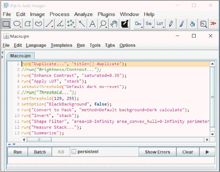
Generated macro showing the steps for quantifying image segments in Fiji.
acb-56-2-236-app-f5.tif
Table 1
Modified Johnsen scoring criteria
Spermatogenesis level Johnsen’s score
Many spermatozoa in the tubule lumen 10
Many elongated spermatids in the apical region of the epithelium 9
Many elongated spermatids still embedded in the Sertoli cell membrane 8
Few elongated spermatids, many round spermatids 7
No elongated spermatids, few round spermatids 6
No spermatids, many spermatocytes 5
Few spermatocytes 4
Spermatogonia cell only 3
Sertoli cells only 2
No seminiferous epithelial cells 1
Table 2
Mean testicular morphometry and component area fractions of testes from rats treated with A, cART, and both
Animal groups Control A cART A+cART
Testis morphometry
Testis weight (g) 1.91±0.05a 1.78±0.04a 1.85±0.03a 1.78±0.09a
Testis volume (mm3) 1.88±0.09a 1.83±0.08a 1.83±0.08a 1.67±0.11a
Testis size (mm) 1,493±40.52a 1,491±24.19a 1,378±46.83a 1,409±43.89a
AF (%)
EAF 74.25±0.50a 71.43±0.60b 71.82±0.53b 70.59±0.47b
LAF 8.56±0.29a 8.64±0.27a 7.38±0.24b 9.80±0.30c
CTAF 11.97±0.31a 13.66±0.35b 15.11±0.32c 15.26±0.33c
ISAF 4.93±0.12a* 5.73±0.17b 5.77±0.16b 4.99±0.12c*

For all parameters: Values in the same row with similar superscripts are not significantly different. Whilst values in the same row with different superscripts are significantly different except when the different superscripts are both marked with an asterisk (*). A, alcohol; cART, combination antiretroviral therapy; AF, area fractions; EAF, epithelial; LAF, luminal; CTAF, connective tissue; ISAF, interstitial space. EAF of A, cART, and A+cART groups significantly decreased relative to control (bP=0.0012, bP=0.0078 and bP<0.0001). LAF of cART group significantly decreased relative to control and A (bP=0.0123 and bP=0.0059) and A+cART significantly increased relative to control, A, and cART (cP=0.0141, cP=0.0271, and cP<0.0001). CTAF of A, cART, and A+cART groups significantly increased relative to control (bP=0.0017, cP<0.0001, and cP<0.0001) and cART and A+cART significantly increased compared to A (cP=0.0110 and cP=0.0038). ISAF of A and cART groups significantly increased compared to control (bP=0.0008 and bP=0.0003), but A+cART significantly decreased compared to A and cART (cP=0.0025 and cP=0.0010).

Table 3
Mean for seminiferous tubule, germinal epithelium cell count, and Leydig cell morphometry of testes from rats treated with A, cART, and both
Animal groups Control A cART A+cART
Tubule measurements
TA (µm2) 78,939±692a 68,854±773b 65,972±739c 72,069±803d
TD (µm) 310.2±1.30a 286.5±1.54b 283.0±1.52b 293.2±1.56c
EH (µm) 97.51±0.62a 93.34±0.71b 92.52±0.72b 89.46±0.78c
LD (µm) 97.97±3.54a 105.90±2.67a* 84.95±2.06b 116.9±3.38c*
Epithelium cell count
Spermatogonia 59.20±0.62a 67.93±1.50b 66.43±1.18b* 61.97±1.33a*
Spermatocytes 88.62±0.80a 86.00±1.00a* 84.33±0.71b* 83.72±0.95b*
Round spermatids 92.30±0.95a 80.41±1.47b 84.25±1.43b 83.79±1.18b
Elongated spermatids 102.9±1.02a 83.20±1.13b 85.93±1.48b 85.75±0.80b
Sertoli 23.61±0.24a 22.56±0.38a 22.75±0.28a 22.54±0.31a
Leydig cell nuclear
Diameter (µm) 5.85±0.04a 5.37±0.04b 5.48±0.05b 5.42±0.04b
Volume (µm3) 110.00±2.46a 85.95±2.18b 91.73±2.28b 88.03±2.13b

For all parameters: Values in the same row with similar superscripts are not significantly different. Whilst values in the same row with different superscripts are significantly different except when the different superscripts are both marked with an asterisk (*). A, alcohol; cART, combination antiretroviral therapy; TA, tubule area; TD, tubule diameter; EH, epithelium height; LD, luminal diameter. TA of A, cART, and A+cART groups significantly decreased compared to control (b,c,dP<0.0001), cART significantly decreased compared to A (cP=0.0413), and A+cART significantly increased compared to A and cART (dP=0.0155 and dP<0.0001). TD of A, cART, and A+cART groups significantly decreased compared to control (b,cP<0.0001) and A+cART significantly increased compared to A and cART (cP=0.0089 and cP<0.0001). EH of A, cART, and A+cART groups significantly decreased compared to control (bP=0.0002, bP<0.0001, and cP<0.0001) and A+cART significantly decreased compared to A and cART (cP=0.0007 and cP=0.0143). LD of cART group significantly decreased compared to control (bP=0.0340), A (bP=0.0004), and A+cART (bP<0.0001), whilst A+cART increased significantly (cP=0.0013) compared to control. Spermatogonia count of A and cART groups significantly increased compared to control (bP<0.0001 and bP=0.0002 respectively), and A+cART significantly decreased compared to A (aP=0.0032). Spermatocytes count of cART and A+cART groups significantly decreased compared to control (bP=0.0043 and bP=0.0007). Round and elongated spermatids significantly decreased (bP<0.0001) in treated groups A, cART, and A+cART compared to control. Leydig cell diameter and volume significantly decreased in A, cART, and A+cART groups compared to control (bP<0.0001).

Table 4
Median Johnsen’s score and histological changes observed and their score range in testes of rats treated with A, cART, and both
Animal groups Control A cART A+cART
Johnsen’s score 8.81a 8.29b* 8.50a* 8.38b*
Histological changes
Lifting of epithelium 0.06±0.01a 0.18±0.04a 0.09±0.02a 0.15±0.02a
(0–1) (0–2) (0–1) (0–2)
Widened intercellular space 0.06±0.02a 0.37±0.14a* 0.29±0.09a* 0.82±0.17b*
(0–1) (0–2) (0–2) (0–4)
Karyolysis 0.00±0.00a 0.11±0.02a 0.17±0.04b 0.13±0.03b
(0–0) (0–1) (0–1) (0–1)

For all parameters: Values in the same row with similar superscripts are not significantly different. Whilst values in the same row with different superscripts are significantly different except when the different superscripts are both marked with asterisk (*). A, alcohol; cART, combination antiretroviral therapy. Johnsen’s score of A and A+cART treated groups significantly decreased compared to control (bP=0.0115 and bP=0.0256). Widened intercellular space of A+cART group significantly increased compared to control (bP=0.0029). Karyolysis of cART and A+cART groups significantly increased compared to control (bP=0.0081 and bP=0.0346 respectively).

Appendix

Appendix

Image segmentation

Ilastik software (v1.3.3; https://www.ilastik.org) was used for image segmentation. Images were segmented into 3, 3’-diaminobenzidine tetrachloride (DAB) positive area and background [84, 85]. A pixel classifier was trained with eight sample images selected from all animal groups (two images per group). The available features i.e., intensity/color, texture, and edge at a sigma of 0.3–10 pixels were included in the classification algorithms. To train the classifier an appropriate brush was used to annotate a few DAB-positive areas (yellow color), and representative areas not stained with DAB were annotated as the background (blue color). To place the annotations precisely, the images were zoomed out. The prediction was inspected after adding each annotation. Then more annotations were added to all training images to refine the classifier and increase its applicability to large training naïve image sets. Once the prediction was of a good standard across the sample images, the trained classifier was applied to image sets, and their simple segmentations were exported for quantification (Fig. 1).

Image quantification

Segmented images were quantified using Fiji software (v1.52e; https://imagej.net/Fiji) into numerical values for statistical analysis [85]. The ilastik exported image segments were imported into Fiji through the ilastik plugin. Then, the Fiji scale was adjusted according to the magnification of the image. The parameters to measure (area and area fraction) were selected and limited to the threshold. The segments were analyzed using a newly generated macro shown in Fig. 2. The percentage area of expression for antibodies was calculated using the formula below.
TOOLS
Similar articles