

 PDF
PDF Citation
 Print
Print


Introduction
The increasing interest in individual facial and physical appearance has resulted in increased demand for cosmetic surgery procedures. In dermatology, the procedures, including neurotoxin injection and dermal filler injections are conducted for both facial rejuvenation and contouring as insignificantly invasive methodologies [1]. Owing to the increase in the number of cosmetic procedures, there have been reports of numerous complications. Predominantly, several cases of tissue necrosis resulting from direct occlusion of the arteries as a result of the particles of injected filler present chronic side effect [2]. The infraorbital, transverse facial, and facial arteries are the key blood supplies of the face [3]. Though the facial artery performs a major blood supply function on the face [4], it presents divergent inter-individual variations in humans.
Earlier studies, mostly those restricted to cadaveric researches, demonstrated the existence of discrepancies in facial arteries between individuals, mainly in terms of the branches, and also in angular branches [5, 6]. The comprehension of the detailed facial artery anatomy is, therefore, vital for the performance of facial surgery and other types of cosmetic procedures to avert the complications linked to facial arteries. Further, the infraorbital artery originates from the maxillary artery and mainly supplies the face’s infraorbital region [7]. Variations have also been observed in the infraorbital artery, particularly within the artery’s end branch and at the distal ophthalmic artery, and the facial artery’s angular branch [8]. The acquisition of more knowledge regarding the variations found in the infraorbital artery is important to the comprehension of the arterial face supply and to the reduction of potential complications resulting from cosmetic procedures and surgeries. The present study has utilized conventional angiography as the most appropriate assessment tool to aid in evaluating variations in the facial arteries. The choice of conventional angiography is informed by the observation that it has been widely employed in the assessment smaller vascular anatomy owing to the perfect spatial resolution and portrayal of vascular anatomy [9]. Conventional angiography has also been chosen owing to its attribute of occurring in real time, thereby enabling the assessment of dominant arteries related to the vessel territory by the operator in the course of performing the procedure [10]. As a technique, the conventional angiography’s digital subtraction methods enables the overcoming of the challenge of metal artefact than when the computed tomography (CT) angiography and ultrasonography is used. Also, the present study is among a limited number of researches that have evaluated the variations in facial arteries terminations through the use of conventional angiography.
Go to : 
Materials and Methods
For this study, the methodology entailed approval by the institutional review board and the subsequent waiver of the need to obtain informed consent owing to the research’s retrospective study design. The study involved the analysis of the anatomy of the infraorbital and facial arteries among patients undergoing carotid angiography for effective assessment of congenital anomalies, cerebral vascular related malformations, and intra-arterial procedures, including brain cerebral artery aneurysm coil embolization, and tumour embolization, carotid artery stent graft, and cerebral artery thrombolysis. To conduct the evaluation of variations in the facial arteries, the researcher also utilized a biplane angiography as it enabled the performance of external angiography.
Study participants
For the study, a total of 86 participants were recruited to partake in external carotid angiography examination. Of the participants, 64 were examined and underwent carotid angiography while 22 participants were excluded from the study owing to the poor quality of their angiography images, as they would not enable accurate examination of their infraorbital and facial arteries. Moreover, the 22 participants were also excluded from the study as a result of their images not having adequate fields for effectual discrimination of the different kinds of infraorbital and facial arteries. The 64 participants were further required to undergo a bilateral external carotid angiography, out of which 36 indicated intact facial arteries devoid of anomalies or variations.
Analysis of the images
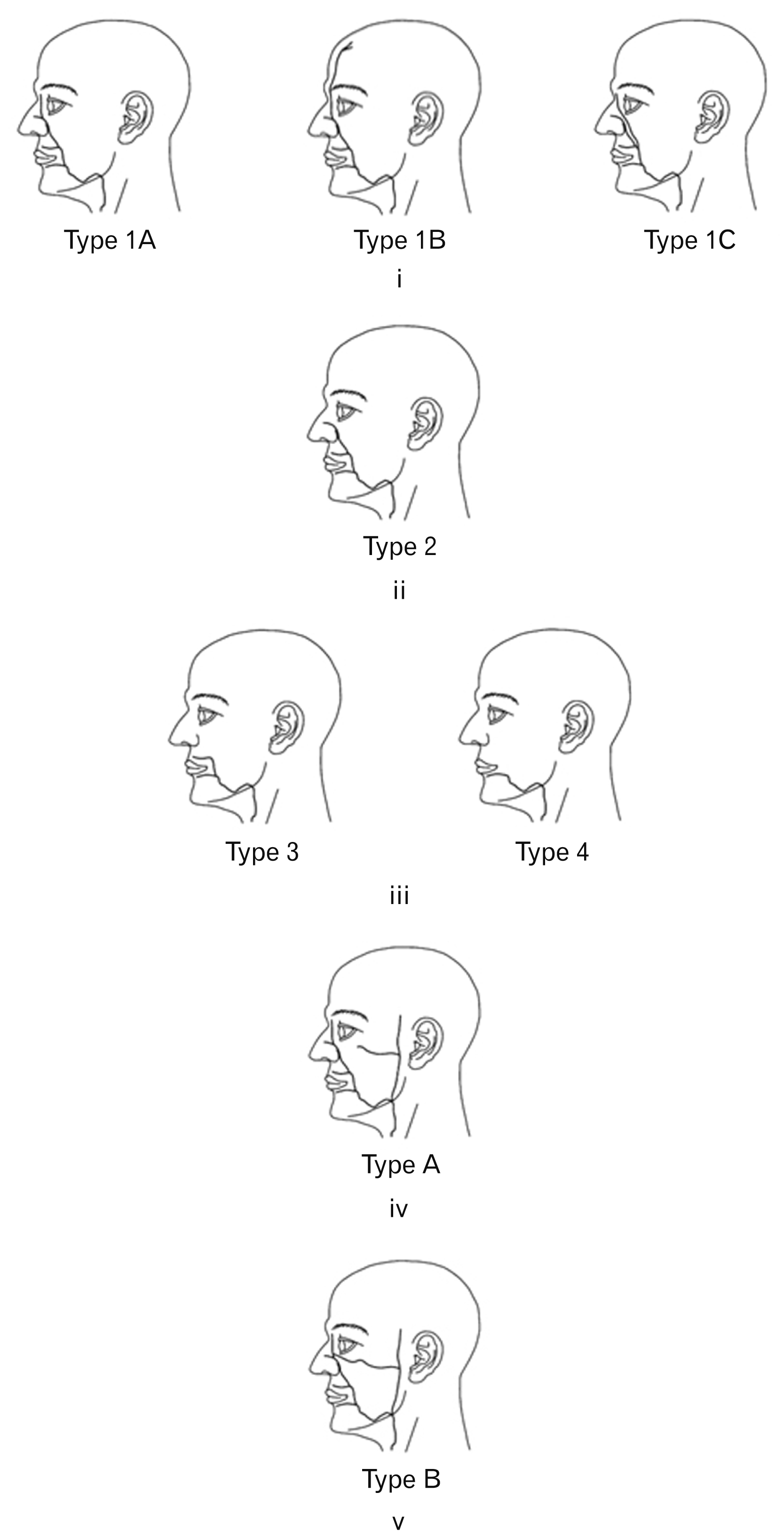
For the analysis, the research sought the help of two radiologists for a retrospective external carotid angiographies examination and interpretation. The reviewers arrived at the final decisions based on consensus on existence of dissonant cases. In this regard, the facial arteries courses were further placed into for distinct categories and the categorizations was mainly based on the final branching of the arteries. The distinct categories of facial arteries based on the arteries final branching have been aptly indicated in Fig. 1 [11, 12]. Further, the first category (Type 1) comprised facial arteries with angular branching that the final branch’s orbit midline as indicated in Fig. 2. The second category (Type 2) entailed facial arteries whose lateral nasal arteries whose final branching had or did not have alar branches as indicated in Fig. 3. The third category (Type 3) entailed facial arteries whose final branching comprised superior labial branches as indicated in Fig. 4. The last category of type 4 entailed facial arteries who, unlike type 3, had their final branching consisting inferior labial branches as indicated in Fig. 5.
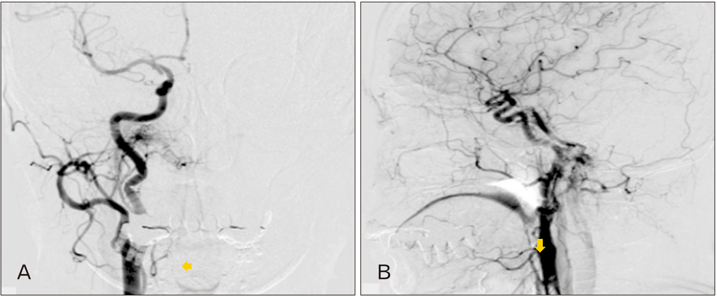
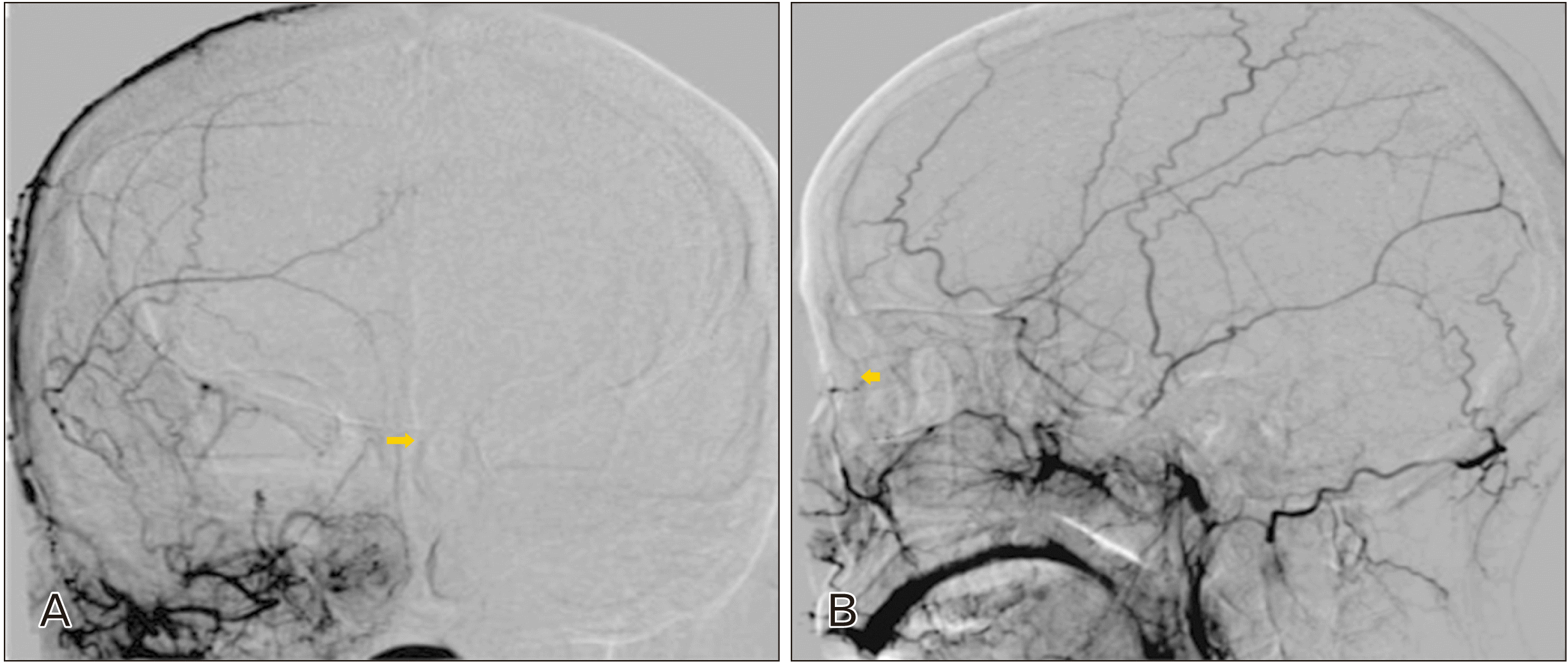 | Fig. 2Type 1A Facial artery and external carotid artery angiography of a cerebral infarction patient (female) who is 44 years old, with an angular branching of the facial artery observed without supratrochlear or a duplex branch. (A) Anteroposterior view (yellow arrow). (B) Lateral view (yellow arrow).
|
 | Fig. 3Type 2 facial artery variation and external carotid artery angiography of a female moyamoya disease patient aged 40 years. (A) Anteroposterior view (yellow arrow). (B) Lateral view (yellow arrow).
|
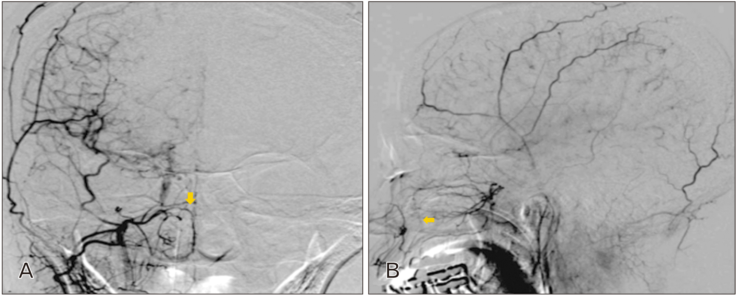
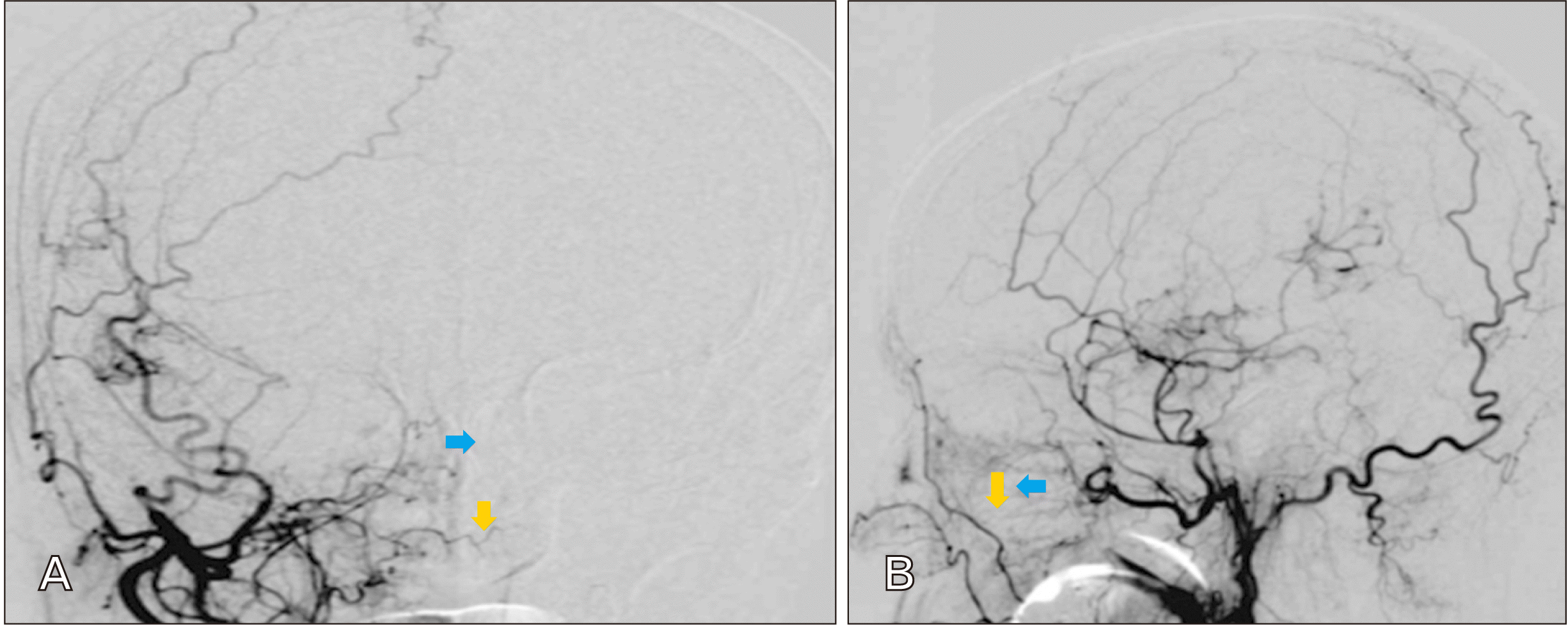
 | Fig. 4Type 3 Facial artery variation and am external carotid angiography of a 41 year old female cavernous fistula patient. For this patient, the facial artery’s superior labial branch, indicated using a yellow arrow, marks the end branch. (A) Anteroposterior view. (B) Lateral view.
|
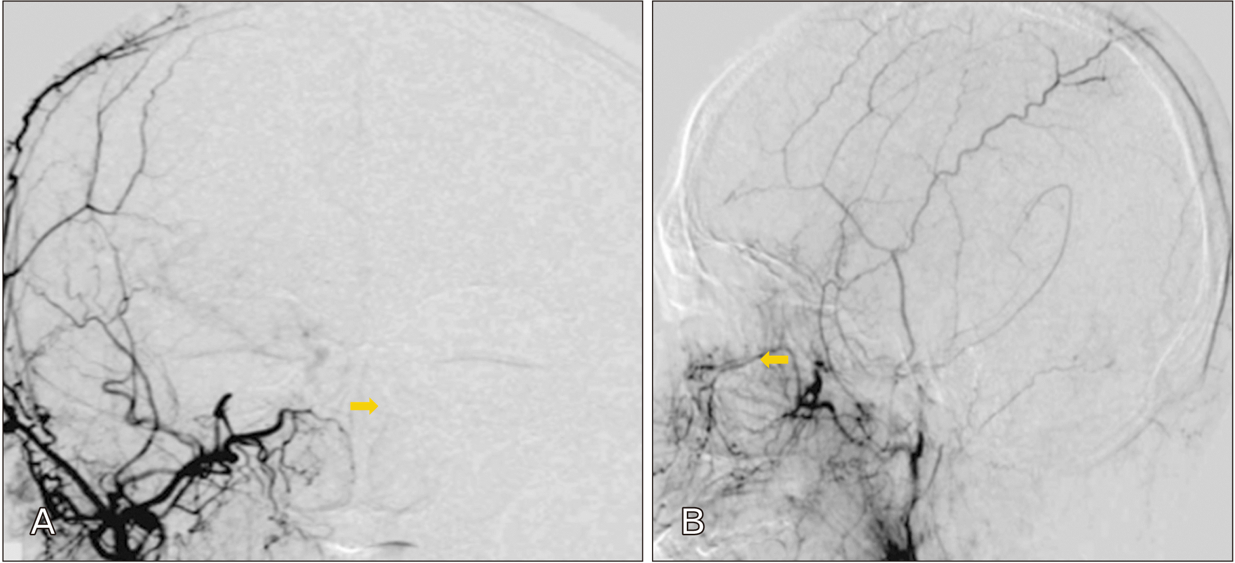
 | Fig. 5Type 4 facial artery variation and external carotid artery angiography of a dural arteriovenous fistula patient aged 57 years old. For this patient, only the facial artery’s inferior labial artery branches, indicated by the yellow arrow, can be observed, devoid of other branches. (A) Anteroposterior view. (B) Lateral view.
|
The first category (Type 1) was further categorized into three additional types, name Type 1A, Type 1B, and Type 1C. Thus, Type 1a category comprised facial arteries with angular branching that run along a traditional course and devoid of angular artery duplex and supratrochlear branching while Type 1B category comprised angular branching facial arteries with extensive supratrochlear branching that runs to the frontal bones as depicted in Fig. 6. Lastly, Type 1C category entailed facial arteries with angular branching with two duplexes occurring at the proximal facial arteries as indicated in Fig. 7.
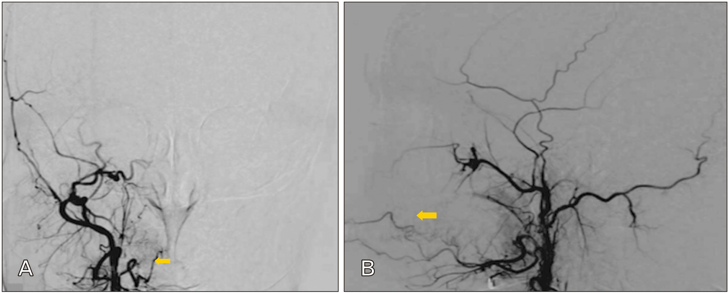
 | Fig. 6Type 1C Facial artery variation and external carotid artery angiography of a female spontaneous intracranial haemorrhage patient aged 65 years. Blue arrows indicate the angular artery’s duplex, which has been separated from the facial artery’s lateral nasal branch indicated using the yellow arrows. (A) Anteroposterior view. (B) Lateral view.
|
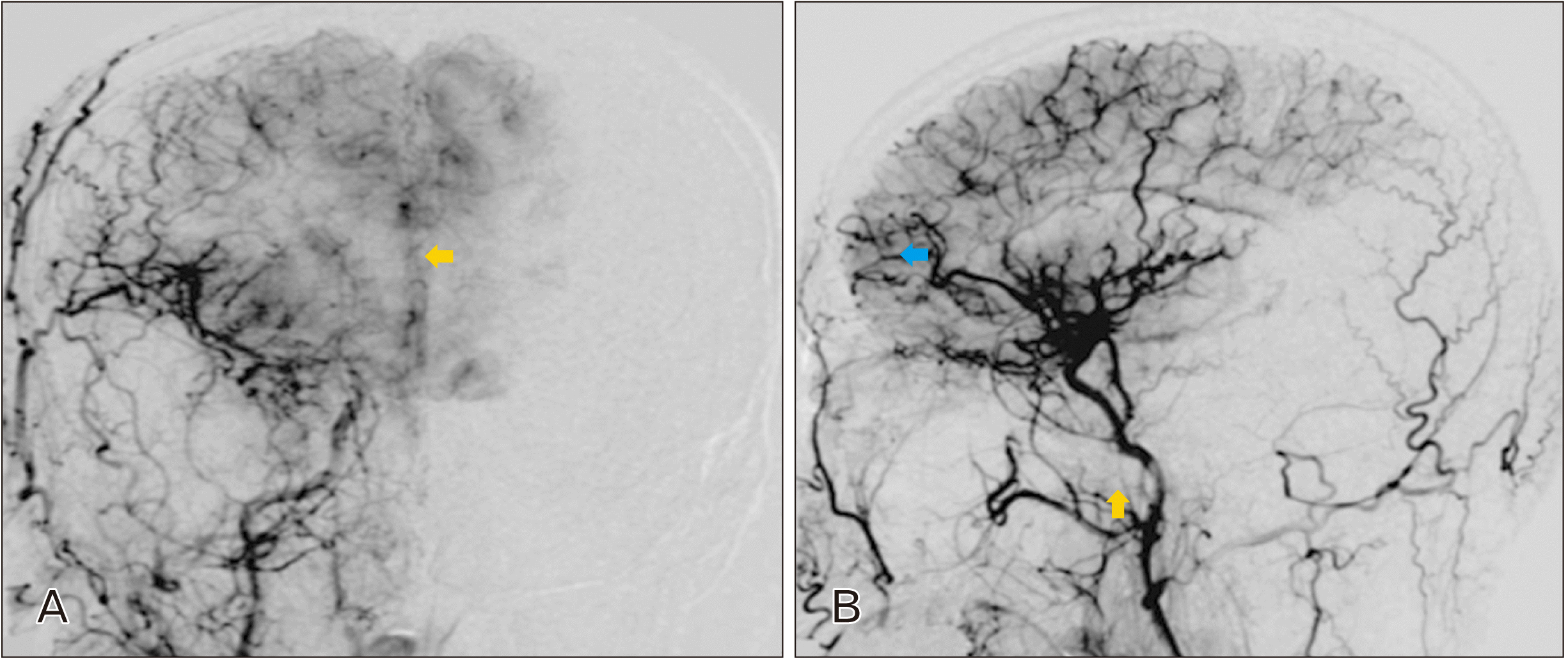
 | Fig. 7Type 2 facial artery variation and external carotid artery angiography photo of a moyamoya disease patient aged 70 years, and with maxillary artery (yellow arrow) indicating an infraorbital branching that has extended to the angular artery territory. (A) Anteroposterior view. (B) Lateral view.
|
The facial arteries’ symmetry was additionally characterized and the possession of comparable bilateral facial artery types was viewed as a symmetric pattern even as the possession of divergent facial artery type was perceived as being asymmetric. Further, the infraorbital artery courses were divided and categorized into two distinct kinds, namely Type A and Type A, with Type A being infraorbital arteries with anastomoses that further comprising extensive angular arteries that run towards the orbit’s anterior portion that is the angular artery’s territory as indicated in Fig. 7. Consequently, in Type B, the infraorbital arteries lacked anastomoses with angular branching and were not extensive as they were terminated and did not run towards the angular artery’s territory as indicated in Fig. 8.
 | Fig. 8Type 2 facial artery variation and external carotid artery angiography of a male cerebral infarction patient aged 55 years old. The blue arrow indicates the angular artery’s Supratrochlear branch extended to the frontal bone, and is categorized as Type 1B, even as the infraorbital artery, indicated by the yellow arrow ends prior to reaching the angular artery’s territory and has been categorized as Type 2. (A) Anteroposterior view. (B) Lateral view.
|
Go to :
Results
The facial arteries’ pattern of branching has been captured in Fig. 1. Of the 28 facial arteries variations examined, 35.7% (10 patients) were categorized as being Type 1, as their facial arteries termination occurred at the angular branching. The other 50.0% (14 patients) of the 64 cases were categorized as Type 2, given that the point of their facial artery termination was the lateral nasal artery and either had or did not have alar branching. Consequently, 7.1% (2 patients) of the cases were categorized as being Type 3, given that their facial artery termination points were the superior labial branching, even as 7.1% (2 patients) of the cases were categorized as Type 4 as they presented inferior labial branching.
Of the Type 1 category facial arteries, 10.0% (1 patient) was observed to meet Type 1B categorization, as they presented extensive supratrochlear branches that ran towards the frontal bones, even as another 10.0% (1 patient) was categorized as Type 1C, given his/her presentation of angular artery duplex. The remaining 80.0% (8 patients) presented a conventional angular artery course, and were categorized as Type 1A, even as they did not have angular artery duplex and supratrochlear branching. Of the 10 patients that were subjected to bilateral external carotid angiography, 70.0% (7 patients) present symmetric pattern facial artery type while 30.0% (7 patients) presented asymmetric pattern facial artery type. With regard to the symmetric facial artery type, of the 28 patients, 32.1% (9 patients) were categorized as Type 1, 57.1% (16 patients) were categorized as Type 2, while 7.1% (2 patient) were categorized as Type 3, and the remaining 3.6% (1 patient) classified as Type 4. Further, regarding the infraorbital arteries, it was observed that 57.1% (16 patients) of the cases were categorized as Type A, given that they presented anastomoses with extensive angular artery running towards the angular artery territory as indicated in Fig. 1 (Type A). Furthermore, the other 42.9% (12 patients) of the cases were categorized as Type B, given their lack of correlations with angular artery as indicated in Fig. 1 (Type B).
Go to :
Discussion
The increment in cosmetic procedures in the recent times has resulted in increased complications ranging from mild and short-term to severe and permanent, such as infection, tissue necrosis, and erythema, blindness due to arterial occlusion due to injection of filler particles, nodular masses, and swelling. In this regard, several studies have maintained that visual loss often occurred as a result of a retrograde arterial occlusion in the ophthalmic artery from either the dorsal nasal artery or supratrochlear [13, 14]. Reducing such complications risk require the attending physicians to have an in-depth knowledge and understanding of anatomy, including the precise course, variations, and locations of the different facial arteries prior to carrying out any kind of cosmetic procedure. Regardless of the numerous earlier researches on facial artery anatomy, the locations, and courses, there has been a great and considerable divergence in the results and there has not been any consensus among researchers [15, 16]. Furthermore, a number of the earlier researches have been limited owing to their focus on cadaveric researches [6, 17, 18].
An initial research conducted with regard to facial arteries distributions among Japanese citizens disclosed that the ending of facial arteries was angular in 16% of the study population, and lateral in 64% of the population [17]. A comparable research performed by dissecting cadavers belonging to adult French citizens disclosed that only 4% of the study population presented angular facial artery ending while 78% of the study population had a lateral nasal ending [19]. On the contrary, a comparable research conducted on adult British citizens cadaver specimens disclosed angular facial artery ending in 68% of the study population even as 26% of the study population presented lateral nasal artery endings [20]. Still, a cadaveric specimen based Korean research disclosed that 41% of the study population presented angular facial artery ending while 44% of the study population had lateral nasal facial artery ending [6]. Also, in studying variations with regard to the facial arteries branching distributions, various studies have disclosed that the ratio angular ending facial arteries was 51%, 22%, and 20%, correspondingly [4, 18].
Numerous potential causes with regard to the observed considerable facial artery anatomical variations in earlier studies. Though the variations were initially perceived as due to divergences in races, the divergent distribution patterns of facial arteries have failed to reflect racial dissimilarities owing to the disclosure that the facial arteries distribution of mongoloids, particularly the Japanese and Koreans, was different [6]. Nevertheless, the findings of the present research are comparable to those of a research conducted by Wang et al. [21], using cadaver specimens of Indians, in comparison to studies with British, Japanese, and French specimen. In addition to the facial artery variations being attributed to racial divergences, it is anticipated that the environment also plays a key role in the variations. The present angiographic study is, however, divergent from earlier researches that utilized cadaveric specimen. For instance, the angiographic study offer a research methodology that enabled the researcher to observe the improved artery’s inner lumen as a result of the contrast agent injection (intravenous) while cadaveric researches are not only static but also restricted to surface anatomy [5]. As such, the researcher opted for angiography owing to its aptitude to detect deeper and superficial arterial branches, even as the contrast is able to disclose the blood vessels with smaller diameters making it possible to differentiate using naked eyes. Further, anatomic dissection of cadavers may not offer precise reflection of the vascular status as well as physiological blood flows, and in the course of contrast injection, occlusion of vessels resulting from extant air bubbles and blood clots in the arteries are unlikely to correspond. Also, factors that include the injection force have the ability to alter the diameter of an artery, even as the liquid dye might have an impact on the study findings owing to its thickness.
A recent evaluation of the facial artery branching through the use of tools for non-invasive imaging, including CT angiography, disclosed considerable divergences from the findings of cadaveric researches [22]. In this regard, the present study disclosed that the percentage of angular branch ending facial arteries like lateral nasal branch was comparatively lower at 26%, even as termination occurred at the inferior and superior labial branching, and this was comparatively high in relation to findings of earlier similar studies. A comparable study that utilized CT Angiography was conducted by Koziej et al. [23], and disclosed a higher percentage of angular branching facial arteries at 44%, compared to the finding of the study conducted by Furukawa et al. [5], and lower proportion of inferior and superior labial branching facial artery terminations at 56%. The differences observed with regard to the percentage in the facial artery types between the present study and early studies that utilized CT angiography can be attributed to divergence in classification. Nevertheless, in earlier studies, such as those conducted by Koziej et al. [23] and Furukawa et al. [5], there was no separation of lateral nasal arteries, as they were considered to have angular branching even though the present research has distinguished them from angular branching as initially done in cadaveric researches. The observed variations might be attributed to dissimilar specificities and sensitivities for the examination of the peripheral arteries between CT angiography and conventional angiography [24]. The conventional angiography has, however, been used as the gold standard of references for evaluation of the peripheral vessels owing to its higher rate of precision alongside spatial resolution with regard to detection of the diseases of the peripheral vessels [5].
The symmetry of facial arteries had been studied initially, including in studies conducted by Perrini et al. [25], and Niranjan [20] that reported 68% symmetry, and studies by Furukawa et al. [5] and Lohn et al. [4] reported 53% symmetry correspondingly. Further, it is worth noting that in the present study, the facial artery’s symmetric distribution was noted in 70% of the participants. A study conducted by Koh et al. [6] reported angular artery ending in 50% of symmetric facial arteries, and a further lateral nasal artery ending in 46% of the symmetric facial arteries. In instances where asymmetry was observed, it was noted that while one side had a lateral nasal artery ending, the other presented a superior labial artery ending. Consequently, in instances where asymmetric facial arteries were observed, a larger proportion of the cases had lateral nasal artery endings while the opposite side had contralateral superior labial artery endings. Such findings corroborates the outcomes of the study conducted by Lohn et al. [4], which concluded that the developmental divergences in individuals are likely to have impacted the facial arteries patterns.
Still, arterial blood to the soft tissue muscles and organs found in the neck and head regions is mainly supplied by the maxillary artery alongside its different branches. At certain points, the masseter and infraorbital arterial branches anastomose with the facial arteries’ angular branching. Further, occasional connection of the maxillary artery’s infraorbital branching to distal ophthalmic artery was also observed by Park et al. [10]. In the present study 58% of the cases with infraorbital branching located within the maxillary arteries share both the anastomoses and the supplying territory with angular arteries as observed in the external carotid angiography conducted on the participants. Under such instances of infraorbital branching, partly compensated supply of blood originating from maxillary artery may be anticipated under circumstances where injury occurs to the facial artery during procedures that include the injection of dermal filler. Consequently, the maxillary artery’s infraorbital branch may be the probable route leading to occlusion of the ophthalmic artery as a result of particles of dermal filler [26]. Also, owing to the extant differential variations within angular arteries, both the angular region and the infraorbital region might turn out to be increasingly dangerous region as a result of the potential for occlusion of the ophthalmic artery owing to the facial artery’s variable anastomotic regions that have retrograde flows [27].
The present study experienced two key limitations, including the use of retrospective research design and the inability of the readers to identify the smaller vessels that received inadequate contrast dye from facial arteries. Nonetheless, such underestimation of the research findings were minimized through allocation of adequate time for performing angiography, which was conducted till such a time that the external carotid artery’s draining veins became perceptible to the operator.
In summary, the present study was conducted with the objective of identifying variations in the facial arteries branching patterns through the use of CT angiography, and disclosed significant findings. The findings of the present research are important given that they will aid surgeons and other physicians to effectively avoid extreme vascular complications in the course of facial procedures. Further, the present study forms an important basis for future studies on facial arteries and other arteries through the use of various tools, including CT angiography, MRI, and ultrasonography tools.
Go to :
 XML Download