Journal List > Anat Cell Biol > v.56(2) > 1516083075

Bansal, Jain, Budhiraja, Swami, and Gupta: A cadaveric study of arteriovenous trigone of heart: the triangle of Brocq and Mouchet

Abstract

Left coronary artery divides into anterior interventricular branch and circumflex branch. As both the arteries run in their corresponding grooves, an arteriovenous trigone is formed between conus arteriosus and left auricle called triangle of Brocq and Mouchet. The triangle base is formed by great cardiac vein. This study aims to describe the frequency of triangle and its type and relationship between various boundaries and content of triangle and to supplement the existing knowledge of clinicians. This observational and descriptive study was conducted on 40 formalin fixed cadaveric hearts in department of anatomy, Kalpana chawla government medical college. The triangle was found in 92.5% of specimen with most common type being closed (51.3%) which is followed by inferiorly open in 35.1%, superiorly open in 8.1% and completely open in 5.4% hearts. Most frequent content of triangle was median artery followed by diagonal branches of anterior interventricular and circumflex branches. The mean area of the triangle was 246.3 mm2. Relationship of vein with two arterial branches was either superficial or deep. The knowledge of different patterns of existence will be required for angiographic procedures. Further the triangle is a potential epicardial access route to left fibrous ring. Thus detailed knowledge of variations will help cardiologist to achieve better outcome in interventional procedures with minimal complications.

Introduction

Great cardiac vein (GCV) is the main tributary of coronary sinus. It starts from cardiac apex, run in anterior interventricular groove either left or right to the anterior interventricular branch (AIB) to the base of ventricles. It then curves around the left margin of heart and reach to coronary sulcus. After merging with oblique vein of left atrium it forms coronary sinus which drains into right atrium. When the GCV crosses the left border of heart it forms the base of an arteriovenous triangle called triangle of Brocq and Mouchet. The other two boundaries of this triangle are formed by AIB and circumflex branch (CB), branches of left coronary artery (LCA). It is topographically located between conus arteriosus and left auricle. This arteriovenous triangle is classified into four types depending on the arrangement of its three boundaries. Closed, inferiorly open, superiorly open, completely open type [1]. Rarely triangle may be absent. The trigone bears variable relationship with its boundaries. Sometimes GCV is superficial or deep to both AIB and CB or one of them. At times when it is deep, chances of compression by atherosclerotic coronary arteries are higher and impede the venous return and aggravates cardiomyopathy [2]. Contents of this triangle are variable usually median branch arising from LCA or diagonal branches arising from AIB and CB. The contents may pass superficial or deep to GCV. The importance of knowing the variable pattern of existence of this triangle lies in the fact that it is important while doing surgical procedures on heart and interpreting radiology and catheter based procedures. The triangle is commonly approached while performing intravascular ultrasound of coronary arteries. The initial segments of LCA bifurcation belongs to the limits of trigone, an area full of small caliber vessels and subjected to frequent surgical procedures and there is significant risk of injury. The purpose of this clinical anatomy research is to supplement the existing knowledge of clinicians regarding the incidence of triangle and frequency of type of triangle, conformation and content of aforementioned triangle.

Materials and Methods

This observational and descriptive study was conducted in department of anatomy, Kalpana chawla government medical college, Karnal. Forty human cadaveric hearts were obtained in the department as a part of routine undergraduate teaching schedule. The cadavers were procured as a part of voluntary body donation program. The study was exempted from the institutional ethical clearance as all the specimens were collected from voluntarily donated cadavers to the department, obtained with prior consent from the immediate kin regarding the use of body for dissection and research purposes. All the specimens had been fixed in 10% formalin solution. Since the collected specimens were used, we could not document the exact age and gender of source cadaver.

Procedure

After procuring, the specimens were washed in running tap water and microdissection was done to remove the epicardial fat along the course of two terminal branches of LCA. GCV was traced from apex of heart to its termination in coronary sinus. During the dissection, utmost care was taken not to disturb the normal anatomy of triangle. All the heart specimens with intact atria and ventricles and preserved coronary vasculature were included in study. Specimens with grossly evident cardiomyopathy and disturbed coronary vasculature were excluded from the study.
Following observations were noted:
1. Incidence of trigone
2. Frequency of type of trigone
3. Content of trigone
4. Area of trigone
- The relationship of GCV with AIB and CB
The length of boundaries of complelety closed trigone were measured with the help of a digital vernier caliper and area of trigone was calculated using the Heron’s formula.
A=PPa.Pb.Pc
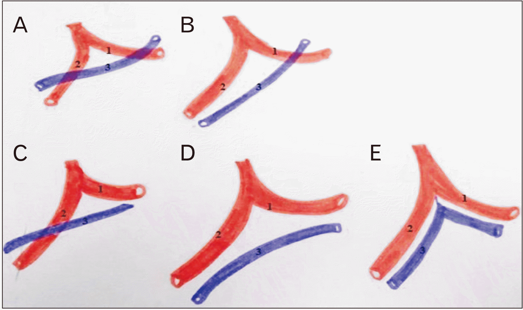
Where P represents the semi-perimeter (sum of the sides divided by two) and a, b, c represents the extensions of the sides of the trigone. All the specimens were photographed and later the data obtained was analyzed and compared with previous studies. The triangle so formed is further classified while following the recommendations given by previous studies [1, 3] and a schematic is drawn in Fig. 1.
Closed: If the GCV crosses AIB in its ascending trajectory and then crosses CB in coronary sulcus.

Inferiorly open

If GCV lies to the left of AIB in its ascending trajectory and crosses the CB either superficial or deep.
Superiorly open: If the GCV crosses only AIB (superficial or deep) during its course in interventricular groove and lies inferior to CB without crossing it.
Completely open: If the GCV lies to the left of AIB in anterior interventricular groove and while curving to the left, it lies inferior to LCA and then inferior to CB.
Absence of the triangle: When the GCV lies side by side with AIB and while curving the left border of heart ascends to the bifurcation of LCA and then accompanies the CB.

Results

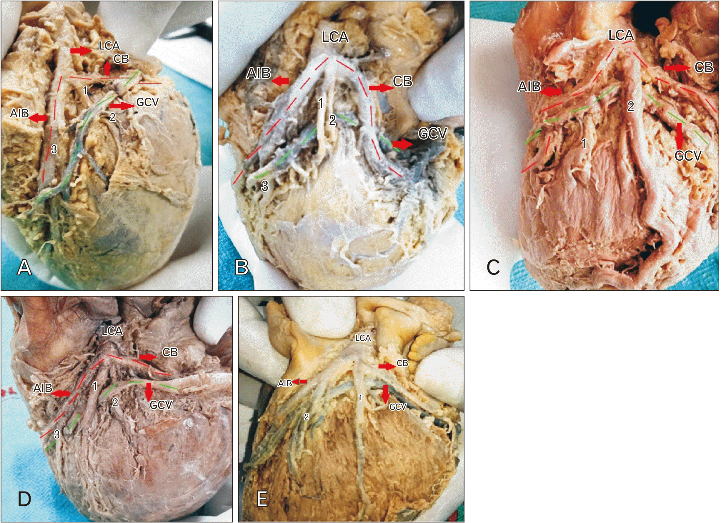
Forty cadaveric hearts were observed for the presence of triangle of Brocq and Mouchet. The triangle was found in 37 hearts (92.5%) with no such formation seen in 3 (7.5%) hearts. Regarding the type of triangle the pattern of distribution was found to be closed in 19 (51.3%), inferiorly open in 13 (35.1%), superiorly open in three (8.1%) and completely open in two (5.4%) hearts as shown in Fig. 2. Regarding the content of triangle, in 37 hearts where the triangle was present, diagonal artery is the content in 100% cases which arose either from the trunk of LCA, or as a branch from AIB or CB. Diagonal artery originated from LCA was referred as Median Artery [4] and branch which arose from AIB or CB were referred as diagonal artery. In present study out of 37 triangles, median artery was found in 20 (54.1%) hearts and diagonal arteries were found in 17 hearts. Out of 20 hearts one median was found in 15 (40.5%) and two median in 5 (13.5%) hearts. Out of 17 hearts, in 12 cases (32.4%) one diagonal arose from AIB and in 4 (10.8%) one diagonal each from AIB and CB and in one (2.7%) heart two diagonal from AIB were seen. The mean area of completely closed triangles was calculated and found to be 246.3 mm2, ranging from 659.5 mm2 to 7.3 mm2. The relationship of GCV with AIB and CB were observed as. In all 51.3% of closed trigone, GCV was superficial to both AIB and CB in 37.8% and deep to both in 13.5% hearts. The pattern observed in superiorly open type of trigone was, GCV crossed AIB anteriorly in 8.5% hearts. GCV crossed CB posteriorly in 35.1% inferiorly open type of trigone.

Discussion

The trigone is the site of arteriovenous confluence, formed as GCV ascends and crosses the branches of LCA and forming the triangle of Brocq and Mouchet. The triangle of Brocq and Mouchet was present in 37 (92.5%) hearts and absent in 3 (7.5%) hearts. Above incidence when compared with previous studies which all were cadaveric in nature, it was found to be similar with kulkarni and Ramesh [5], Kharbuja et al. [4], Roy and Dubey [6] with a slight difference with studies done by Sousa-Rodrigues et al. [1] and Andrade et al. [7] as shown in Table 1. The difference in incidence might be due to difference in sample size. Regarding the frequency of type of trigone, pattern found was closed in 19 (51.3%) hearts, followed by inferiorly open in 13 (35.1%), superiorly open in 3 (8.5%) and least was completely open in 2 (5.4%) hearts. Present study was comparable with the studies done by Bharathi and Sathyamurthy [8] and Roy and Dubey [6] which also found closed type of triangle in maximum cases followed by inferiorly and superiorly opens and least was completely open. However it got a difference in observation with the studies done by Sousa-Rodrigues et al. [1] Ortale et al. [2], Kulkarni and Ramesh [5], Suma and Shanthini [9] and Kharbuja et al. [4] as shown in Table 2. This might be explained by the fact that sometimes during the removal of epicardial fat and clearing the course of vessels, normal anatomy get disrupted and that results in difference in frequency of each type of triangle. Since the incidence of presence of triangle was 92.5% and frequency of closed type of trigone was most common, it will give an fair idea to the clinician before the procedure on the area whether for implanting bypass grafts, or repairing the left fibrous ring as the shortest distance between coronary venous circle and the left fibrous ring is 5.2 mm at the level of anterolateral commissure of mitral valve. It is the same distance where GCV forms the base of triangle of Brocq and Mouchet. This may be the site of adherence of GCV and myocardium of left ventricular wall. The knowledge of the triangle is essential for coronary sinus cannulation for catheter treatment of arrhythmia and left ventricular pacing as GCV may flex or kink during the procedure [10]. Regarding the content of trigone, one median artery was found in 15 (40.5%) and two median in 5 (13.5%) hearts. Diagonal artery in the form of median artery was present as the content of the triangle in 39.3% cases reported by Kharbuja et al. [4], 40% by Kalpana [11] and 65.3% by Kulkarni and Ramesh [5]. In present study, in 12 (32.4%) hearts one diagonal arose from AIB and in 4 (10.8%) one diagonal each from AIB and CB and in one (2.7%) heart two diagonals from AIB were seen. Kharbuja et al. [4] reported diagonal branches from CB were present in eight (28.6%) and diagonal branches from both AIB and CB in eight (28.6%) hearts varying in number from 1 to 2 diagonal branches from each. Relationship of vein with arteries forming the triangle had been studied by various authors. Present study found that GCV to be superficial to both AIB and CB in 37.8% and deep to both in 13.5% in all 51.3% of closed trigone. The pattern observed in superiorly open type of trigone was GCV crossed AIB anteriorly in 8.5% hearts. GCV crossed CB posteriorly in all inferiorly open type of trigone 35.1% as shown in Table 3. However Kharbuja et al. [4] found GCV was not crossing any arterial branches in 6.7% (absence of triangle), GCV superficial to both branches in eight (33.3%), GCV deep to both branches in four (16.7%), superficial to one and deep to another in 12 (50.0%), parallel to AIB and crossed CB in four (14.3%), parallel to CB and crossed AIB in one (3.6%). Andrade et al. [7] found that GCV is parallel to AIB and cross the CB anteriorly in 6 (26.0%) hearts. Sousa-Rodrigues et al. [1] reported that GCV is parallel to AIB and crossed CB anteriorly in 12 (52.0%) cases, GCV is posterior to AIB and cross CB anteriorly in 5 (22.0%) cases and no heart was found where GCV crossed both the branches. Nevertheless on comparison relationship differs between the studies that indicate further studies to be conducted to know the relationship between GCV and both arterial branches. If the position of GCV lies deep to either of rigid wall of AIB or CB, it may cause compression of the vein resulting in altered venous return. Gerber et al. [12] suggested that percutaneous coronary artery bypass grafting is not suitable in individuals with crossing of AIB and GCV since the site of overlap can mask the calcification of artery. After observing the different pattern of relationship of GCV with AIB and CB in a trigone, authors formed following classification and a schematic is drawn in Fig. 3.
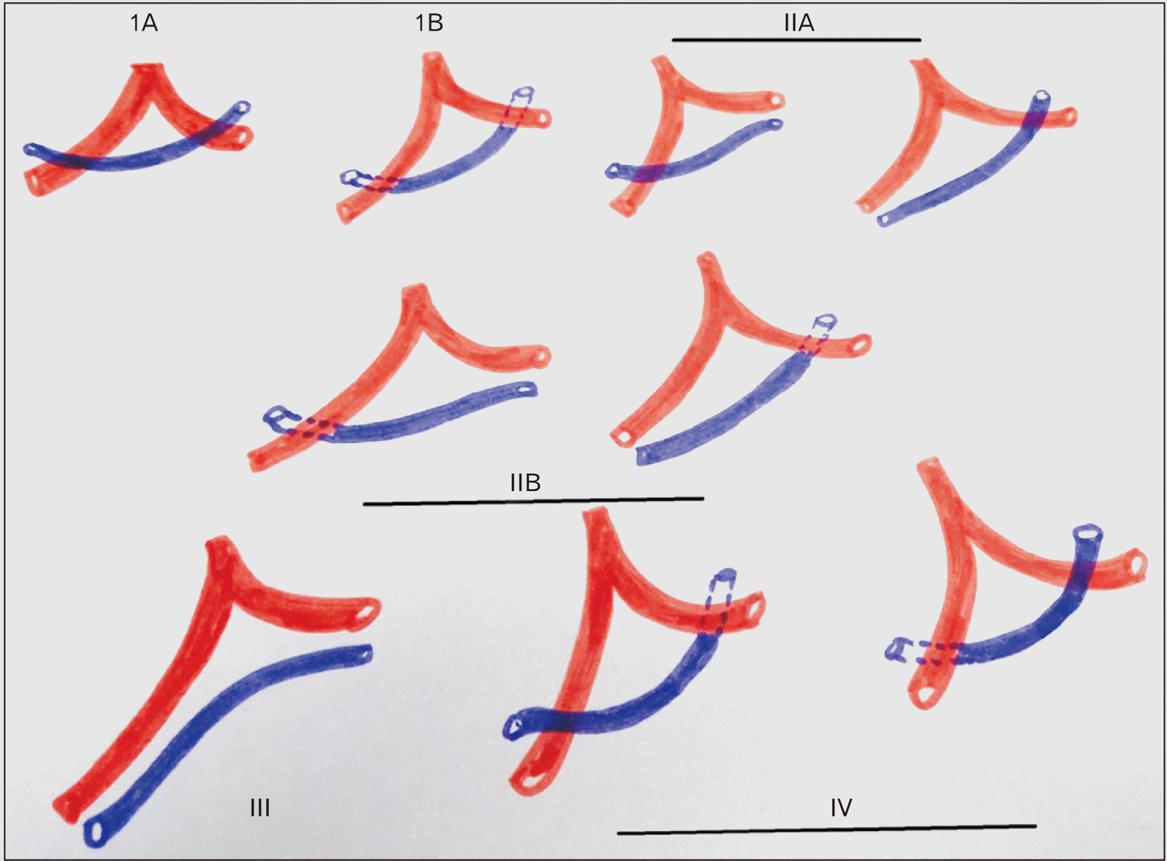
Type IA: Anteriorly crossing both AIA and CA
Type IB: Posteriorly crossing both AIA and CA
Type IIA: Anteriorly crossing one of branch either AIA and CA and parallel to another
Type IIB: Posteriorly crossing one of branch either AIA and CA and parallel to another
Type III: Not crossing any branch either AIA and CA
Type IV: Anterior to one and posterior to another
According to above classification maximum pattern observed in present study was type IA followed by type II A and type II B and least was type III. After comparing the relationship of GCV with both arterial branches, present study studied the relationship between GCV and content of trigone i.e. diagonal branches. Authors found three type of arrangement 1) GCV is anterior to median artery 2) GCV is posterior to median artery 3) GCV is anterior to some and posterior to other diagonal branches. In majority of cases in present study GCV is deep to median artery and the median artery was too of big lumen and it other cases where there were small diagonal branches, then mostly GCV is anterior to contents. When comparing with previous studies von Lüdinghausen [13] indicated that in 70% of the cases the GCV passes anterior to the diagonal artery and that in 29% it does so posteriorly. Sousa-Rodrigues et al. [1] reported that MCV crossed anteriorly and posteriorly to the diagonal artery in 57% and 43%, respectively. James [14] reported that when minor ventricular branches were present inside the trigone, they were crossed anteriorly by the GCV in a greater percentage (52%) than posteriorly (26%) means anterior crossing is frequent. The content of triangle was median artery in all the hearts. The specialty of median artery is that it does not run in any cardiac groove and has unusual course in human beings. The median artery is source of anterior septal branches and to anterior papillary muscle of left ventricle. Its presence reduces risk of atherosclerosis of left anterior interventricular artery and circumflex coronary artery. Median artery is a constant feature of rabbit, monkey, squirrel monkey and pig hearts [15]. In conclusion the incidence of closed type of trigone with median artery as a predominant content is found in maximum hearts. The mean area of the triangle was 246.3 mm2. The results of this research will complement the anatomical knowledge of the vascular trigone in question and therefore will be a contribution to the surgical anatomy of this important organ. Thus detailed anatomical knowledge of the triangle will help cardiologist to achieve results with least complications.

Acknowledgements

We would like to express our sincere gratitude to all the staff of Anatomy Department for their valuable support and encouragement.

Notes

Author Contributions

Conceptualization: SB, RJ, VB. Data acquisition: SB, SS, RG. Data analysis or interpretation: SB, RJ. Drafting of the manuscript: SB. Critical revision of the manuscript: SS, RG. Approval of the final version of the manuscript: all authors.

Conflicts of Interest

No potential conflict of interest relevant to this article was reported.

References

1. Sousa-Rodrigues CF, Alcantara FS, Silva WNV, Alcantara FS, Olave E. 2004; Arterio-venous trigone of the heart (Brocq & Mouchet trigone). Int J Morphol. 22:291–6. DOI: 10.4067/S0717-95022004000400009.
2. Ortale JR, Gabriel EA, Iost C, Márquez CQ. 2001; The anatomy of the coronary sinus and its tributaries. Surg Radiol Anat. 23:15–21. DOI: 10.1007/s00276-001-0015-0. PMID: 11370136.
3. Pejkovic B, Bogdanovic D. 1992; The great cardiac vein. Surg Radiol Anat. 14:23–8. DOI: 10.1007/BF01628039. PMID: 1589843.
crossref
4. Kharbuja R, Basnet L, Dhungel S. 2020; Anatomical study of triangle of Brocq and Mouchet in human cadaveric heart. Nepal Med Coll J. 22:111–7. DOI: 10.3126/nmcj.v22i3.32626.
crossref
5. Kulkarni V, Ramesh BR. 2014; Incidence of triangle of Brocq and Mouchet and median artery as its content in south Indian cadaveric hearts. Clin Res. 6:4–9.
6. Roy SS, Dubey A. 2016; Triangle of Brocq and Mouchet: an anatomical study in human cadaveric heart and its clinical significance. Int J Anat Res. 4:2266–8. DOI: 10.16965/ijar.2016.192.
crossref
7. Andrade FM, Ribeiro DC, Babinski MA, Cisne R, Goes ML. 2010; Triangle of Brocq and Mouchet: anatomical study in Brazilian cadavers and clinical implications. J Morphol Sci. 27:127–9.
8. Bharathi D, Sathyamurthy B. 2013; An anatomical study of triangle of Brocq & Mouchet in human cadaveric heart & its clinical relevance. J Dent Med Sci. 8:12–5. DOI: 10.9790/0853-0821215.
crossref
9. Suma HY, Shanthini S. 2019; Relationship of great cardiac vein in the triangle of Brocq and Mouchet- a corrosion cast study. Int J Anat Res. 7:6437–42. DOI: 10.16965/ijar.2019.137.
crossref
10. Kavimani CF, Jebakani CF. 2014; Coronary sinus. World J Med Sci. 10:61–4. DOI: 10.53347/rid-32061.
11. Kalpana R. 2003; A study on principal branches of coronary arteries in humans. J Anat Soc India. 52:137–40.
12. Gerber TC, Sheedy PF, Bell MR, Hayes DL, Rumberger JA, Behrenbeck T, Holmes DR Jr, Schwartz RS. 2001; Evaluation of the coronary venous system using electron beam computed tomography. Int J Cardiovasc Imaging. 17:65–75. DOI: 10.1023/A:1010692103831. PMID: 11495511.
13. von Lüdinghausen M. 1987; Clinical anatomy of cardiac veins, Vv. cardiacae. Surg Radiol Anat. 9:159–68. DOI: 10.1007/BF02086601. PMID: 3120334.
crossref
14. James TN. 1965; Anatomy of the coronary arteries in health and disease. Circulation. 32:1020–33. DOI: 10.1161/01.CIR.32.6.1020. PMID: 5846099.
crossref
15. Nikolić V, Blagojević Z, Stijak L, Mališ M, Parapid GT, Stanković G, Spasojević G, Filipović B. 2011; The third branch of the main trunk of the left coronary artery in Cercopithecus aethiops sabaeus. Is the nonhuman primate model appropriate? Anat Rec (Hoboken). 294:1506–10. DOI: 10.1002/ar.21445. PMID: 21809460.
crossref

Fig. 1
Schematic classification of triangle of Brocq and Mouchet, where 1, circumflex branch; 2, anterior interventricular branch, 3, great cardiac vein. (A) Closed, (B) inferiorly open, (C) superiorly open, (D) completely open, (E) absence of triangle.
acb-56-2-205-f1.tif
Fig. 2
Light photographs of adult cadaveric human hearts showing various types of triangle of Brocq and Mouchet. (A) Left view of Heart; completely closed triangle, GCV crosses both AIB and CB. 1, diagonal artery arising from CB. (B) Inferiorly open triangle. GCV (green dotted line) crosses CB only. 1, 2, median artery arises from LCA; 3, diagonal artery arises from AIB. (C) Superiorly open triangle, GCV (green dotted line) crosses AIB only. 1, diagonal artery from AIB; 2, median artery from LCA. (D) Completely open triangle. GCV neither cross AIB nor CB. 1, median artery is superficial to GCV; 2, another median artery which is deep to GCV; 3, diagonal artery from AIB. (E) Absent triangle. GCV is reaching to root of LCA bifurcation. 1, median artery from LCA; 2, diagonal from AIB. GCV, great cardiac vein; AIB, anterior interventricular branch; CB, circumflex branch; LCA, left coronary artery.
acb-56-2-205-f2.tif
Fig. 3
A schematic diagram showing various possibilities of relationship between great cardiac vein with anterior interventricular branch and circumflex branch.
acb-56-2-205-f3.tif
Table 1
Comparing the incidence of triangle of Brocq and Mouchet from previous studies
Authors Sample size Presence of triangle (%) Absence of triangle (%)
Kharbuja et al. [4] 30 93.3 6.7
Kulkarni and Ramesh [5] 52 93.3 3.0
Andrade et al. [7] 23 86.9 11.1
Sousa-Rodrigues et al. [1] 26 88.0 12
Roy and Dubey [6] 30 93.3 6.7
Present study 40 92.5 7.5
Table 2
Comparing the frequency of type of triangle of Brocq and Mouchet from different studies
Classification of the triangle Sample size Population Closed (%) Superiorly open (%) Inferiorly open (%) Complelety open (%) Absent
Sousa-Rodrigues et al. [1] 26 Brazil 35 4 52 9 12
Ortale et al. [2] 37 Brazil 18 3 64 15
Bharathi et al. [8] 30 South India 50 10 20 6.7 13.3
Roy and Dubey [6] 30 North India 46.4 14.3 28.6 10.7 6.7
Kulkarni and Ramesh [5] 52 South India 38 10 37 12 3
Suma and Shanthini [9] 104 South India 6.7 0.9 87.5 1.9 2
Kharbuja et al. [4] 30 Nepal 24 1 3 0 6.7
Present study 40 North India 51.3 8.5 35.1 5.4 7.5
Table 3
Relationship of GCV with AIB and CB
Variable pattern of GCV with AIB and CB Value (n=37)
GCV anterior to AIB and CB 14 (37.8)
GCV posterior to AIB and CB 5 (13.5)
GCV anterior to AIB (not crossing CB) 3 (8.1)
GCV posterior to CB (not crossing AIB) 13 (35.1)
GCV not crossing AIB or CB (completely open triangle) 2 (5.4)

Values are presented as number (%). GCV, great cardiac vein; AIB, anterior interventricular branch; CB, circumflex branch. Excluding 3 cases where triangle was not formed.

TOOLS
Similar articles