

 PDF
PDF Citation
 Print
Print


Introduction
An accurate understanding of the dental morphology, especially root canal morphology, is vital when administering dental treatments. Some mandibular incisors, including 0.4%–40% of mandibular central incisors and 5%–35% of mandibular lateral incisors, reportedly have two root canals [1-6].
India ink specimens or transparent root canal specimens of extracted teeth have been studied previously [1]. Although, micro-computed tomography (micro-CT) provides accurate, high-resolution images, it is mostly used for in vitro studies considering the cumulative radiation [7]. Cone beam computed tomography (CBCT) has recently been employed in dental diagnosis, aiding observation of the internal structure of hard tissues [2-6, 8-14]. Further, the effective dose of CBCT is lower than that of multislice CT [15]. In a previous study, CBCT helped detect an isthmus in the apical 1 to 3 mm of mandibular molars in more than 75% of the sample [7]. Although the accuracy of CBCT images is not superior to that of micro-CT scans, CBCT is valuable in endodontic treatment. However, CBCT images are subject to the effects of scattered radiation, and a reduced imaging range results in the appearance of more artifacts [16-19]. In such cases, CBCT scans may have low reliability [19]; therefore, other methods have been employed to observe the morphology of hard tissues [8-14]. Age estimation based on age-related internal changes of the tooth is performed in forensic odontology [20]. Although root canal morphology is considered to alter with aging [21-23], there is a lack of quantitative reports on such morphological changes.
In the present study, the images of extracted mandibular permanent incisors acquired by CBCT were compared with those obtained by micro-CT to assess the accuracy of morphological measurements. In addition, the mandibular incisor root canal morphology of Japanese people of different ages was studied using CBCT.
Go to : 
Materials and Methods
Evaluation using CBCT and micro-CT
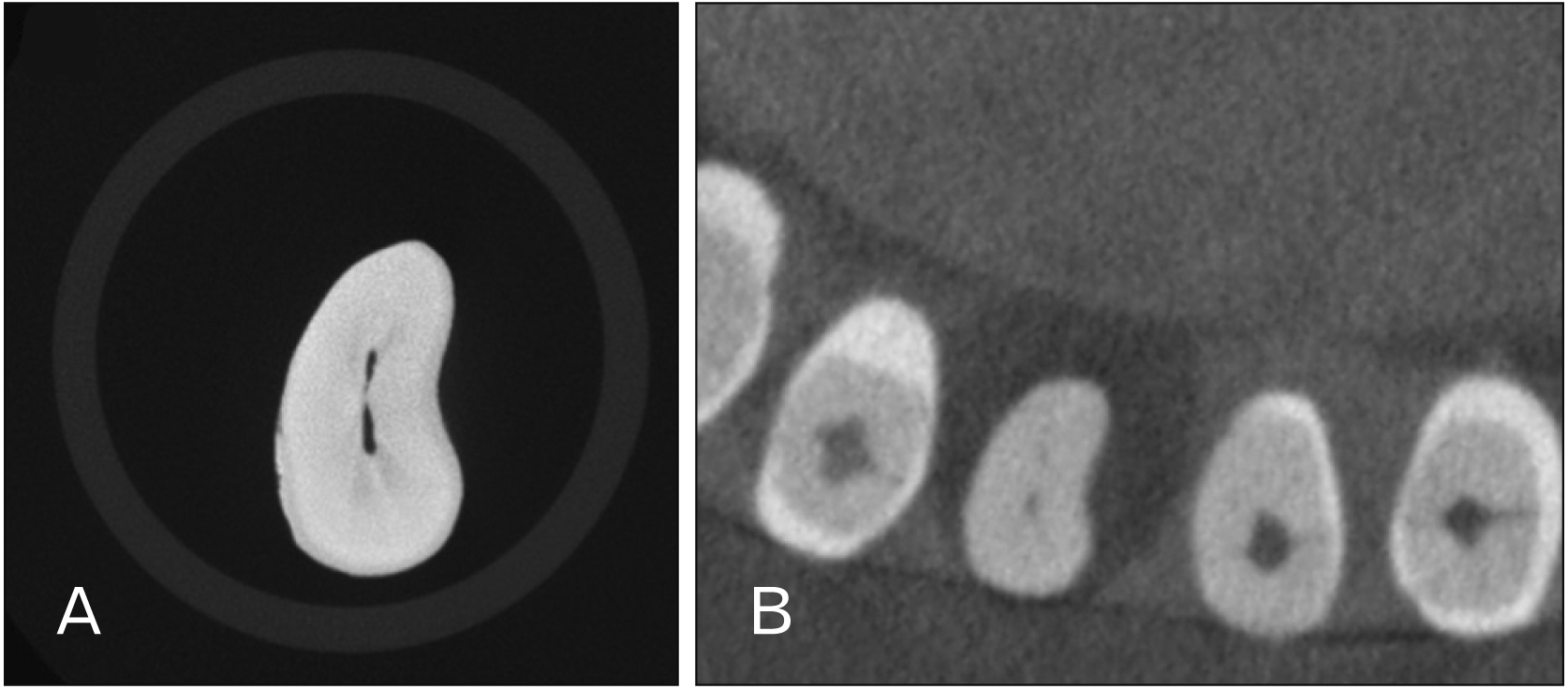
Tooth extraction specifications for a dental radiological phantom head (PH76; Kyoto Kagaku) were used to image six extracted permanent mandibular incisors of Japanese individuals (Fig. 1). The mandibular body and maxillary alveolar bone of the phantom head are removable. In addition, the maxillary/mandibular incisors, maxillary premolars, and maxillary molars are removable, and sample teeth can be inserted and fixed in the extracted areas. Images were obtained with scanning performed for 18 seconds at 80 kV and 5 mA using CBCT (MORITA 3DX Multi Image Micro CT FDP; Morita). An image software (One Volume Viewer; Morita) was used to reconstruct images perpendicular to the tooth axis, and slice images were created at the cervical third, center, and apical third of the root. Next, the extracted teeth were imaged with micro-CT (TXS-CT300; Tesco). The imaging conditions were as follows: tube current, 140 kV; tube voltage, 150 µA; and voxel size, 16 µm. A 0.1-mm copper filter was used to suppress beam hardening during imaging. A multifunctional display software (TomoShop; Midorino Research) was used to create micro-CT slice images from the micro-CT image data at the cervical third, root center, and apical third of the root (Fig. 2).
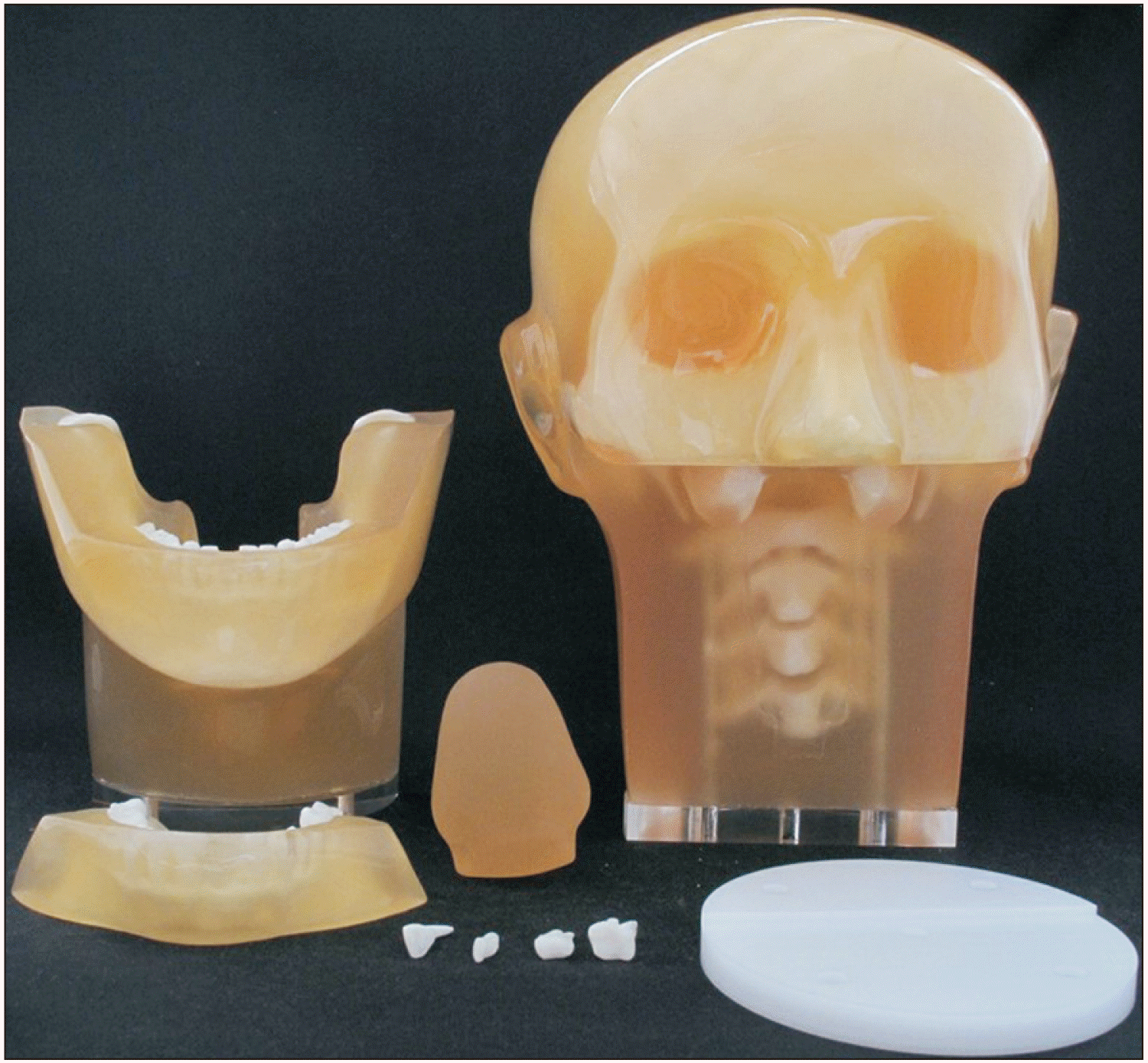
Two dentists evaluated micro-CT images of the extracted permanent teeth, and the number of root canals was counted.
Evaluation using CBCT
CBCT (MORITA 3DX Multi Image Micro CT FDP) of mandibular central and lateral incisors of 20 male and 61 female (mean age, 36.7 years) were obtained from the database of Ohu University Hospital and retrospectively studied.
The selection criteria for these images were as follows [13]:
The entire mandibular incisor was contained within the imaging area.
The root was complete.
There was no root resorption, and transmission images were observed at the apex.
The tooth had no root canal filling, post, or crown.
Clear and complete images were available for use.
The exclusion criteria were the existence of dental caries, root canal filling, or post and crown restoration.
An image software program (Morita One Volume Viewer) was used to reconstruct images perpendicular to the tooth axis, and slice images were created at the cervical third, center, and apical third of the root. Once the images were adjusted, two dentists evaluated the number of root canals in the teeth of interest. In case of disagreement, the final evaluation was reached by consensus.
Yoza et al. [13] had divided their study participants into three groups: those younger than 30 years, those between 30–50 years, and those over 50 years. They found differences in the root canal shape between the three age groups. In this study, the subjects were divided into two groups: those aged <30 years and those aged ≥30 years. The incidence of two root canals in the mandibular incisors was then compared between these groups. All images were evaluated by two dentists, and the presence of two root canals was determined based on consensus between them. This study protocol was approved by the Ethics Committee of Ohu University (No. 267).
Go to :
Results
On comparing the CBCT and micro-CT slice images of the extracted teeth, the same number of root canals were evaluated at 17 of 18 sites (94.4%) (Table 1).
Using CBCT, the mandibular central incisors were found to have two root canals in the cervical third of the root in five cases (6.2%), in the middle third in four cases (4.9%), and in the apical third in three cases (3.7%). Two root canals were observed in either cross-section in 9.9% of the mandibular central incisors. In the mandibular lateral incisors, two root canals were observed in the cervical third of the root in eight cases (9.9%), in the middle third in nine cases (11.1%), and in the apical third in two cases (2.5%). Two root canals were observed in either cross-section in 12.3% of the mandibular lateral incisors. The incidence of two root canals was higher in the lateral than in the central incisors (Table 2).
Table 2
Number of root canals in the mandibular incisors
![]()
In terms of root canal morphology according to age, two root canals in the mandibular central incisor were observed in two subjects (6.3%) <30 years old and in six subjects (12.2%) ≥30 years old. In case of the mandibular lateral incisors, two root canals were found in one subject (3.1%) <30 years old and nine subjects (18.4%) ≥30 years old. In CBCT images, there was a high incidence of two root canals in the mandibular incisors of people ≥30 years old (Fig. 3, Table 3).
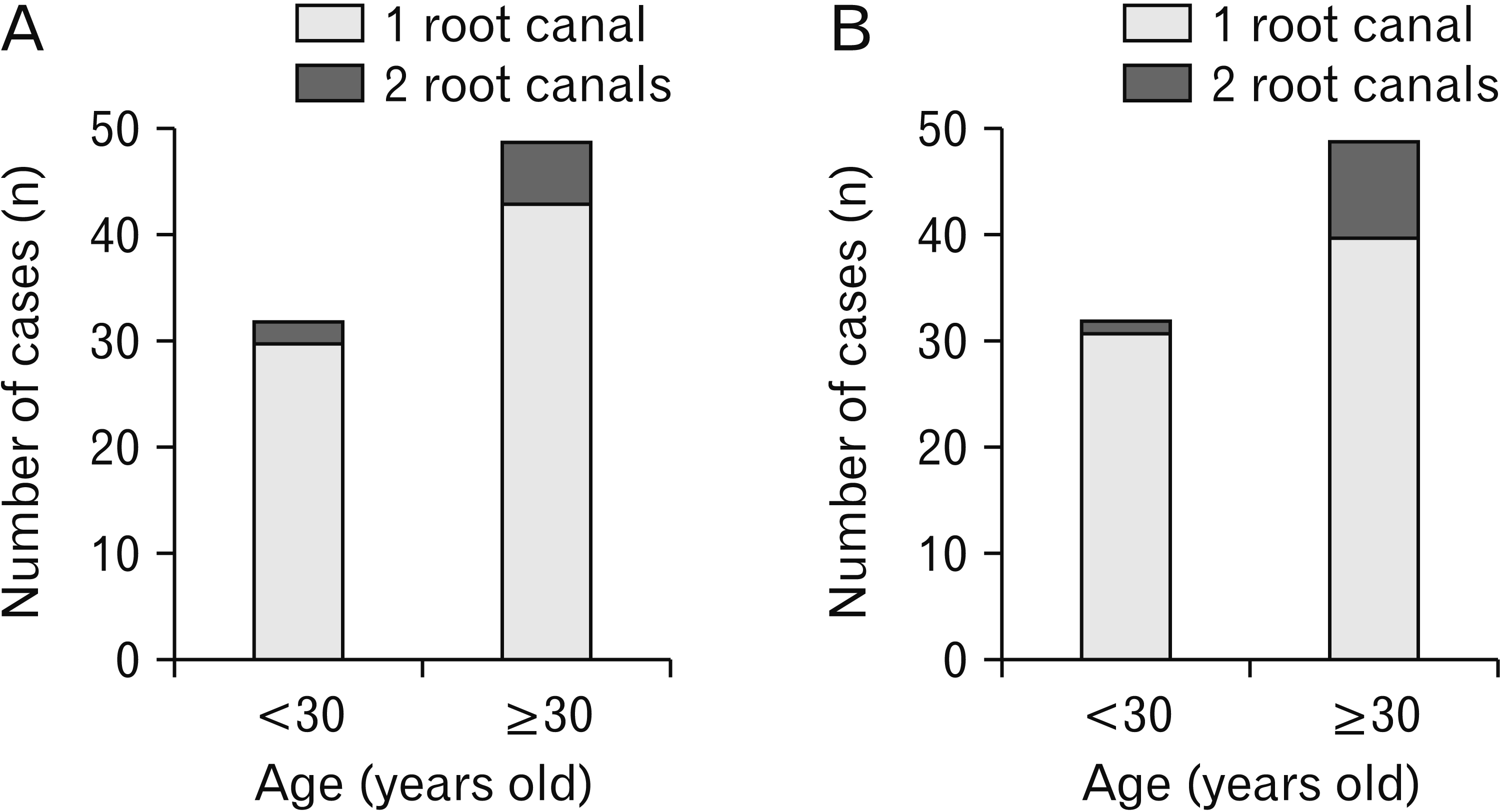 | Fig. 3Incidence of root canal morphology by age (<30 and ≥30 years old) mandibular central incisors (A) and mandibular lateral incisors (B).
|
Table 3
Root canal morphology by age (<30 and ≥30 years old) in mandibular incisors
![]()
Go to :
Discussion
CBCT is useful in identifying the internal structure of hard tissues with a lower effective dose than that of multislice CT. Tissue resorption can be measured based on the Hounsfield units with medical multislice CT, since the field of view (FOV) encompasses the entire subject, including the head [16]. CBCT, however, is affected by scattered radiation [17], and therefore the CT value is unstable in CBCT [18].
The “scatter to primary ratio” (SPR) is calculated by dividing the quantity of scattered radiation by the quantity of irradiation. The SPR is 0.05–0.15 with fan-beam or spiral CT but between 0.4–2.0 with CBCT [19]. CBCT ends up including ≥40% scattered radiation. For three-dimensional imaging using CBCT, the X-rays must pass through the hard tissue outside the region of interest. In the anterior teeth, the bilateral molars and the cervical spine overlap twice with the FOV, which may affect the density of the images. The smaller the FOV, the larger the artifacts, possibly due to the hard tissue outside the region of interest. Increasing the imaging area improves image resolution but also increases the exposure dose [16].
According to the American Association of Endodontics and the American Academy of Oral and Maxillofacial Radiology, CBCT is a useful diagnostic imaging tool for complex root canal morphology or non-standard root canal morphology during pulp extirpation [24]. Appropriate FOV settings would reduce the exposure dose and enable suitable diagnostic imaging of the root canal morphology.
CT values in CBCT have low reliability [19], and various other imaging modalities for morphological measurements have been reported by [8-14] taking multiple measurements, testing the reliability of the data with a κ-test [8], including observations by a single evaluator [9, 10], reaching consensus across multiple evaluators [11, 12], or image processing [13, 14]. Image analysis is performed using various methods, and selection of an appropriate method is necessary.
In a study on the incidence of a second mesiobuccal root canal in the maxillary first and second molars, extracted pig teeth were immobilized and imaged using CBCT. An oral and maxillofacial radiologist and endodontist reached a consensus regarding the number of root canals in these teeth. There was a high degree of correlation between CBCT slice images and cut cross-sections [10].
Similarly, the present study involved immobilizing extracted mandibular incisors in a phantom head and acquiring CBCT images to reproduce the effects of the surrounding hard tissue. The comparison of the evaluations of the two dentists of CBCT and micro-CT slice data showed a high degree of correlation regarding root canal morphology. The evaluated number of root canals was identical for 17 of 18 sites.
CBCT, which has become widely applied in the past 10 years, could be used in endodontics training programs [25], in which imaging using a phantom head could help simulate clinical conditions. At present, the precision of CBCT is not superior to that of micro-CT; however, improvement of the analysis software may help enhance CBCT precision.
Using transparent specimens, Vertucci [1] observed that 70% of mandibular central incisors and 75% of mandibular lateral incisors had one root canal. The ethnicity of the people whose extracted teeth were used in that study, however, is unknown. In a study on extracted mandibular incisors of Japanese people, 87.6% had one root canal [26]. A study with CBCT in Germany found that 77.4% and 75.7% of the specimens had one root canal, respectively [4]. In a study performed on an Indian subpopulation, 68.3% of mandibular central incisors and 65% of mandibular lateral incisors had one root canal [5]; in a subpopulation of Iraq, 70.4% and 70.8% of mandibular central and lateral incisors, respectively, had one root canal [6].
The number of root canals in the mandibular incisors of Japanese people according to CBCT in the present study was two for 9.9% of mandibular central incisors and one for 90.1%. For the mandibular lateral incisors, 12.3% had two root canals, and 87.6% had one root canal. The results from CBCT observation were largely identical to those previously obtained using transparent specimens of extracted teeth of Japanese people [26]. A meta-analysis of CBCT studies conducted among Chinese people showed that 5.6% of mandibular central incisors and 14.1% of mandibular lateral incisors had two root canals [4]. Similar to these findings, the present study showed that the mandibular lateral incisors had two root canals more frequently than did mandibular central incisors. The root canal has a complex configuration, and CBCT is a valuable tool for these observations.
Although it is impossible to distinguish between restored dentin formation following stimulation and secondary dentin formation following aging [22], secondary dentin continues to form for life, leading to narrowing of the pulp cavity [23]. However, there are almost no reports on the relationship between root canal morphology and age. On comparing radiographic images of the root canals of various teeth of individuals aged ≤25, 25–35, and >55 years old, Pineda and Kuttler [21] found that root canals clearly decreased in diameter with age. However, their study did not involve measurements or comparison of root canal morphology.
A CBCT study on aging-related changes in the root canal morphology of maxillary first premolars of individuals aged <30, 30–50 and >50 years showed an increasing ratio of two root canals to one with aging [13]. Herein, the comparison of mandibular incisors of individuals aged <30 and ≥30 years showed that people ≥30 years old have a higher incidence of two root canals in both central and lateral incisors.
In conclusion, the present comparison of root canal morphology observed on CBCT and micro-CT images of extracted mandibular incisors showed a high correlation between the two modalities. Among the mandibular incisors, the lateral incisors more frequently showed two root canals. Comparison between the two age groups showed that for both central and lateral incisors, people aged ≥30 years had two root canals more often than those aged <30 years. The micro-CT and CBCT findings coincided in 94.4% of the cases. These results show that CBCT is useful for endodontic preoperative assessment or training as it allows accurate evaluation of complex root canal morphologies.
Go to :
 XML Download