

 PDF
PDF Citation
 Print
Print


Introduction
Functional endoscopic sinus surgery through transnasal approach is a common modality of treatment for disorders of the nasal cavity, paranasal sinuses as well as cranial cavity [1]. The variations in morphology of the paranasal sinuses may alter the results during minimally invasive procedures and therefore anatomical considerations of ethmoid sinus (ES) were reported as early as in 1929 by great Mosher that ‘If the ES were placed in any other part of the body, it would be an insignificant and harmless collection of bony cells. In the place where nature has put it, it has major relationships so diseases and surgery of the labyrinth often lead to tragedy. Any surgery in this region should be simple but has proven one of the easiest ways to harm a patient’ [2].
The ethmoid bone has a delicate cribriform plate, which separates the nasal cavity from the anterior cranial fossa and is the thinnest most variable part of the cranial base. The olfactory fossa (OF) is located along the superior aspect of cribriform plate which varies in shape and depth. This variable measurement of the depth of OF or the height of ethmoid roof is mostly responsible for a greater risk of intracranial infiltration during endoscopic procedures in and around the nasal cavity [3-5]. Previous authors and Kero’s described high risk cases where the depth of OF was between 7–16 mm and categorized them as type III category on paranasal sinus CT scans. Further, currently there is limited research on measurements of OF [5-7].
The morphology of frontal and ES and frontoethmoid recess vary from simple to complex. The frontal sinus (FS) is located in the bone of the same name, posterior to the superciliary arches, and drains through frontonasal duct to ethmoid infundibulum which opens into the semilunar hiatus of middle nasal meatus. The size of FS is directly related to prevalence of sinusitis. The medium and larger FS are more prone to infection in comparison to smaller FS. These are the only paranasal sinuses that are absent at birth, and usually, these sinuses do not extend up into the frontal bone until the age of six years. As the left and right FS develop independently, a significant asymmetry between FS can be observed in the same individual [8]. The ES are the only paranasal air sinuses that are formed of multiple thin walled cavities in the ethmoid labyrinth. Clinically the ES are categorized into anterior and posterior groups on each side by a tortuous barrier [9]. Although, there is continuous progress in three-dimensional imaging techniques during past two decades but with lack of necessary depth of perception [3-6]. This cadaveric study is planned to improve the ability of the otolaryngologist, radiologist, and anatomist to understand the possible morphological variations and plan steps of less invasive “precision surgery” to have a safe and complication free procedures and communicate these complexities when teaching or reporting outcomes.
Materials and Methods
The study was carried out on 37 cadavers available in the department of Anatomy, AIIMS Bhopal, India. Permission through letter number IHEC-LOP/2018/IM0175 from the Institutional Ethical Committee of AIIMS, Bhopal was taken before starting the project. The cadavers were fixed in 10% neutral formalin and were partially dissected by medical students. The anatomic specimens belonged to subjects with an age range from 31 to 80 years (mean age, 63 years). All cadavers included were free of any gross signs of nasal and paranasal sinus deformity. The specimens with pathological appearances such as deformity, fracture, cysts, or tumors were excluded from the study.
OF
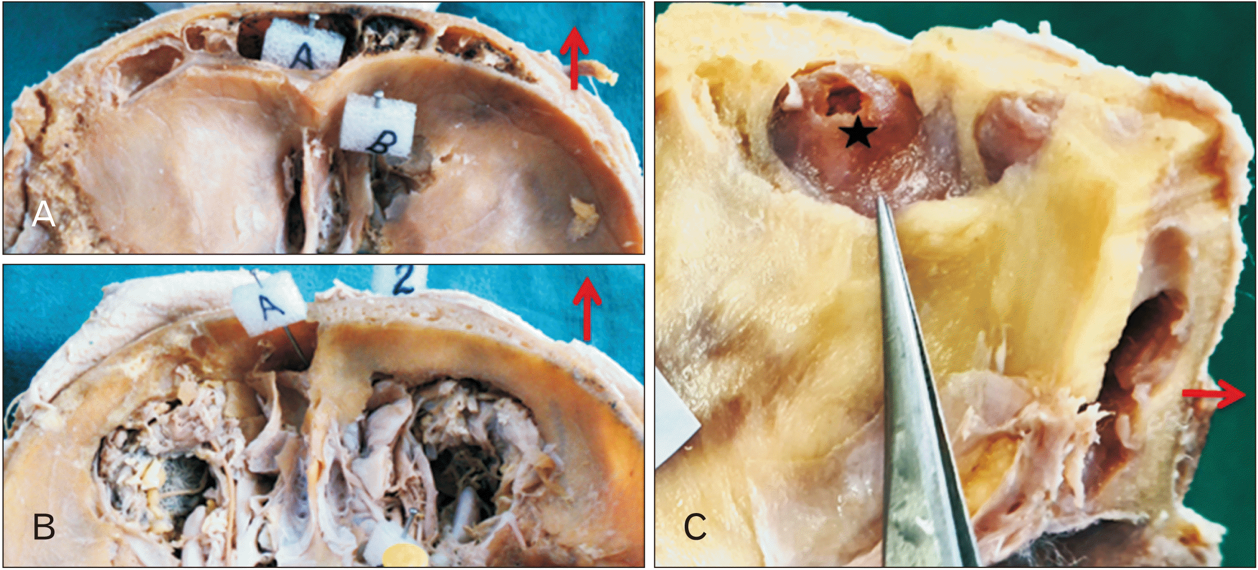
In the selected head and neck specimens, the horizontal section was taken through a point 1 cm above the glabella anteriorly, and the external occipital protuberance posteriorly. The brain was removed from anterior cranial cavity and the OF) was cleaned from any remaining tissue. The OF is located in the anteromedial part of anterior cranial fossa forming roof of the nasal cavity. Its floor is formed by the cribriform plate and lateral lamina of the ethmoid bone. The crista galli lies medially. The shape and dimensions of OF were noted. In present study, for classification of OF Modified Kero’s classification was used. According to measurements of depth of OF, different groups were formed as Modified Keros type I (0–3.99 mm), Modified Keros type II (4–7.99 mm), Modified Kero’s type III (8–16 mm) respectively.
FS
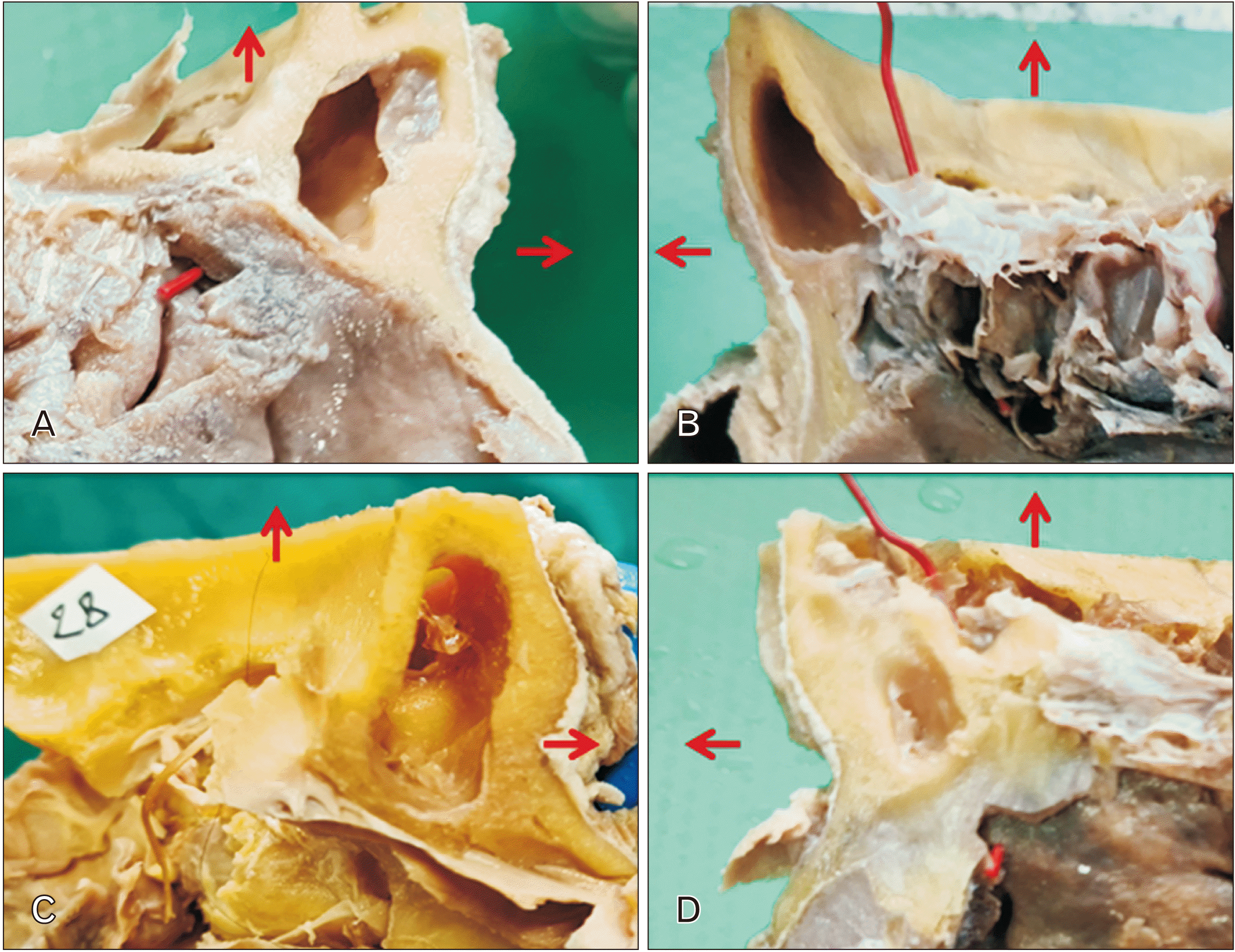
After taking a sagittal section, deviation of the nasal septum was noted. The frequency of occurrence of FS in both horizontal and sagittal sections was noted. The presence of septa in FS was also observed. The orientation and morphology of FS septa was recorded. Then different dimensions, antero-posterior, transverse, and depth of FS were recorded with the help of a venier caliper to the nearest 0.1 mm. The shape of (FS) was noted in four categories absent, comma shaped, oval and irregular as shown in Fig. 1. Degree of pneumatization of FS was observed. It was graded as small, medium sized and large when medial 1/3, 2/3, and the whole of the orbital roof pneumatized respectively. FS drainage pathway was ascertained by passing a probe from the FS to the FS ostium and another from ES to ES ostium toward the frontal recess meticulously to explore and find the drainage pattern. After locating the drainage pattern of FS, the entire drainage pathway of the FS was exposed by careful dissection.
Ethmoid sinus
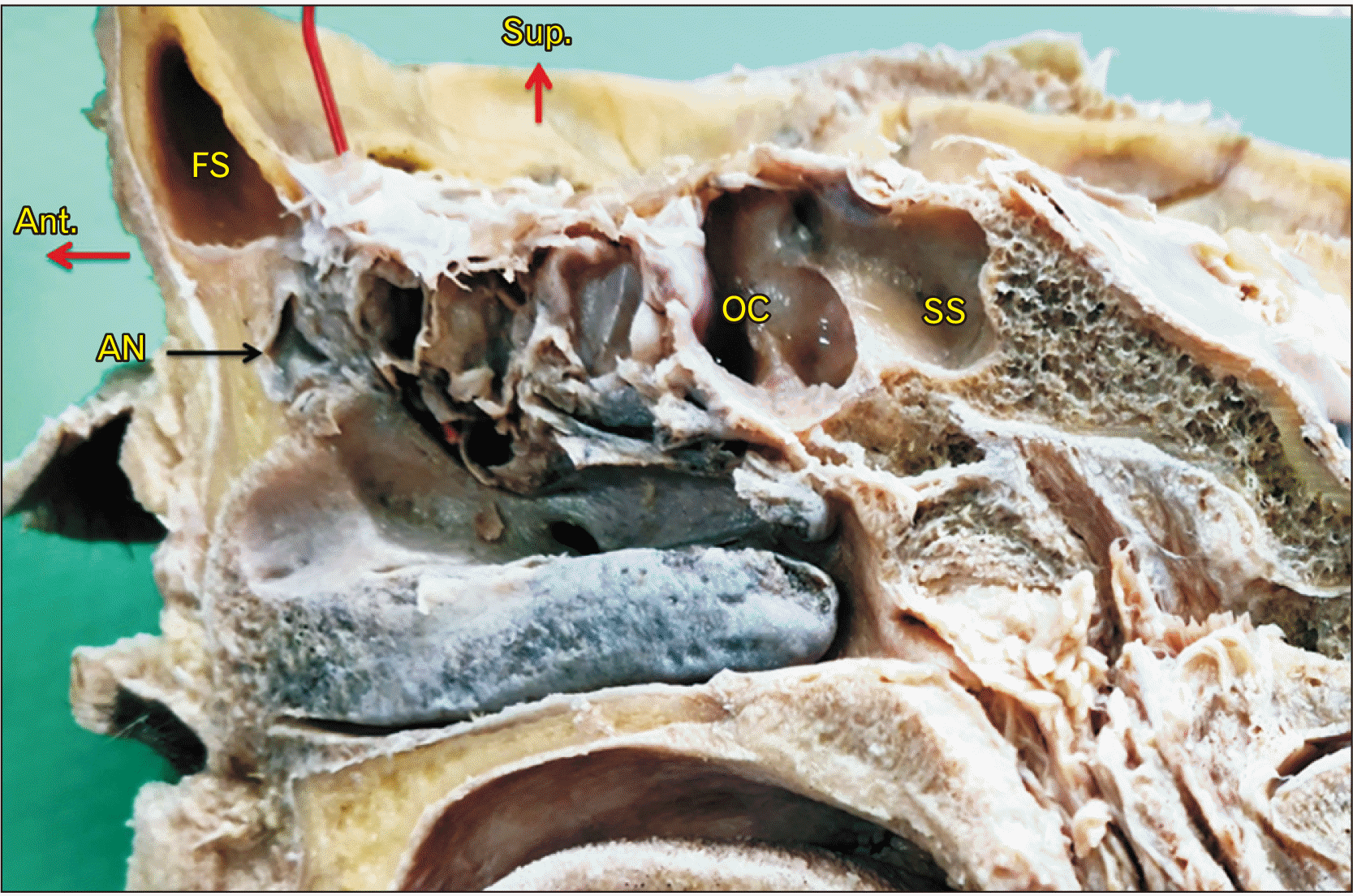
The middle and inferior nasal conchae along the lateral wall of the nasal cavity were dissected carefully to preserve the complex ES. The septa between the honey combed like anterior and posterior group of ES was identified and recorded. The number of cells on left and right side of anterior and posterior ES were recorded after confirming the drainage of each of the sinus cavity. We also noted the extensions of ES to adjoining region as the agger nasi cells and Onodi cells in the specimens.
Results
The OF
In the present study, asymmetric OF between the right and left sides were found in all 37 specimens. The incidence of type of OF in accordance with modified Kero’s classification in present study is tabulated in Table 1. The most common type of OF was observed in 60.8% of specimens having depth 4–7.99 mm.
Deviated nasal septum (DNS)
We found that DNS was a common variant seen in 33 cases (89.2%). The nasal septum was deviated to left side in 19 (57.6%) and in 14 (42.4%) cases towards right side.
The morphological variations of FS
The FS was found well developed in 67 sides out of 74 FS studied. The FS was observed to be small size in 46 sides (62.2%), medium in 14 (18.9%) and large in 7 (9.5%) sides and absent in 7 (9.5%) sides. In 3 specimens FS was absent bilaterally & in one it was unilaterally absent on right side as shown in Fig. 2B.
The shape of FS was noted in four categories as absent, comma shaped, oval and irregular and shown as in Table 2. The most common shape was comma shaped found in 55.4% of cases. The mean antero-posterior 2.9 cm and transverse 4.9 cm dimensions and depth 4.9 cm of FS was observed and details of these dimensions along with range is shown in Table 3. There was no significant difference between the median (interquartile range) value of the dimensions on the right and left side of the available cadaveric specimens by Mann Whitney U-test. Intra sinus septa were observed in 30 (44.8%) FS out of well developed 67 FS. Eccentric septa as shown in Fig. 2A were more common and seen in 26 (38.8%) FS while central septa in 4 (6.0%) sinus only.
The morphological variations of ES
In the meticulously dissected specimens, macroscopically we found one to seven cells of ES, some of them were incompletely divided by bony septa. The roof of the ethmoid labyrinth is mainly formed by the orbital part of the frontal bone, which separates the ethmoid cells from the anterior cranial fossa and its contents. The ES was present in all 74 (37 right and 37 left sides) specimens and distribution of their overall number and sub stratification into anterior and posterior ethmoid cells were shown in Table 4 across the side (right/left) of the specimens. We observed the agger nasi cells and Onodi cells in the specimens as shown in Fig. 3. There were 6 (8.1%) Onodi cells present and 11 (14.9%) agger nasi cells. There was no retro maxillary extension observed on either side.
Discussion
Functional endoscopic sinus surgical procedures on one hand enhanced the outcomes of surgery in and around paranasal air sinuses for almost three decades and on the other side has also made it essential to have precise knowledge about the anatomy of paranasal air sinuses and floor of anterior cranial fossa [1, 3, 5]. The depth of OF was studied extensively by Kero’s P. The author categorized the OF into three categories where the depth of the OF was estimated as type 1 (1–3 mm), type 2 (4–7 mm), and type 3 (8–16 mm) [6]. We found that in the original Kero’s classification the measurements between 0–0.9, 3.1–3.9, and 7.1–7.9 mm were difficult to classify in given types and there was a breach of almost 0.99 mm between the mentioned first, second and third categories. To maintain the continuity in measurements of the depth of OF, this Modified Kero’s classification is proposed and applied in the present study. The Modified Kero’s classification was applied and measurements were grouped as modified Keros type I (0–3.99 mm), type II (4–7.99 mm), type III (8–16 mm) respectively. In Kero’s study, Type I was described in 26.3% of population, versus 29.7% in our study. Keros Type II OF described in 73.3% of the population, versus 60.8% in this study. The OF of Keros III was described in 0.5% of the population, versus 9.5% in our study. Almushayti et al. [4] also found the type II category as the most common followed by type I then type III. Few authors reported that type one was the most common followed by type two then type three [7, 10, 11]. This significant difference in the result between the present study and the previous ones may be attributed to the difference in the populations, and variation in techniques of measurement (radiographic CT study versus cadaveric specimen) and variation in grouping the measurements into different types. In Table 5 we have compared the results of Kero’s classification by various authors from different countries [10-16].
An absence of pneumatization in the frontal bone results in FS aplasia. Bilateral absence of the FS has been reported in 2% to 33%, whereas the incidence of a unilateral absence had been reported to be between 0.8% and 7.4% [17-19]. In the present study absence of FS is observed in 9.46% cases. Krogman [20] had observed its absence in 5% adults, while Gulisano et al. [21] observed its absence in 24.7% of the cases. Ozdemir et al. [22] found frontal sinusitis more common in medium and large FS which we encountered in 19% and 9.5% of specimens respectively.
Agger nasi cell is the anterior most ethmoid air cell, located below the FS forming a significant part of the anterior wall of the frontal recess. Agger nasi cells may impinge upon the FS drainage tract, extending infero-laterally to lacrimal fossa and located antero-superior to the hiatus semilunaris. AN incidence was reported at different rates in different studies. Kayalioglu et al. [23] reported AN incidence as 7.8%. In contrast, in another study, Bradley and Kountakis [24] reported this incidence very high, 93%. Özdemir and Arslan [22] found the incidence of AN cells was 51.9%, and bilateral AN cells 25.2%. We found 14.8% agger nasi cells during the study.
The anatomical variation of posterior most ethmoid cells is called Onodi cells (spheno-ethmoid cells), seen extending posterior, lateral and superior to the sphenoid sinus, medial to the optic nerve. Extensive pneumatization can expose the circumference of optic nerve, surrounded by air spaces. In presence of Onodi cell, both the internal carotid artery and optic nerve may be exposed within the posterior ethmoid cells and result in most serious surgical complications. Driben et al. [25] noted the prevalence of Onodi cell in 39% on endoscopic examination in cadavers vs. 7% on an axial view of CT scans. Weinberger et al. [26] informed that the prevalence of the Onodi cell was 14% on endoscopic examination in cadavers versus 8% on a coronal view of CT scans. Arslan et al. [27], studied CT scans using two views (axial and coronal) and reported prevalence as 12%. Yeoh et al. [28] and Thanaviratananich et al. [29] found the prevalence as 51% (95% CI: 41.4–60.5) and 60% (95% CI: 47.9–71.0) respectively on endoscopic examinations of the posterior ES. In our study, we found 8% Onodi cells.
In conclusion, an attempt has been made to identify and reclassify the depth of OF as modified Kero’s classification as few dimensions were ignored in original Kero’s classification. Study of different shapes, dimensions, septa in FS and number of cells along with Onodi cell and agger nasi cell of ES presented in study provide more precise information.
 XML Download