Journal List > Anat Cell Biol > v.56(2) > 1516083071

Latiff and Olateju: Morphometry of the patellar tendon using a simple tracing method: a gold standard for anterior cruciate ligament reconstruction

Abstract

The anterior cruciate ligament (ACL) is mostly damaged in sporting activities. To reconstruct the damaged ACL, a patellar tendon (PT) is often the most preferred graft due to its fast healing and bone integration i.e. bone-patellar tendon-bone graft. Suitability of the PT often depends on the morphometric profile of the tendon. This study reported on the harvestable surface area (SA) of the tendon using a simple tracing method. The PT of 79 adult formalin–fixed cadavers of South Africans of European Ancestry were dissected, and the margins of the PT were traced on a wax paper before the tracings were scanned. The SA, straight proximal width (SPW), curved proximal width (CPW), straight distal width (SDW), curved distal width (CDW) and length of tendon (LOT) from the digitized image of the PT was measured. In addition, the length of the lower limbs was measured to normalize the measurements. The results showed no significant side differences, and the measurements were not sexually dimorphic. A strong correlation was reported for SA vs. LOT, SPW vs. CPW and SDW vs. CDW for both sexes and sides. The presented morphological profile provides additional information on the usability of the graft and with respect to healing and recovery.

Introduction

The anterior cruciate ligament (ACL) contributes to knee function and stability. It can perform these functions due to its ability to become taut during flexion or extension thus helping to stabilize the knee by preventing knee hyperextension [1-3]. Failure to do so would damage the ACL, and this is often seen in some contact sports e.g., rugby and football [4-6]. When an ACL is injured, it is necessary to eliminate instability and prevent any further damage to the knee structures by performing a reconstruction surgery using a graft [6]. The choice of graft often depends on factors such as the dimension of harvestable graft, location, and post-surgery recovery etc. [7-11]. At the same time, the choice of graft often depends on the surgeon’s preference based on surgical training, experience, and success rates. The choice of graft should also be determined by the graft morphometries and other considerations such as age, body size, activity level, concomitant injuries, donor-site morbidity, and return to active sport [6, 12, 13] as well as morphological differences and variations amongst individuals of different race, ethnicity, and sexes [14-17].
The present study explored the morphometries of the patellar tendon (PT). The PT autograft is considered the gold standard for ACL reconstruction [18-20] due to its high tensile strength and possibility of re-vascularization by the inferior lateral or inferior medial genicular artery as well as being able to provide a good bone integration where bone parts are harvested with the tendon as a graft [2, 21, 22]. The PT serves as an anchor for the quadriceps tendon after blending with the periosteum of the patella. It extends from the apex of the patella to the tibial tuberosity where the PT inserts [23-25]. Through this connection, the PT and the patella improve the way the quadriceps femoris muscles pull on the tibia [25] while the patella in this case is acting as a fulcrum at the knee joint [25, 26].
Factors such as length, harvestable tendon area, thickness, tensile strength, ease of graft harvesting and bone–to–bone healing after reconstruction are important factors to consider before deciding on a graft choice but unfortunately all these factors are not obtainable in a single tendon [21] thus reaching a consensus on the best graft for ACL reconstruction is often a challenge. Other factors of importance are rapid healing, low donor site morbidity, immediate rigid fixation (i.e., ‘ligamentizing’ once implanted), and similar structural properties that mimic the mechanical properties of ACL [21, 27]. In all, it is imperative that the knee function of the graft site is not in any way compromised [21].
Even though the PT is considered the gold standard for ACL reconstruction [20], information on the extent of the harvestable surface area (SA) of the tendon is lacking in the literature. Thus, this study presents data on the morphometric profiles (i.e., the SA, length and width) of the PT in both limbs of the male and female cadavers of South Africans of European Ancestry using a simple tracing method. Understanding these profiles will contribute to knowledge and will assist surgeons on the choice of autografts preoperatively to determine whether the PT of an individual is adequate for an ACL reconstruction in the same individual [7]. In addition, this study also explored relationships between different paired measurements and emphasizes on healing and recovery at the harvest site which are crucial to the return of knee functions. It is envisaged that the outcomes of this study will provide additional information on the usability and suitability of the PT as an autograft, and which will better inform the choice of graft when performing an ACL reconstruction.

Materials and Methods

Demographics of sample

Data on the PT morphometry was collected from the same lower limbs of adult formalin–fixed cadavers of South Africans of European ancestry from a similar study on the morphometry of the quadriceps tendon [28]. To determine the sample size required for this study, a mean of 62.66±7.89 mm in the female and a mean of 69.74±7.94 mm in the male for the length of PT were assumed as the measurement from the apex of the patella to the tibial tuberosity [26]. At 80% statistical power and a significance level of 5%, the minimum sample size required was 42 cadavers (i.e., 21 cadavers per sex). Thus, the final sample size considered in this study was 40 female (79 lower limbs assessed; age range: 46–96) and 39 male (77 lower limbs assessed; age range: 53–96). The cadavers were housed in the School of Anatomical Sciences at the University of the Witwatersrand, Johannesburg, South Africa for teaching purposes. Approval to conduct this study was granted by the Human Research Ethics Committee of the University of the Witwatersrand (Ethics Number: W-CJ-140604-1). The PT on both limbs in each cadaver was measured and a cadaver limb or PT with any obvious physical scar or deformity was excluded from the study.

Dissections

Similar dissection described by Latiff and Olateju [28, 29] was utilized, the PT on the lower limb was exposed by making a midline longitudinal incision on the anterior surface of the lower limb from midway of the thigh to midway of the leg. Transverse incisions perpendicular to the midline incisions were made proximally in the thigh and distally in the leg to expose the PT and allow for adequate space for observation and morphometric measurements. The skin, subcutaneous fascia, fat, fascia lata, and crural fascia were carefully removed without altering the morphology of the PT.

Morphometry of PT

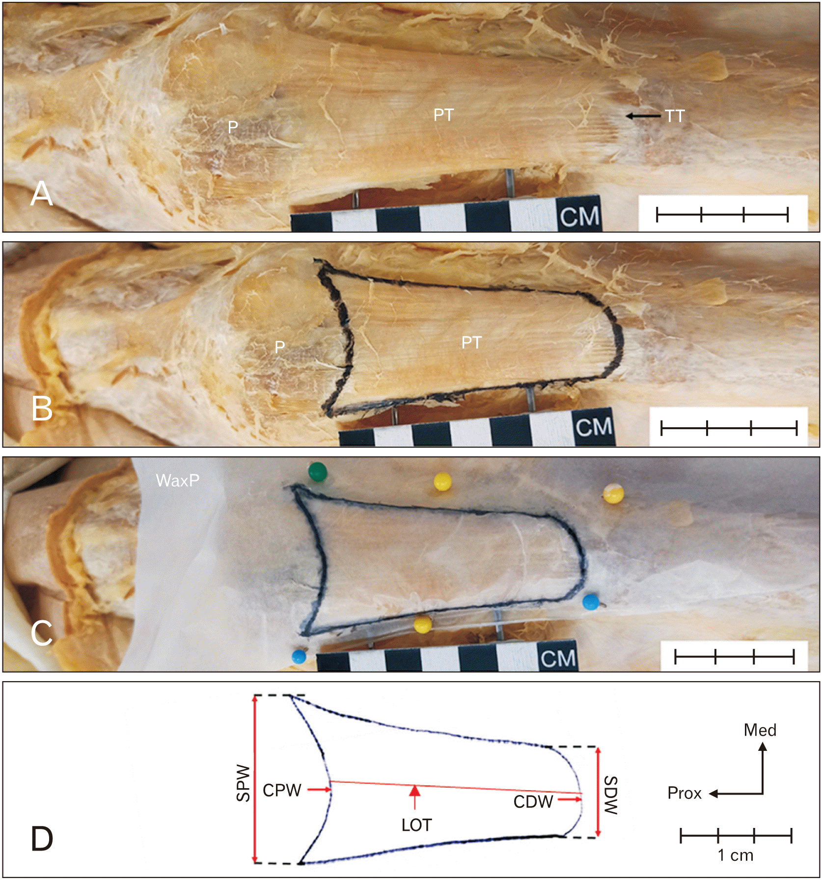
The knee was flexed at an angle of about 45° and then the PT was carefully wiped with a dry absorbent cloth. Thereafter, a non-elastic transparent wax paper (dimension: 5 cm×5 cm×19 µm; Superior wax paper–donated by Superhaze Trading Co.) was carefully placed in such a manner that it assumed the curvatures of the underlying PT (Fig. 1) and the wax paper was firmly secured with pins. The margins of the PT were pre-marked with a permanent marker before placing the wax paper onto the tendon to enhance visibility and accuracy of the tracing of the tendon margins on the wax paper [28].
After the tracing, the wax paper was removed and then a known scale bar was drawn on it. Then the wax paper with its inscribed scale bar was then scanned at 300 dpi using an Epson workforce DS-50000 scanner. From the digitized image, the SA, straight proximal width, curved proximal width, straight distal width, curved distal width and length of tendon (LOT) for the PT (Table 1) were measured using an ImageJ 1.47v software (National Institutes of Health). The measured length of each lower limb at full extension (from the anterior superior iliac spine to the medial malleolus) [16, 30] was used to normalize the data by dividing the raw (actual) values by the length of the corresponding lower limb (LLL) similar to a study on the quadriceps tendon morphometry [28].

Statistical analysis

To test for reliability of all measurements, an intra–observer reliability test was conducted. A Lin’s Concordance test (ρc) was performed to determine the level of precision and accuracy between the measurements [31]. Normality test was conducted for each measured parameter using Shapiro-Wilk test. For data that was parametric, a Student’s t-test was used to perform side and sex differences for each measured parameter. For a non-parametric data, a Mann-Whitney U-test was used. To determine a possible correlation between any two paired measurements, a Pearson’s correlation was used for normally distributed data or a Spearman’s correlation for non-parametric data. All statistical analyses were performed using IBM SPSS Statistics for Windows (version 22.0; IBM Co.). Statistical difference of 5% was regarded as significant for all the statistical analyses.

Results

Test of reliability of measurements

The ρc ranged from 0.931 for CDW to 0.997 for LOT (Table 2). This indicates a high degree of reproducibility and the data analysed in this study are considered to have an error low enough to be acceptable.

Morphometry of the PT

The descriptive statistics of the raw data and the normalized data for each side and according to sex is shown in Table 3. In addition, the data of the length of limbs (i.e., for normalization of raw data) of cadavers used in this study is the same as that used for the quadriceps tendon morphometry [28]. For all the measurements in the female or male (Table 3), there were no statistically significant differences in sides (i.e., left vs. right). There was also no significant difference when the measurements in both sexes were compared. Thus, none of the measurements in the PT exhibited a side dominance nor was sex dimorphic. Despite the non-significant difference between the measurements from both limbs, the measurements of the SA of one limb were observably different from the other limb in most individual cadavers assessed (indicated by red arrows in Fig. 2). This is also an indication that the SA of the PT in both limbs are morphometrically different in some individuals compared to the similarity of the LLL measures (for illustrative purposes) in both limbs (Fig. 2).

Correlation analyses on the measured parameters of the PT

Only paired parameters that revealed moderate (range 0.4–0.69) or strong correlations (range 0.7–1.0) [32] are presented in Table 4. A strong correlation (R ≥0.7) was observed between the paired parameters of SA vs. LOT, SPW vs. CPW and SDW vs. CDW in both limbs of the male and female. Other paired parameters showed a positive moderate correlation as shown in Table 4. When LLL was paired with other parameters, only a moderate correlation (R=0.4) was observed in the female between LLL vs. SA or LLL vs. LOT of the left limb and between LLL vs. LOT of the right limb.

Discussion

The harvesting of PT as an autograft is considered a gold standard for ACL reconstruction. This study presents data on the morphometry of the harvestable area of PT which is suggested to be an important parameter to consider in preoperative surgical planning of ACL reconstruction when considering recovery and limb functionality at the donor and reconstruction sites [33, 34]. An important step in the methodology of the present study is that the actual measurements (in situ tendon tracing) were used to obtain a more realistic measurement [33, 35]. However, the present study acknowledges the possible influences of tissue fixatives on morphometry, the extent of which has not yet been elucidated [26, 36].
In the present study, the dimension of the SA in the population group assessed was similar in both limbs despite the observable differences in the SA in both limbs of most of the individuals. Similarly, the SA was not significantly different between the sexes. Unfortunately, there is no report in the literature to directly compare the measurements. Even though the reason for the variations between the limbs could not be ascertained, it is assumed that daily activities such as standing, sitting and/or postural stance when exercising may impact the morphology and morphometry of the muscle or its tendon because of a possible gradient in the muscle tone in both limbs [34]. In addition, one implication of the observable differences in the dimension of SA is that one or both PT may not be suitable as an autograft in an individual (bone pathologies being additional reasons) which is similar to the observations in the morphometries of the quadriceps tendon [28]. It is therefore important to consider whether adequate amount of tissue remains at the harvest site to ensure that knee function is not compromised in the limb from which the graft was removed. This reaffirms that the advantages of a PT being able to incorporate bone grafts at its two ends (i.e., a bone-patellar tendon-bone [BPTB] graft) is not the only important parameter to consider when choosing the PT as a graft.
In the present study, the length of PT was similar in both limbs and in both sexes. The recorded tendon length for the present population group was approximately 51.1 mm in the female and 56.8 mm in the male compared to similar studies on the PT of South Africans of European ancestry e.g. Van Zyl et al. [15] reported an average of 45.7 mm for a male population while Olateju et al. [26] reported an average of 65.9 mm for both sexes with a significantly higher tendon length in the male. Similarly, Xerogeanes et al. [14] reported a significantly higher tendon length in the males of an American population. The reported mean tendon length in a Swiss population (51.9 mm) [37] and in the males of a South Indian population (47.8 mm) [34] are within the range that was observed in the present study. The differences in the mean LOT across the different studies may also be due to the differences in methodologies, age, race or ethnicity [16].
Considering the average length of the PT observed in this study (which is lower than the required 70 mm graft length required for an ACL reconstruction [38]), with the incorporation of bone blocks it means that most individuals may still be considered suitable recipients of a PT autograft. The benefit associated with using the BPTB graft is that it incorporates and heals at a much faster rate within the bone tunnels thus affirming its popularity in ACL reconstruction [39, 40]. Apart from the benefits of a BPTB, graft site morbidity and return of tendon and knee functions are the other important factors to consider before deciding on harvesting tissues e.g. from the contralateral limb. It must be emphasized that harvesting a graft from the contralateral side is more advantageous than harvesting from the ipsilateral side because it encourages a faster restoration of motion range and strength and return to full sporting capacity with no compromised stability [41, 42].
However, a BPTB autograft may be inadequate in a surgical approach like in an all-inside ACL reconstruction where it becomes difficult to appropriately shorten the graft length [43]. An all-inside ACL reconstruction is less invasive which utilizes closed socket tunnels as opposed to the traditional complete bone tunnels which is prone to tunnel expansion and compromised healing as the graft movement activity increases, and which may create problems in the subject [44]. Usually only one tendon is required which may be quadrupled, thus an all-inside technique provides sufficient graft (no need to make the tendon longer) as there is a relatively shorter distance to insert the graft [45]. Graft augmentation (i.e., making the graft larger in size by using an allograft, a hybrid construct or harvesting another tendon) is also not a requirement but this is often the case in a traditional ACL reconstruction technique where a surgeon may not be able to accurately pre-determine the size of the preferred graft or in a case of non-envisaged complication or discovery during surgery [45]. An all-inside technique also offers less morbidity as there is no need to harvest bone blocks which may lead to donor site morbidity, patellar instability, or fracture as is the case for a BPTB [45, 46]. However, an all-inside technique requires a single–use special equipment for the retrograde drilling of the sockets thus it is relatively more expensive [44] and special arthroscopic skills are required [45] which often makes the PT a preferred graft.
For the widths of the PT, the present study reported no significant differences between the limbs and between sexes for the proximal or distal widths. A common morphology of the PT is that the proximal width is wider than the distal and this is often attributed to the convergence of fascicles towards the midline of the tendon [33, 34, 47]. There was similarly no significant difference between the curved and straight measurements of the widths suggesting that one or both measurements may be adequate for preoperative planning when considering the PT as an autograft. In comparison to other studies, patellar width of about 25.5 mm reported in an Arabian population [21] is within the range reported in the present study. In a study using CT scans from cadavers of a Japanese population, Oikawa et al. [48] reported a mean proximal width of 29.9 mm and a mean distal width of 25.0 mm while Aithal Padur et al. [34] reported no significant difference in the proximal width (about 35.3 mm) or in the distal width (about 25.5 mm) of both limbs of a male South Indian population.
Another important consideration is that the remaining tissue left at the harvest site must be adequate to ensure that the knee function is not hindered. Considering the width of the PT and after simulating a harvest of a 10 mm wide graft from the population assessed, on average 71% of the proximal and 61% of the distal widths remained. In comparison to Xerogeanes et al. [14], about 56% of the PT remained in an American population. It must be emphasized that after harvesting a portion of the PT, the remaining tendon at the harvest site are crucial for maintaining knee function, reducing complications, and producing satisfactory post-surgery outcomes. This is also dependent on the way tendons are harvested [49] as problems can arise from the harvest site. Harvesting the middle third of the PT could cause patellar tendonitis (i.e., kneeling pain) [39, 49]. Similarly, in a BPTB graft, the removal of an inferior part of the patella bone could weaken the patella and induce donor-site morbidity and in rare cases, causes a fracture of the patella bone from a blunt trauma [50]. These complications require an urgent need for another surgical repair, and which may further worsen the knee functions. In addition, excessive scarring in the knee area after harvesting a PT graft is common as the body begins the healing process. This often hinders normal motion in the knee, causes inflammation and the weakening of the PT thus increasing the risk of a tendon rupture. Usually, these problems of the PT could also affect the strength and function of the quadriceps tendon [50].
The association between the paired measurements of the PT was tested to determine their relationships. A strong correlation was found between the straight and the curved proximal or distal widths in both limbs and for both sexes. This relationship further shows that one or both measurements is adequate for preoperative investigation of the PT as a possible autograft. However, correlation between proximal and distal widths (e.g., CPW vs. CDW or SPW vs. SDW) for both limbs and sexes range from weak to moderate positive correlation. Similarly, a positive moderate correlation was reported in a male South Indian population between the distal and proximal widths [34]. Interestingly, a strong correlation was found between the SA and the LOT in both limbs and for the two sexes assessed in the present study. This relationship also indicates that the SA of the PT can be used to predict the length of the PT when considering the use of this tendon for ACL reconstruction. Interestingly, none of the measurements paired with the LLL revealed a strong correlation suggesting that the LLL is not a good predictor of any measurements of the PT in the population group assessed similar to the relationships between measurements of the quadriceps tendon paired with the LLL [28].
In conclusion, despite the limitations of cadaveric studies such as a limited sample size and the possible effects of formalin fixation on tissues, cadaveric studies have proven to be advantageous and reliable in that it provides an in situ measurement [26, 51]. The PT autograft provides a faster healing and graft integration owed to the bone blocks harvested with the PT compared to other tendons such as the quadriceps or the semitendinosus tendon. However, the harvest site of the PT may create unintended problems if a proper evaluation of the PT and its morphometry was ignored. In addition, a PT graft may become undesirable and non-usable in an all–inside procedure. PT graft may also not be desirable in an individual with osteoporosis, osteological degenerative diseases, patellar tendinopathy, or inadequate contralateral tendon. In a situation where the PT (e.g., contralateral tendon) is undesirable, an alternate graft may be considered (e.g., semitendinosus or quadriceps tendon). Aside from the problems that could make a PT graft undesirable, it still provides better bone integration and healing outcome as highlighted in the present study.

Acknowledgements

The authors sincerely thank those who donated their bodies to science so that anatomical research could be performed. Results from such research can potentially increase mankind's overall knowledge that can then improve patient care. Therefore, these donors and their families deserve our highest gratitude. We are grateful to the School of Anatomical Sciences of the University of the Witwatersrand for giving access to the Human Collections. We are grateful to Dr Gomes for proofreading this manuscript.

Notes

Author Contributions

Conceptualization: OIO. Data acquisition: SL. Data analysis or interpretation: SL, OIO. Drafting of the manuscript: SL, OIO. Critical revision of the manuscript: SL, OIO. Approval of the final version of the manuscript: all authors.

Conflicts of Interest

No potential conflict of interest relevant to this article was reported.

References

1. Zhu J, Zhang X, Ma Y, Zhou C, Ao Y. 2012; Ultrastructural and morphological characteristics of human anterior cruciate ligament and hamstring tendons. Anat Rec (Hoboken). 295:1430–6. DOI: 10.1002/ar.22527. PMID: 22807249.
crossref
2. Cerulli G, Placella G, Sebastiani E, Tei MM, Speziali A, Manfreda F. 2013; ACL reconstruction: choosing the graft. Joints. 1:18–24. PMID: 25606507. PMCID: PMC4295687.
3. Markatos K, Kaseta MK, Lallos SN, Korres DS, Efstathopoulos N. 2013; The anatomy of the ACL and its importance in ACL reconstruction. Eur J Orthop Surg Traumatol. 23:747–52. DOI: 10.1007/s00590-012-1079-8. PMID: 23412211.
crossref
4. Quatman CE, Hewett TE. 2009; The anterior cruciate ligament injury controversy: is "valgus collapse" a sex-specific mechanism? Br J Sports Med. 43:328–35. Erratum in: Br J Sports Med 2021;55:e6. DOI: 10.1136/bjsm.2009.059139. PMID: 19372087. PMCID: PMC4003572.
crossref
5. Webb J, Corry I. 2000; Injuries of the sporting knee. Br J Sports Med. 34:227–8. DOI: 10.1136/bjsm.34.3.227. PMCID: PMC1763269.
crossref
6. Hurley ET, Calvo-Gurry M, Withers D, Farrington SK, Moran R, Moran CJ. 2018; Quadriceps tendon autograft in anterior cruciate ligament reconstruction: a systematic review. Arthroscopy. 34:1690–8. DOI: 10.1016/j.arthro.2018.01.046. PMID: 29628380.
crossref
7. Hamada M, Shino K, Mitsuoka T, Abe N, Horibe S. 1998; Cross-sectional area measurement of the semitendinosus tendon for anterior cruciate ligament reconstruction. Arthroscopy. 14:696–701. DOI: 10.1016/S0749-8063(98)70096-9. PMID: 9788365.
crossref
8. Shelton WR, Fagan BC. 2011; Autografts commonly used in anterior cruciate ligament reconstruction. J Am Acad Orthop Surg. 19:259–64. DOI: 10.5435/00124635-201105000-00003. PMID: 21536625.
crossref
9. Reboonlap N, Nakornchai C, Charakorn K. 2012; Correlation between the length of gracilis and semitendinosus tendon and physical parameters in Thai males. J Med Assoc Thai. 95(Suppl 10):S142–6.
10. Janssen RP, Scheffler SU. 2014; Intra-articular remodelling of hamstring tendon grafts after anterior cruciate ligament reconstruction. Knee Surg Sports Traumatol Arthrosc. 22:2102–8. DOI: 10.1007/s00167-013-2634-5. PMID: 23982759. PMCID: PMC4142140.
crossref
11. Sun J, Wei XC, Li L, Cao XM, Li K, Guo L, Lu JG, Duan ZQ, Xiang C, Wei L. 2020; Autografts vs synthetics for cruciate ligament reconstruction: a systematic review and meta-analysis. Orthop Surg. 12:378–87. DOI: 10.1111/os.12662. PMID: 32180364. PMCID: PMC7189042. PMID: 66884f9852f1451ba8de4b08e150d934.
crossref
12. Mall NA, Matava MJ, Wright RW, Brophy RH. 2012; Relation between anterior cruciate ligament graft obliquity and knee laxity in elite athletes at the National Football League combine. Arthroscopy. 28:1104–13. DOI: 10.1016/j.arthro.2011.12.018. PMID: 22421564.
crossref
13. Macaulay AA, Perfetti DC, Levine WN. 2012; Anterior cruciate ligament graft choices. Sports Health. 4:63–8. DOI: 10.1177/1941738111409890. PMID: 23016071. PMCID: PMC3435898.
crossref
14. Xerogeanes JW, Mitchell PM, Karasev PA, Kolesov IA, Romine SE. 2013; Anatomic and morphological evaluation of the quadriceps tendon using 3-dimensional magnetic resonance imaging reconstruction: applications for anterior cruciate ligament autograft choice and procurement. Am J Sports Med. 41:2392–9. DOI: 10.1177/0363546513496626. PMID: 23893419.
crossref
15. Van Zyl R, Van Schoor AN, Du Toit PJ, Louw EM. 2016; Clinical anatomy of the anterior cruciate ligament and pre-operative prediction of ligament length. SA Orthop J. 15:47–52. DOI: 10.17159/2309-8309/2016/v15n4a7.
crossref
16. Gupta R, Malhotra A, Masih GD, Khanna T. 2017; Equation-based precise prediction of length of hamstring tendons and quadrupled graft diameter by various anthropometric variables for knee ligament reconstruction in Indian population. J Orthop Surg (Hong Kong). 25:2309499017690997. DOI: 10.1177/2309499017690997. PMID: 28228049. PMID: 62a74879e4ce4daab083ca17fd770ac0.
crossref
17. Vadgaonkar R, Prameela MD, Murlimanju BV, Tonse M, Kumar CG, Massand A, Blossom V, Prabhu LV. 2018; Morphometric study of the semitendinosus muscle and its neurovascular pedicles in South Indian cadavers. Anat Cell Biol. 51:1–6. DOI: 10.5115/acb.2018.51.1.1. PMID: 29644103. PMCID: PMC5890011.
crossref
18. Hadjicostas PT, Soucacos PN, Berger I, Koleganova N, Paessler HH. 2007; Comparative analysis of the morphologic structure of quadriceps and patellar tendon: a descriptive laboratory study. Arthroscopy. 23:744–50. DOI: 10.1016/j.arthro.2007.01.032. PMID: 17637410.
crossref
19. Shani RH, Umpierez E, Nasert M, Hiza EA, Xerogeanes J. 2016; Biomechanical comparison of quadriceps and patellar tendon grafts in anterior cruciate ligament reconstruction. Arthroscopy. 32:71–5. DOI: 10.1016/j.arthro.2015.06.051. PMID: 26382635.
crossref
20. Eriksen CS, Svensson RB, Gylling AT, Couppé C, Magnusson SP, Kjaer M. 2019; Load magnitude affects patellar tendon mechanical properties but not collagen or collagen cross-linking after long-term strength training in older adults. BMC Geriatr. 19:30. DOI: 10.1186/s12877-019-1043-0. PMID: 30704412. PMCID: PMC6357404. PMID: 02f97037f173495898e92225318c9926.
crossref
21. Hijazi MM, Khan MA, Altaf FMN, Ahmed MR, Alkhushi AG, Sakran AMEA. 2015; Quadriceps tendon and patellar ligament; a morphometric study. Prof Med J. 22:1192–5. DOI: 10.29309/TPMJ/2015.22.09.1135.
crossref
22. DeFroda S, Fice M, Tepper S, Bach BR Jr. 2021; Our preferred technique for bone-patellar tendon-bone allograft preparation. Arthrosc Tech. 10:e2591–6. DOI: 10.1016/j.eats.2021.08.002. PMID: 34868866. PMCID: PMC8626798.
crossref
23. Ilan DI, Tejwani N, Keschner M, Leibman M. 2003; Quadriceps tendon rupture. J Am Acad Orthop Surg. 11:192–200. DOI: 10.5435/00124635-200305000-00006. PMID: 12828449.
crossref
24. Slone HS, Romine SE, Premkumar A, Xerogeanes JW. 2015; Quadriceps tendon autograft for anterior cruciate ligament reconstruction: a comprehensive review of current literature and systematic review of clinical results. Arthroscopy. 31:541–54. DOI: 10.1016/j.arthro.2014.11.010. PMID: 25543249.
crossref
25. Drake RL. Drake RL, Wayne Vogl A, Mitchell AWM, editors. 2010. Lower limb. Gray's Anatomy for Students. 2nd ed. Churchill Livingstone;Philadelphia: p. 558–64. DOI: 10.1016/B978-0-443-06952-9.00011-4.
crossref
26. Olateju OI, Philander I, Bidmos MA. 2013; Morphometric analysis of the patella and patellar ligament of South Africans of European ancestry. S Afr J Sci. 109:6. DOI: 10.1590/sajs.2013/20130069. PMID: fd9c07ea5f9d4b39b8d956e843ad5839.
crossref
27. Goldblatt JP, Fitzsimmons SE, Balk E, Richmond JC. 2005; Reconstruction of the anterior cruciate ligament: meta-analysis of patellar tendon versus hamstring tendon autograft. Arthroscopy. 21:791–803. DOI: 10.1016/j.arthro.2005.04.107. PMID: 16012491.
crossref
28. Latiff S, Olateju OI. 2022; Morphometry of the harvestable surface area of quadriceps tendon using a simple tracing method: a common ACL autograft. Eur J Anat. 26:679–90. DOI: 10.52083/TDBN2622.
crossref
29. Latiff S, Olateju OI. 2022; Quantification and comparison of tenocyte distribution and collagen content in the commonly used autografts for anterior cruciate ligament reconstruction. Anat Cell Biol. 55:304–10. DOI: 10.5115/acb.22.005. PMID: 35668478. PMCID: PMC9519766.
crossref
30. Sabharwal S, Kumar A. 2008; Methods for assessing leg length discrepancy. Clin Orthop Relat Res. 466:2910–22. DOI: 10.1007/s11999-008-0524-9. PMID: 18836788. PMCID: PMC2628227.
crossref
31. Landis JR, Koch GG. 1977; The measurement of observer agreement for categorical data. Biometrics. 33:159–74. DOI: 10.2307/2529310. PMID: 843571.
crossref
32. Schober P, Boer C, Schwarte LA. 2018; Correlation coefficients: appropriate use and interpretation. Anesth Analg. 126:1763–8. DOI: 10.1213/ANE.0000000000002864. PMID: 29481436.
33. Milankov M, Obradović M, Vranješ M, Budinski Z. 2015; Bone-patellar tendon-bone graft preparation technique to increase cross-sectional area of the graft in anterior cruciate ligament reconstruction. Med Pregl. 68:371–5. DOI: 10.2298/MPNS1512371M. PMID: 26939302.
crossref
34. Aithal Padur A, Kumar N, Lewis MG, Sekaran VC. 2021; Morphometric analysis of patella and patellar ligament: a cadaveric study to aid patellar tendon grafts. Surg Radiol Anat. 43:2039–46. DOI: 10.1007/s00276-021-02837-z. PMID: 34570285. PMCID: PMC8536615.
crossref
35. Toritsuka Y, Horibe S, Mitsuoka T, Nakamura N, Hamada M, Shino K. 2003; Comparison between the cross-sectional area of bone-patellar tendon-bone grafts and multistranded hamstring tendon grafts obtained from the same patients. Knee Surg Sports Traumatol Arthrosc. 11:81–4. DOI: 10.1007/s00167-003-0349-8. PMID: 12664199.
crossref
36. Abuhaimed AK, Almulhim AM, Alarfaj FA, Almustafa SS, Alkhater KM, Al Yousef MJ, Al Bayat MI, Madadin M, Menezes RG. 2020; Histologic reliability of tissues from embalmed cadavers: can they be useful in medical education? Saudi J Med Med Sci. 8:208–12. DOI: 10.4103/sjmms.sjmms_383_19. PMID: 32952513. PMCID: PMC7485655.
crossref
37. Stäubli HU, Schatzmann L, Brunner P, Rincón L, Nolte LP. 1996; Quadriceps tendon and patellar ligament: cryosectional anatomy and structural properties in young adults. Knee Surg Sports Traumatol Arthrosc. 4:100–10. DOI: 10.1007/BF01477262. PMID: 8884731.
crossref
38. van Eck CF, Illingworth KD, Fu FH. 2010; Quadriceps tendon: the forgotten graft. Arthroscopy. 26:441–2. author reply 442–3. DOI: 10.1016/j.arthro.2010.02.021. PMID: 20362820.
crossref
39. Frank RM, Higgins J, Bernardoni E, Cvetanovich G, Bush-Joseph CA, Verma NN, Bach BR Jr. 2017; Anterior cruciate ligament reconstruction basics: bone-patellar tendon-bone autograft harvest. Arthrosc Tech. 6:e1189–94. DOI: 10.1016/j.eats.2017.04.006. PMID: 29354416. PMCID: PMC5621981.
crossref
40. Mehran N, Skendzel JG, Lesniak BP, Bedi A. 2013; Contemporary graft options in anterior cruciate ligament reconstruction. Oper Tech Sports Med. 21:10–8. DOI: 10.1053/j.otsm.2012.10.005.
crossref
41. Shelbourne KD, Urch SE. 2000; Primary anterior cruciate ligament reconstruction using the contralateral autogenous patellar tendon. Am J Sports Med. 28:651–8. DOI: 10.1177/03635465000280050501. PMID: 11032219.
42. de Souza Borges JH, Oliveira M, Junior PL, de Souza Machado R, Lima R, Ramos LA, Cohen M. 2022; Is contralateral autogenous patellar tendon graft a better choice than ipsilateral for anterior cruciate ligament reconstruction in young sportsmen? A randomized controlled trial. Knee. 36:33–43. DOI: 10.1016/j.knee.2022.03.015. PMID: 35468330.
crossref
43. Slone HS, Ashford WB, Xerogeanes JW. 2016; Minimally invasive quadriceps tendon harvest and graft preparation for all-inside anterior cruciate ligament reconstruction. Arthrosc Tech. 5:e1049–56. DOI: 10.1016/j.eats.2016.05.012. PMID: 27909674. PMCID: PMC5124375.
crossref
44. Connaughton AJ, Geeslin AG, Uggen CW. 2017; All-inside ACL reconstruction: how does it compare to standard ACL reconstruction techniques? J Orthop. 14:241–6. DOI: 10.1016/j.jor.2017.03.002. PMID: 28360487. PMCID: PMC5360217.
crossref
45. Jones PE, Schuett DJ. 2018; All-inside anterior cruciate ligament reconstruction as a salvage for small or attenuated hamstring grafts. Arthrosc Tech. 7:e453–7. DOI: 10.1016/j.eats.2017.11.007. PMID: 29868418. PMCID: PMC5984294.
crossref
46. Yang YT, Cai ZJ, He M, Liu D, Xie WQ, Li YS, Xiao WF. 2022; All-inside anterior cruciate ligament reconstruction: a review of advance and trends. Front Biosci (Landmark Ed). 27:91. DOI: 10.31083/j.fbl2703091. PMID: 35345323. PMID: 37821e1077db4880818f6fa64ebcca65.
crossref
47. Yoo JH, Yi SR, Kim JH. 2007; The geometry of patella and patellar tendon measured on knee MRI. Surg Radiol Anat. 29:623–8. DOI: 10.1007/s00276-007-0261-x. PMID: 17898923.
crossref
48. Oikawa R, Tajima G, Yan J, Maruyama M, Sugawara A, Oikawa S, Saigo T, Takahashi H, Doita M. 2019; Morphology of the patellar tendon and its insertion sites using three-dimensional computed tomography: a cadaveric study. Knee. 26:302–9. DOI: 10.1016/j.knee.2018.12.002. PMID: 30635153.
crossref
49. Hardy A, Casabianca L, Andrieu K, Baverel L, Noailles T. 2017; Complications following harvesting of patellar tendon or hamstring tendon grafts for anterior cruciate ligament reconstruction: systematic review of literature. Orthop Traumatol Surg Res. 103(8S):S245–8. DOI: 10.1016/j.otsr.2017.09.002. PMID: 28888527.
50. Barié A, Sprinckstub T, Huber J, Jaber A. 2020; Quadriceps tendon vs. patellar tendon autograft for ACL reconstruction using a hardware-free press-fit fixation technique: comparable stability, function and return-to-sport level but less donor site morbidity in athletes after 10 years. Arch Orthop Trauma Surg. 140:1465–74. DOI: 10.1007/s00402-020-03508-1. PMID: 32504178. PMCID: PMC7505888.
crossref
51. Latiff S, Bidmos MA, Olateju OI. 2021; Morphometric profile of tendocalcaneus of South Africans of European ancestry using a cadaveric approach. Folia Morphol (Warsz). 80:196–203. DOI: 10.5603/FM.a2020.0026. PMID: 32159844.
crossref

Fig. 1
Illustrations of the PT tracings on the anterior compartment of the thigh showing (A) the exposed PT after removing the superficial structures, (B) the highlighted PT margins (i.e., its extent) with a permanent marker in order to improve visibility and tracing accuracy, (C) the PT tracing on a superimposed wax paper secured by coloured pins to assume the curvature of PT and (D) the digitized image of the traced tendon from which parameters were measured. PT, patellar tendon; P, patella; TT, tibial tuberosity; WaxP, wax paper; LOT, length of tendon; SPW, straight proximal width; CPW, curved proximal width; CDW, curved distal width; SDW, straight distal width; Prox, proximal; Med, medial.
acb-56-2-191-f1.tif
Fig. 2
Superimposed line graphs showing the pattern of variations between the SA or the LLL of both limbs for each individual cadaver. Red arrows indicate where patterns of the measurements in both limbs of an individual are not visibly identical compared to the measurements of the LLL for both limbs of same individual. SA, surface area; LLL, length of lower limb.
acb-56-2-191-f2.tif
Table 1
Parameters for the morphometry of the PT
Parameter Acronym Description
Surface area SA Defined by the borders of the tendon i.e. superior border – extent of PT attachment on the apex of patella; inferomedial and inferolateral borders of PT tendon; inferior border – at its insertion i.e. tibial tuberosity.
Straight proximal width SPW Measurement taken as ‘a crow flies’ at the superior border.
Curved proximal width CPW Measurement taken along the curvature of the superior border.
Straight distal width SDW Measurement taken as ‘a crow flies’ at the inferior border.
Curved distal width CDW Measurement taken along the curvature of the inferior border.
Length of PT tendon LOT Maximum height extending from the halfway of the CPW (i.e., proximal) to the halfway of the CDW (i.e., distal)
Table 2
Lin’s concordance correlation of reproducibility for the measurements in the patellar tendon
Parameter ρc
SA 0.953
LOT 0.997
SPW 0.983
CPW 0.985
SDW 0.981
CDW 0.931

ρc, Lin’s concordance correlation coefficient; SA, surface area; LOT, length of tendon; SPW, straight proximal width; CPW, curved proximal width; SDW, straight distal width; CDW, curved distal width.

Table 3
Comparison of the measurements of the left and right patellar tendon
Parameters Left Right P-value
Raw Normalized Raw Normalized
Limbs assessed Mean (mm) Mean (mm2) Limbs assessed Mean (mm) Mean (mm2)
Female
SA 40 15.68 0.02±0.002 39 15.87 0.02±0.002 0.584
LOT 40 51.01 0.06±0.011 39 51.19 0.06±0.009 0.706
SPW 40 34.09 0.04±0.005 39 34.92 0.04±0.005 0.571
CPW 40 37.14 0.04±0.007 39 38.04 0.04±0.006 0.137
SDW 40 24.83 0.03±0.004 39 25.50 0.03±0.004 0.639
CDW 40 25.95 0.03±0.004 39 26.90 0.03±0.004 0.279
Male
SA 39 17.90 0.02±0.002 38 17.42 0.02±0.002 0.324
LOT 39 57.39 0.06±0.012 38 56.20 0.06±0.011 0.937
SPW 39 37.34 0.04±0.005 38 37.81 0.04±0.005 0.835
CPW 39 41.35 0.05±0.006 38 41.81 0.05±0.005 0.590
SDW 39 27.63 0.03±0.004 38 28.20 0.03±0.004 0.784
CDW 39 29.61 0.03±0.004 38 29.79 0.03±0.005 0.909

Values are presented as number only or mean±SD. SA, surface area; LOT, length of tendon; SPW, straight proximal width; CPW, curved proximal width; SDW, straight distal width; CDW, curved distal width.

Table 4
Correlation coefficient between measurements of the left and right patellar tendon
Parameters Female Male
SA LOT SPW CPW SDW CDW SA LOT SPW CPW SDW CDW
Left
SA 0.7a 0.9a 0.5a
LOT
SPW 0.5a 0.8a
CPW 0.6a 1.0a 0.6a
SDW 0.4c 0.4c
CDW 0.9a 0.9a
LLL 0.4b 0.4c
Right
SA 0.7a 0.5a 0.8a 0.5b
LOT
SPW 0.4c 0.8a 0.4b
CPW 0.9a 0.4c 0.4 0.5b 0.5b
SDW 0.4b
CDW 0.9a 0.4b 0.9b
LLL 0.4c

SA, surface area; LOT, length of tendon; SPW, straight proximal width; CPW, curved proximal width; SDW, straight distal width; CDW, curved distal width; LLL, length of lower limb. aStatistically significant, P<0.001, bStatistically significant, P<0.01, cStatistically significant, P<0.05.

TOOLS
Similar articles