Abstract
The depressor anguli oris (DAO) muscle is a thin, superficial muscle located below the corner of the mouth. It is the target for botulinum neurotoxin (BoNT) injection therapy, aimed at treating drooping mouth corners. Hyperactivity of the DAO muscle can lead to a sad, tired, or angry appearance in some patients. However, it is difficult to inject BoNT into the DAO muscle because its medial border overlaps with the depressor labii inferioris and its lateral border is adjacent to the risorius, zygomaticus major, and platysma muscles. Moreover, a lack of knowledge of the anatomy of the DAO muscle and the properties of BoNT can lead to side effects, such as asymmetrical smiles. Anatomical-based injection sites were provided for the DAO muscle, and the proper injection technique was reviewed. We proposed optimal injection sites based on the external anatomical landmarks of the face. The aim of these guidelines is to standardize the procedure and maximize the effects of BoNT injections while minimizing adverse events, all by reducing the dose unit and injection points.
The depressor anguli oris (DAO) muscle is situated in the most superficial muscle layer of the perioral region [1, 2] The hyperactivity of DAO on both sides can cause the mouth corner to pull inferiorly and laterally, resulting in a droopy appearance. Unilateral contraction of DAO can cause asymmetry of the mouth [3]. The droopy appearance makes one look gloomy and is frequently treated using an injection of botulinum neurotoxin (BoNT), which relaxes the muscle [4]. BoNT prevents DAO contraction when injected into neuromuscular junctions, and hinders the release of acetylcholine from the presynaptic membrane [5]. Since the DAO, depressor labii ineferioris (DLI), and other perioral muscles partially overlap, an injection into the improper depth and location might cause undesirable facial animation [3, 6].
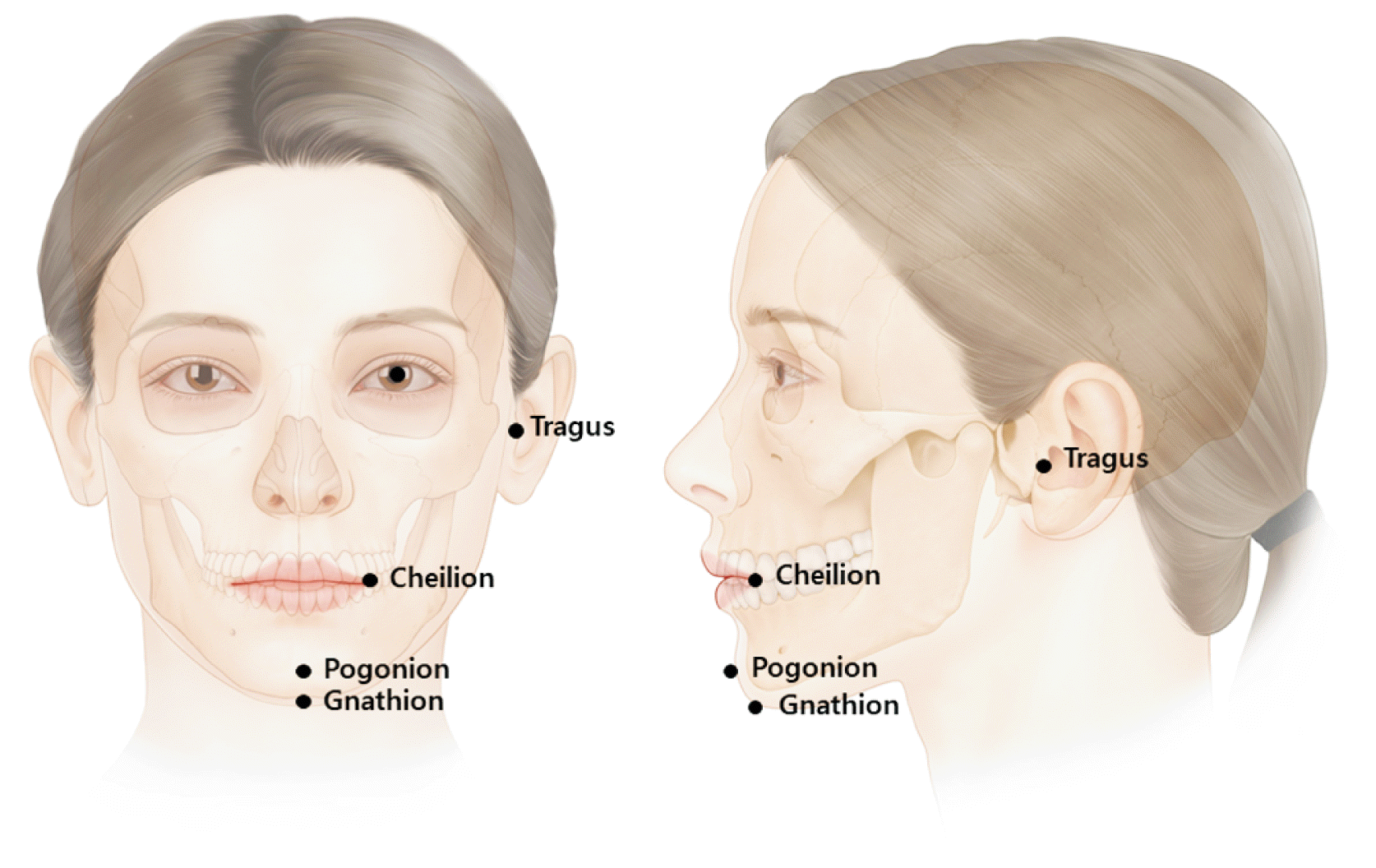
Therefore, consideration of anatomical factors with respect to the DAO and starting the primary treatment with a low dose of BoNT are important. In addition, knowledge of individual muscle variations is crucial for diagnosis [5]. Recent studies on BoNT injections in specific locations have been conducted as part of anatomical assessments of particular muscles on the basis of external anatomical landmarks (Fig. 1) [7-31].
Clinicians treating DAO need to have a thorough understanding of the facial anatomy; however, few anatomical findings in the DAO have been reviewed with clinical application (area, depth, and units). The study narratively reviewed recently published articles of clinical and anatomical perspectives.
The DAO is a triangular-shaped facial expression muscle that originates from the mental tubercle. Its continuation forms an oblique line below and lateral to the DLI muscle, which converges to the narrow fasciculus, blends with the modiolus, and interlaces with the orbicularis oris and the risorius (Fig. 2) [32]. The inferior and medial borders are known to be located 1.5 cm lateral to the mandibular symphysis. The inferior width of the DAO is 3.6 cm on average [3]. The action of the muscle is balanced at the modiolus by other perioral muscles. It dilates the mouth and bilaterally depresses the labial commissures. The depth of the DAO was located 0.25 to 0.4 cm from the skin surface as of our measurement of 20 subjects.
The modiolus is an important anatomical structure in the perioral area. Its thick muscular band is intermingled with the converging fibers around the cheilion, which consists of the zygomaticus major, DAO, levator anguli oris, buccinators, orbicularis oris, and risorius muscles. In the study of histologic sections, the modiolus was observed as a dense tissue with collagen and muscle fibers [33]. The modiolus is the insertion point of the perioral muscles and is heavily involved in lower facial expressions. Contrary to the case of Caucasians, the location of the modiolus in Asians is below the intercheilion line in 58.4% [34].
The injection points can be created by drawing crossing points using two lines: one vertical and one transverse. The vertical line is the line crossing the mid-pupil, and the transverse line is located in the middle of the lower mandibular border and the line that crosses the cheilion and tragus. One-point injection bilaterally, intradermally or subdermally, is proposed with 2–3 units (U) at each point. Since the modiolus is located below intercheilion line in most of the Asian population, the injection point is about the width of the index finger (1.5 cm) in the Caucasians and little finger (1.2 cm) in the Asian population (Fig. 3).
The injection should not be administered medially to the vertical line crossing the cheilion, which might affect inadvertent diffusion of the DLI muscle, leading to an asymmetric smile. If the patient does not have a sufficient effect, additional treatment should be conducted after primary treatment.
The diffusion of BoNT with 2–3 U per point is sufficient for any individual. Because BoNT spreads 1 cm from the injection point, injection into a minimum of three points with 1–2 U of BoNT, totaling 4–6 U, is recommended. Borodic et al. [35] demonstrated that BoNT diffuses up to 4.5 cm from the point of injection in case of 10 U administration into the rabbit longissimus dorsi muscle, and the diffusion gradient of 1 U injection is 1.5 cm to 3 cm.
Choi et al. [2] conducted cadaveric research on 43 hemifaces and discovered that the morphological angle of the DAO (from the modiolus to the lateral border of DAO had a mean of 44.7°±13.7° and that to the medial border was 31.8°±8.5°.
Another three-dimensional evaluation study in 90 hemifaces precisely located the outer landmark of DLI, at the point transversely between the cheilion and mid-sagittal line and vertically between the inferior mandibular border and inferior line of the vermilion border. The DAO was located vertically at the mid-pupillary line and transversely at the middle of the lower face. They described effective guidelines that only targeted DLI and DAO [36].
The anatomical location of the mental foramen has been evaluated by Hur et al. [3], alongside its variation within the DAO and DLI, depending on the individuals. Cadaveric dissection was conducted on 34 hemifaces; 67.7% were covered by DAO, and 22.3% were covered by DLI. Since the mental nerve spreads from the foramen, covered by these muscles, the injection should be administered superficially to avoid neural damage.
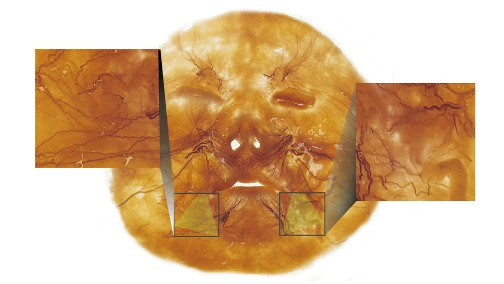
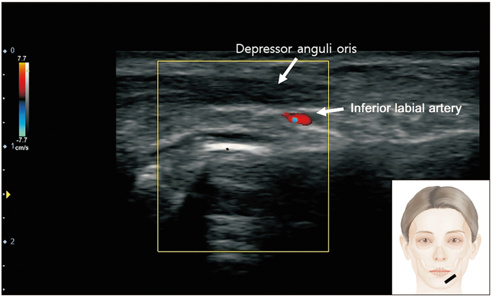
Lee et al. [37] discovered the facial artery runs beside the DAO along the lateral margin of the muscle and inferior labial artery running beneath the muscle. This indicates superficial injection is appropriate for not damaging the artery (Fig. 4). Additionally, we have observed the muscle with whole mount staining technique called Sihler’s method. The intramuscular neural distribution was observed in evenly dispersed throughout the muscle (Fig. 5).
The DAO is a superficially located muscle near the mouth area that overlaps with nearby muscles. It is necessary to identify its detailed anatomical borders and depth when treating perioral asymmetry and a drooping appearance by injecting BoNT [38].
When correcting the depressed mouth corner, the platysma should also be targeted, as the muscle plays an important role in pulling down the mouth corner [39]. The proposal based on anatomically dissected data would be sufficient to support blind injection into the DAO. However, it could be visualized with ultrasound-guided injection. The DAO and its virtual contraction can easily be observed on ultrasonography [40]. A limitation of this study was that it was not clinically applied. Further studies should be conducted in a clinical setting.
Overall, we propose that BoNT injections should be carried out intradermally or subdermally. They should be injected at the crossing point of the mid-pupillary line and at the middle of line crossing the cheilion and tragus and the mandibular border, with 2–3 U for each point.
Acknowledgements
This study was conducted in compliance with the principles of the Declaration of Helsinki. Consent was received from the families of the deceased patients before beginning the dissection. The authors sincerely thank those who donated their bodies to science to enable anatomical research. The results of such research can potentially increase mankind’s overall knowledge, and consequently, improve patient care. Therefore, these donors and their families deserve the greatest gratitude. The authors thank Eun-Byul Yi from the Eonbuk Elementary School for the illustrations.
Notes
Author Contributions
Conceptualization: KHY, JHL. Data acquisition: HWH, YJC. Data analysis or interpretation: KL, HJL. Drafting of the manuscript: KHY. Critical revision of the manuscript: HJK. Approval of the final version of the manuscript: all authors.
Conflicts of Interest
I acknowledge that I have considered the Conflict of Interest statement included in the Author Guidelines. I hereby certify that to the best of my knowledge, no aspect of my current personal or professional situation might reasonably be expected to significantly affect my views on the subject I am presenting.
References
1. Krag AE, Dumestre D, Hembd A, Glick S, Mohanty AJ, Rozen SM. 2021; Topographic and neural anatomy of the depressor anguli oris muscle and implications for treatment of synkinetic facial paralysis. Plast Reconstr Surg. 147:268e–78e. DOI: 10.1097/PRS.0000000000007593. PMID: 33565832.

2. Choi YJ, Kim JS, Gil YC, Phetudom T, Kim HJ, Tansatit T, Hu KS. 2014; Anatomical considerations regarding the location and boundary of the depressor anguli oris muscle with reference to botulinum toxin injection. Plast Reconstr Surg. 134:917–21. DOI: 10.1097/PRS.0000000000000589. PMID: 25347627.

3. Hur MS, Hu KS, Cho JY, Kwak HH, Song WC, Koh KS, Lorente M, Kim HJ. 2008; Topography and location of the depressor anguli oris muscle with a reference to the mental foramen. Surg Radiol Anat. 30:403–7. DOI: 10.1007/s00276-008-0343-4. PMID: 18385924.

4. Moradi A, Shirazi A. 2022; A retrospective and anatomical study describing the injection of botulinum neurotoxins in the depressor anguli oris. Plast Reconstr Surg. 149:850–7. DOI: 10.1097/PRS.0000000000008967. PMID: 35139057.

5. Childers MK. 2004; Targeting the neuromuscular junction in skeletal muscles. Am J Phys Med Rehabil. 83(10 Suppl):S38–44. DOI: 10.1097/01.PHM.0000141129.23219.42. PMID: 15448576.

6. Kaplan SE, Sherris DA, Gassner HG, Friedman O. 2007; The use of botulinum toxin A in perioral rejuvenation. Facial Plast Surg Clin North Am. 15:415–21. v–vi. DOI: 10.1016/j.fsc.2007.07.001. PMID: 18005882.

7. Kim HM, Ree YS, Park MS, Kim JS, Ahn JH, Yi KH. 2022; Clinical guideline: deoxycholic acid injection for submental fat reduction. The Aesthetics. 3:7–11. DOI: 10.46738/Aesthetics.2022.3.2.7.

8. Lee HJ, Lee JH, Yi KH, Kim HJ. 2020; Nov. 25. Sonoanatomy and an ultrasound scanning protocol of the intramuscular innervation pattern of the infraspinatus muscle. Reg Anesth Pain Med. [Epub]. https://doi.org/10.1136/rapm-2022-103682. DOI: 10.1136/rapm-2022-103682. PMID: 36427902.

9. Lee HJ, Lee JH, Yi KH, Kim HJ. 2022; Intramuscular innervation of the supraspinatus muscle assessed using Sihler's staining: potential application in myofascial pain syndrome. Toxins (Basel). 14:3. DOI: 10.3390/toxins14050310. PMID: 35622557. PMCID: PMC9143847. PMID: 34b169cd96874bde9b8c4748508d0448.

10. Rha DW, Yi KH, Park ES, Park C, Kim HJ. 2016; Intramuscular nerve distribution of the hamstring muscles: application to treating spasticity. Clin Anat. 29:746–51. DOI: 10.1002/ca.22735. PMID: 27213466.

11. Yi KH, Cong L, Bae JH, Park ES, Rha DW, Kim HJ. 2017; Neuromuscular structure of the tibialis anterior muscle for functional electrical stimulation. Surg Radiol Anat. 39:77–83. DOI: 10.1007/s00276-016-1698-6. PMID: 27206542.

12. Yi KH, Lee HJ, Choi YJ, Lee JH, Hu KS, Kim HJ. 2020; Intramuscular neural distribution of rhomboid muscles: evaluation for botulinum toxin injection using modified Sihler's method. Toxins (Basel). 12:289. DOI: 10.3390/toxins12050289. PMID: 32375284. PMCID: PMC7291336. PMID: 2a43e00b63b34292bd34bcaa4ce50d09.

13. Yi KH, Lee HJ, Hur HW, Seo KK, Kim HJ. 2022; Guidelines for botulinum neurotoxin injection for facial contouring. Plast Reconstr Surg. 150:562e–71e. DOI: 10.1097/PRS.0000000000009444. PMID: 35759641.

14. Yi KH, Lee HJ, Seo KK, Kim HJ. 2022; Intramuscular neural arborization of the latissimus dorsi muscle: application of botulinum neurotoxin injection in flap reconstruction. Toxins (Basel). 14:107. DOI: 10.3390/toxins14020107. PMID: 35202134. PMCID: PMC8878018. PMID: 0c7534ebd76b4902bff96b4bfe81c6b3.

15. Yi KH, Lee JH, Hu HW, Kim HJ. 2022; Novel anatomical guidelines on botulinum neurotoxin injection for wrinkles in the nose region. Toxins (Basel). 14:342. DOI: 10.3390/toxins14050342. PMID: 35622589. PMCID: PMC9144745. PMID: 95ea3c07c66d4527b5265c5ec719f652.

16. Yi KH, Lee JH, Kim GY, Yoon SW, Oh W, Kim HJ. 2022; Novel anatomical proposal for botulinum neurotoxin injection targeting lateral canthal rhytids. Toxins (Basel). 14:462. DOI: 10.3390/toxins14070462. PMID: 35878200. PMCID: PMC9316553. PMID: 46755210c7064a99b625f2d6b6dc6e0a.

17. Yi KH, Choi YJ, Cong L, Lee KL, Hu KS, Kim HJ. 2020; Effective botulinum toxin injection guide for treatment of cervical dystonia. Clin Anat. 33:192–8. DOI: 10.1002/ca.23430. PMID: 31301235.

18. Yi KH, Lee HJ, Choi YJ, Lee K, Lee JH, Kim HJ. 2021; Anatomical guide for botulinum neurotoxin injection: application to cosmetic shoulder contouring, pain syndromes, and cervical dystonia. Clin Anat. 34:822–8. DOI: 10.1002/ca.23690. PMID: 32996645.

19. Yi KH, Lee HJ, Lee JH, Lee KL, Kim HJ. 2021; Effective botulinum neurotoxin injection in treating iliopsoas spasticity. Clin Anat. 34:431–6. DOI: 10.1002/ca.23670. PMID: 32805076.

20. Yi KH, Lee HJ, Lee JH, Seo KK, Kim HJ. 2021; Application of botulinum neurotoxin injections in TRAM flap for breast reconstruction: intramuscular neural arborization of the rectus abdominis muscle. Toxins (Basel). 13:269. DOI: 10.3390/toxins13040269. PMID: 33918558. PMCID: PMC8070362. PMID: 15169c322e5c4f158142057c4d95936b.

21. Yi KH, Lee HJ, Seo KK, Kim HJ. 2022; Botulinum neurotoxin injection guidelines regarding flap surgeries in breast reconstruction. J Plast Reconstr Aesthet Surg. 75:503–5. DOI: 10.1016/j.bjps.2021.09.081. PMID: 34776389.

22. Yi KH, Lee JH, Hu HW, Kim HJ. 2022; Anatomical proposal for botulinum neurotoxin injection for glabellar frown lines. Toxins (Basel). 14:268. DOI: 10.3390/toxins14040268. PMID: 35448877. PMCID: PMC9032255. PMID: dd131953c6e14366befdc986e94b15dd.

23. Yi KH, Lee JH, Kim HJ. 2022; Intramuscular neural distribution of the serratus anterior muscle: regarding botulinum neurotoxin injection for treating myofascial pain syndrome. Toxins (Basel). 14:271. DOI: 10.3390/toxins14040271. PMID: 35448880. PMCID: PMC9033065. PMID: c4ef8fa6e12442a990c0be9185a526e5.

24. Yi KH, Lee JH, Kim HM, Kim HJ. 2022; The botulinum neurotoxin for pain control after breast reconstruction: neural distribution of the pectoralis major muscle. Reg Anesth Pain Med. 47:322–6. DOI: 10.1136/rapm-2021-102653. PMID: 35039438.

25. Yi KH, Lee JH, Lee DK, Hu HW, Seo KK, Kim HJ. 2021; Anatomical locations of the motor endplates of sartorius muscle for botulinum toxin injections in treatment of muscle spasticity. Surg Radiol Anat. 43:2025–30. DOI: 10.1007/s00276-021-02813-7. PMID: 34378107. PMCID: PMC8354843.

26. Yi KH, Lee KL, Lee JH, Hu HW, Kim HJ. 2022; Guidance to trigger point injection for treating myofascial pain syndrome: intramuscular neural distribution of the quadratus lumborum. Clin Anat. 35:1100–6. DOI: 10.1002/ca.23918. PMID: 35655442.

27. Yi KH, Lee KL, Lee JH, Hu HW, Lee K, Seo KK, Kim HJ. 2021; Guidelines for botulinum neurotoxin injections in piriformis syndrome. Clin Anat. 34:1028–34. DOI: 10.1002/ca.23711. PMID: 33347678.

28. Yi KH, Rha DW, Lee SC, Cong L, Lee HJ, Lee YW, Kim HJ, Hu KS. 2016; Intramuscular nerve distribution pattern of ankle invertor muscles in human cadaver using sihler stain. Muscle Nerve. 53:742–7. DOI: 10.1002/mus.24939. PMID: 26467315.

29. Yi KH, Lee JH, Lee K, Hu HW, Lee HJ, Kim HJ. 2022; Anatomical proposal for botulinum neurotoxin injection targeting the platysma muscle for treating platysmal band and jawline lifting: a review. Toxins (Basel). 14:868. DOI: 10.3390/toxins14120868. PMID: 36548765. PMCID: PMC9783622. PMID: 1fab78f1f2fb436e8f0adab9755ed1d2.

30. Yi KH, Lee JH, Hur HW, Lee HJ, Choi YJ, Kim HJ. 2023; Jan. 6. Distribution of the intramuscular innervation of the triceps brachii: clinical importance in the treatment of spasticity with botulinum neurotoxin. Clin Anat. [Epub]. https://doi.org/10.1002/ca.24004. DOI: 10.1002/ca.24004. PMID: 36606364.

31. Yi KH, Lee JH, Hu HW, Park HJ, Bae H, Lee K, Kim HJ. 2023; Feb. 17. Novel anatomical guidelines for botulinum neurotoxin injection in the mentalis muscle: a review. Anat Cell Biol. [Epub]. https://doi.org/10.5115/acb.22.266. DOI: 10.5115/acb.22.266. PMID: 36796830.

32. Le Louarn C, Buis J, Buthiau D. 2006; Treatment of depressor anguli oris weakening with the face recurve concept. Aesthet Surg J. 26:603–11. DOI: 10.1016/j.asj.2006.08.001. PMID: 19338950.

33. Demiryurek D, Bayramoglu A, Erbil KM, Onderoglu S, Sargon MF, Aldur MM, Korkusuz P, Nazikoglu A. 2003; Three-dimensional structure of the modiolus. A computerized reconstruction study. Saudi Med J. 24:846–9. PMID: 12939669.
34. Kim HJ, Seo KK, Lee HK, Kim J. 2016. Clinical anatomy of the face for filler and botulinum toxin injection. Springer;DOI: 10.1007/978-981-10-0240-3.
35. Borodic GE, Ferrante R, Pearce LB, Smith K. 1994; Histologic assessment of dose-related diffusion and muscle fiber response after therapeutic botulinum A toxin injections. Mov Disord. 9:31–9. DOI: 10.1002/mds.870090106. PMID: 8139603.

36. Choi YJ, We YJ, Lee HJ, Lee KW, Gil YC, Hu KS, Tansatit T, Kim HJ. 2021; Three-dimensional evaluation of the depressor anguli oris and depressor labii inferioris for botulinum toxin injections. Aesthet Surg J. 41:NP456–61. DOI: 10.1093/asj/sjaa083. PMID: 32232427.

37. Lee HJ, Won SY, O J, Hu KS, Mun SY, Yang HM, Kim HJ. 2018; The facial artery: a comprehensive anatomical review. Clin Anat. 31:99–108. DOI: 10.1002/ca.23007. PMID: 29086435.
38. Raspaldo H, Niforos FR, Gassia V, Dallara JM, Bellity P, Baspeyras M, Belhaouari L. 2011; Lower-face and neck antiaging treatment and prevention using onabotulinumtoxin A: the 2010 multidisciplinary French consensus--part 2. J Cosmet Dermatol. 10:131–49. DOI: 10.1111/j.1473-2165.2011.00560.x. PMID: 21649819.
39. Bae JH, Youn KH, Hu KS, Lee JH, Tansatit T, Kim HJ. 2016; Clinical implications of the extension of platysmal fibers on the middle and lower face. Plast Reconstr Surg. 138:365–71. DOI: 10.1097/PRS.0000000000002346. PMID: 27064219.

40. Lee HJ, Kim JS, Youn KH, Lee J, Kim HJ. 2018; Ultrasound-guided botulinum neurotoxin type A injection for correcting asymmetrical smiles. Aesthet Surg J. 38:NP130–4. DOI: 10.1093/asj/sjy128. PMID: 29800051.

Fig. 2
The dissected image of the lower face representing depressor levator inferioris (DLI) and depressor anguli oris (DAO). The DAO originates from the mental tubercle and converges to the narrow fasciculus, blends with the modiolus (M) and interlaces with the orbicularis oris.
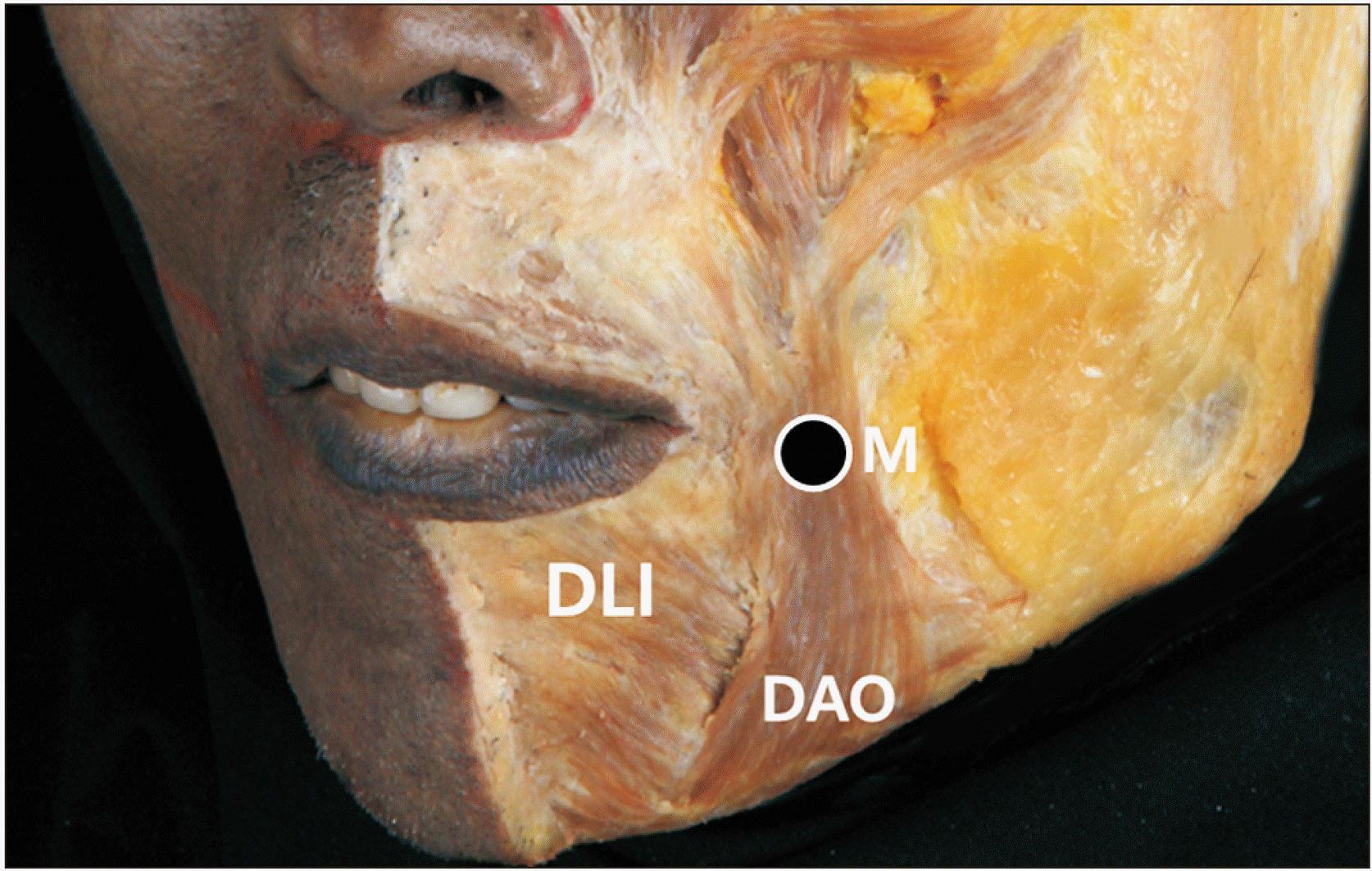
Fig. 3
Injection points of botulinum neurotoxin for targeting depressor angulii oris muscle. The dotted line is drawn vertically at mid-pupil point, mid-pupillary line (MPL), and the transverse dotted line is drawn in middle of line crossing the cheilion and tragus and the mandibular border. The intersection point of dotted lines is injection point (orange colored dot) for the muscle with 2–3 U. This would be easily applied by placing index finger (15 mm width) for caucasions (A) and little finger (12 mm width) for Asians (B) along the lower margin of the mandible.
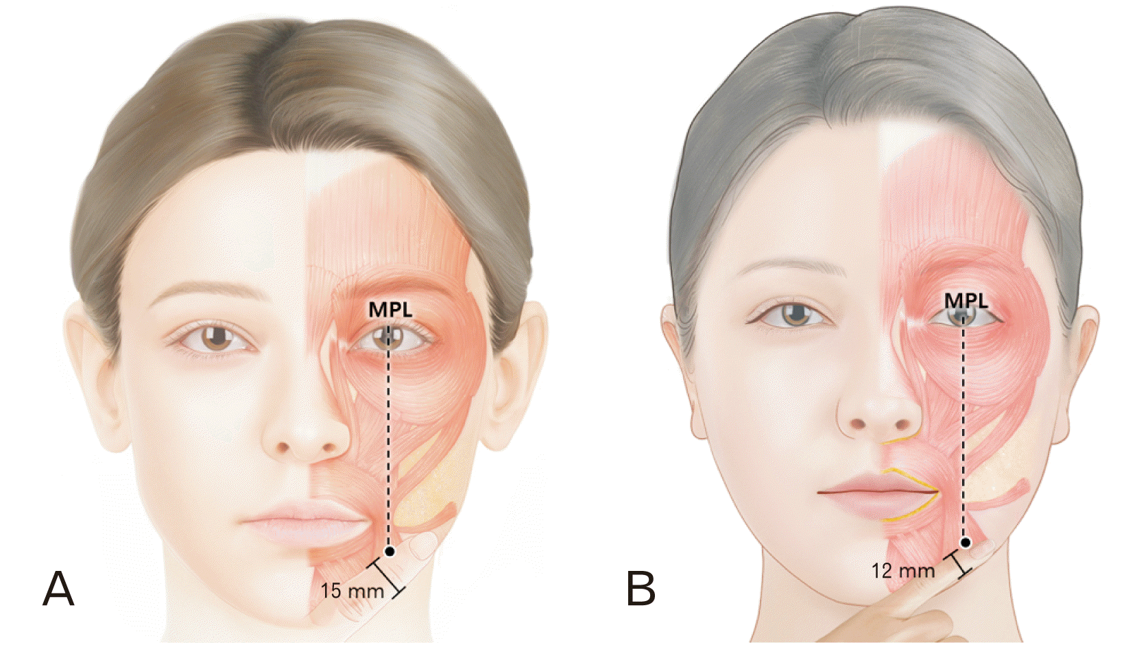
Fig. 5
Neural distribution of the face was revealed with whole mount staining method (Sihler’s method) with motor and sensory distribution. The enlarged panel with depressor anguli oris (yellow shaded) demonstrates neural distribution of the muscle. The nerve distribution seems to evenly dispersed in the muscle region.


 PDF
PDF Citation
 Print
Print


 XML Download