

 PDF
PDF Citation
 Print
Print


I. Introduction
Elongated styloid syndrome (ESS), also referred to as Eagle’s syndrome or styloid process (SP) syndrome in the literature, is commonly associated with anterolateral pain in the neck due to SP elongation or stylohyoid ligament calcification1,2. ESS was initially described in 1937 when Eagle highlighted the classic styloid syndrome with manifestations, including foreign body sensation and pain, as well as a more serious variant of the stylo-carotid-artery syndrome2. ESS also presents various neurological symptoms and pain due to its anatomical relevance to cranial nerves (e.g., VII, VIII, IX, and XI)1.
Despite its early recognition in 1937, the underlying mechanism of ESS has not been thoroughly described1. The etiology of ESS has been attributed to the retention of Reichert’s cartilage tissue in the embryo, calcified or expanded osseous tissues of the stylomandibular ligament, trauma, surgical procedures (e.g., tonsillectomy), and ageing2,3. Epidemiological studies have estimated the mean length of the SP to be 2.5 to 3 cm, with those longer than 4 cm being strongly associated with pain1. The elongated SP is estimated to be present in 4% of the general population, with only 4%-10% of these cases being symptomatic3. Based on their radiographic interpretations, SP elongation is classified into four types with 30 mm as the cut-off level: (i) no elongation detected (type 0), (ii) elongated SP longer than 30 mm (type 1), (iii) pseudo-elongated SP related to the mineralization of the stylohyoid ligament (type 2), and (iv) elongated but segmented/interrupted SP longer than 30 mm (type 3)4. It is generally believed that oral and maxillofacial surgeon practice should include assessing and managing patients with an orofacial pain diagnosis, such as temporomandibular joint disorder (TMJD), especially when conservative management fails or another etiology is considered5,6. Clinicians must be aware of the comorbidities associated with TMJD that can present with distinct oro-facial symptoms, such as earache, tinnitus, fibromyalgia, and Eagle’s syndrome1,5,6. The latter is often presented with heterogeneous clinical presentations. It is often consequently misdiagnosed for several years and managed initially as other craniomandibular disorders including TMJD3,7.
Go to : 
II. Case Report
Informed consent for publication was obtained from the patient, and ethical approval was obtained from the Institutional Review Board of King Saud University (No. 23/0083/IRB). The reporting of the present clinical case is in line with the CARE checklist for case reports (https://www.care-statement.org/).
A 52-year-old male of Arabic ethnicity presented to our institution’s oral medicine clinic in February 2021 with a history of “annoying sounds from my jaws for the past three years.” This was described as a popping sound without bilateral clicking or crepitation. There were no reports of associated pain, parafunctional diurnal or nocturnal activity, or headache in the orofacial region. However, the subject experienced tinnitus and progressive hearing loss in the right ear, which was investigated by the otolaryngology team three years ago. The patient was diagnosed with sensorineural hearing loss and recommended using a hearing-aid device. However, the patient was not amenable to its use. He also complained of a synchronous constant tinnitus intermittently associated with a headache that worsened with a loud noise. Furthermore, the patient experienced dysphagia when drinking liquids. There was no record of otalgia, ear discharge, dizziness, or vertigo. The patient denied any history of weight loss, night sweats, or fever.
The patient visited two general dental practitioners and was informed that his orofacial symptoms could be attributed to temporomandibular joint dysfunction. An oral appliance (night guard) and muscles of mastication exercises were recommended; however, the symptoms persisted. His medical review indicated a history of hypothyroidism, bronchial asthma, and cervical spondylosis on the C4 and C5 vertebrae. There was no history of maxillofacial trauma or hospitalization. He was married with children and worked as a businessman.
According to a physical examination, there was no neurological deficit in cranial nerves VII, VIII, IX, and XI. A temporomandibular joint examination showed no popping or clicking sounds; however, slight mandibular deviation to the right side upon opening and a maximum interincisal opening of 65 mm were observed. Additionally, the right temporalis and masseter muscles were slightly tender on palpation. There was no tenderness on bilateral palpation in the trapezius and sternocleidomastoid muscles. When the patient was asked to turn his head forward, there was an audible bilateral popping sound, with the patient noting the recurrence of an identical sound upon yawning. A hoarse voice (dysphonia) with a high pitch was noted during the consultation. Intraoral examination of hard and soft tissues revealed no abnormalities.
1. Differential diagnosis
TMJDs are often encountered as chronic pain conditions in general medical and dental practice and can significantly impact daily activities and quality of life6. The prevalence of signs and symptoms of TMJD reported in international studies varies widely between 5%-50% owing to differences in study cohorts, such as school or university students, specific patient populations and the general population6,8,9. Based on their level of evidence, these disorders are attributed to altered pain modulation and sensitization, sex, cognitive characteristics (e.g., catastrophizing), and pain comorbidities (e.g., fibromyalgia)6,9. Clinically, TMJD can be defined as pain affecting the pre-auricular area, cheeks, or over the temporal areas with or without functional jaw limitations and sounds6. The working group of the Diagnostic Criteria for temporomandibular disorders (DC/TMD) classifies these disorders according to their relevance to the joint, masticatory muscles, or headache attributions9.
Myalgia is localized pain in the muscles of mastication and is considered the most common diagnosis of orofacial pain6,9. This diagnosis is based on a history of pain modification of jaw function/parafunction9, which was not reported in the present case. Moreover, noise-associated complaints reported among individuals with disc displacement with reduction align with the current complaint of continuous popping sounds for more than 30 days9. However, in this case, jaw opening and closing movements were not clinically associated with clicking or popping sounds. Other pain-associated diagnoses, such as trigeminal and glossopharyngeal neuralgias and vertebral arthritis, were ruled out owing to the lack of pain and physical limitations10-12.
Moreover, there was no history of trauma or surgical procedures that could robustly indicate a diagnosis of ESS1, even though pharyngeal symptoms of dysphagia and foreign body sensation, progressive hearing loss, dysphonia, and popping sounds in the ear have been previously reported with ESS1. In contrast to the demographics of the present patient, females between 40-60 years are more likely to present with ESS13. Moreover, there were no reported complaints of carotid artery compression (e.g., syncope or transient ischemic cardiac events) or jugular vein impingement (e.g., headache, visual disturbance/discomfort, dizziness, and notable cognitive decline) that could implicate its congenital variant (stylohyoid syndrome) or styloidogenic jugular syndrome, respectively1,3,11. Similarly, signs of a cranial nerve or soft palate irritation caused by posterior and anterior process elongation were not observed3.
2. Diagnosis and management
An initial review of the orthopantomogram imaging indicated elongated SP behind the posterior border of the mandibular ramus bilaterally.(Fig. 1) Magnetic resonance imaging (MRI) and cone-beam computed tomography (CBCT) of the temporomandibular joint (TMJ) articular disc complex and full skull assessment were performed. MRI closed and open mouth settings on the right side indicated an abnormal morphological outline of the mandibular condyle with some flattening and irregularity without remarkable attenuation of the joint spaces. However, no apparent bone marrow signal alteration or joint effusion was observed. The MRI open mouth setting for the same side showed normal anterior inferior translation of the mandibular condyle concerning the articular eminence. On the contralateral side (left TMJ), the closed mouth presented a normal morphological outline of the mandibular condyle and temporal glenoid fossa without remarkable attenuation of the joint spaces. There were no signs of erosion, injury, or apparent alterations in bone marrow signal. Similarly, the open-mouth approach indicated no abnormal anterior inferior translation of the mandibular condyle with articular eminence or joint effusion.
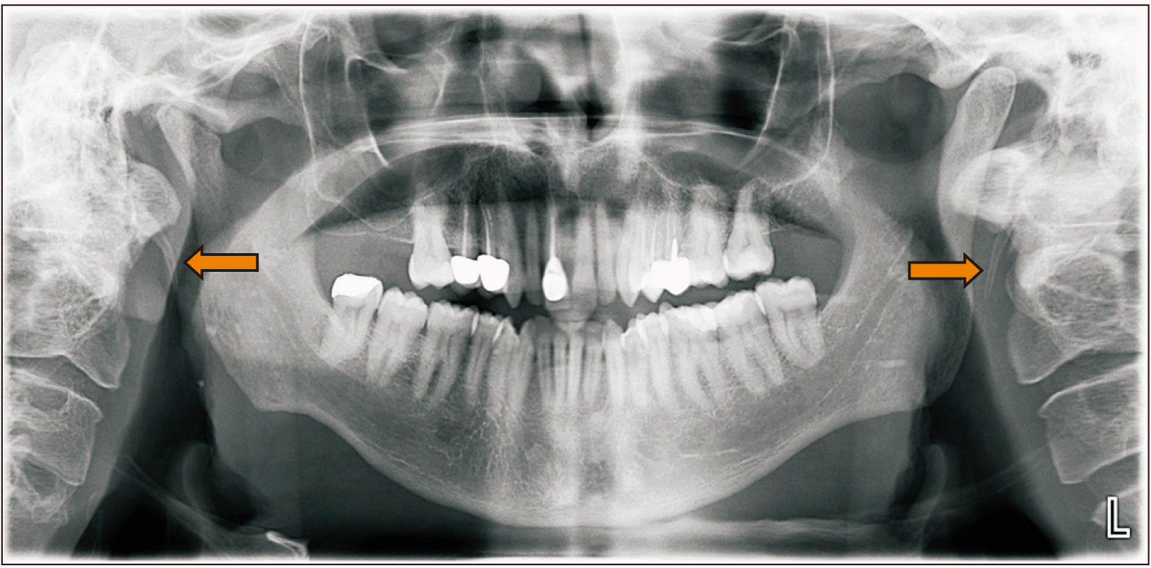
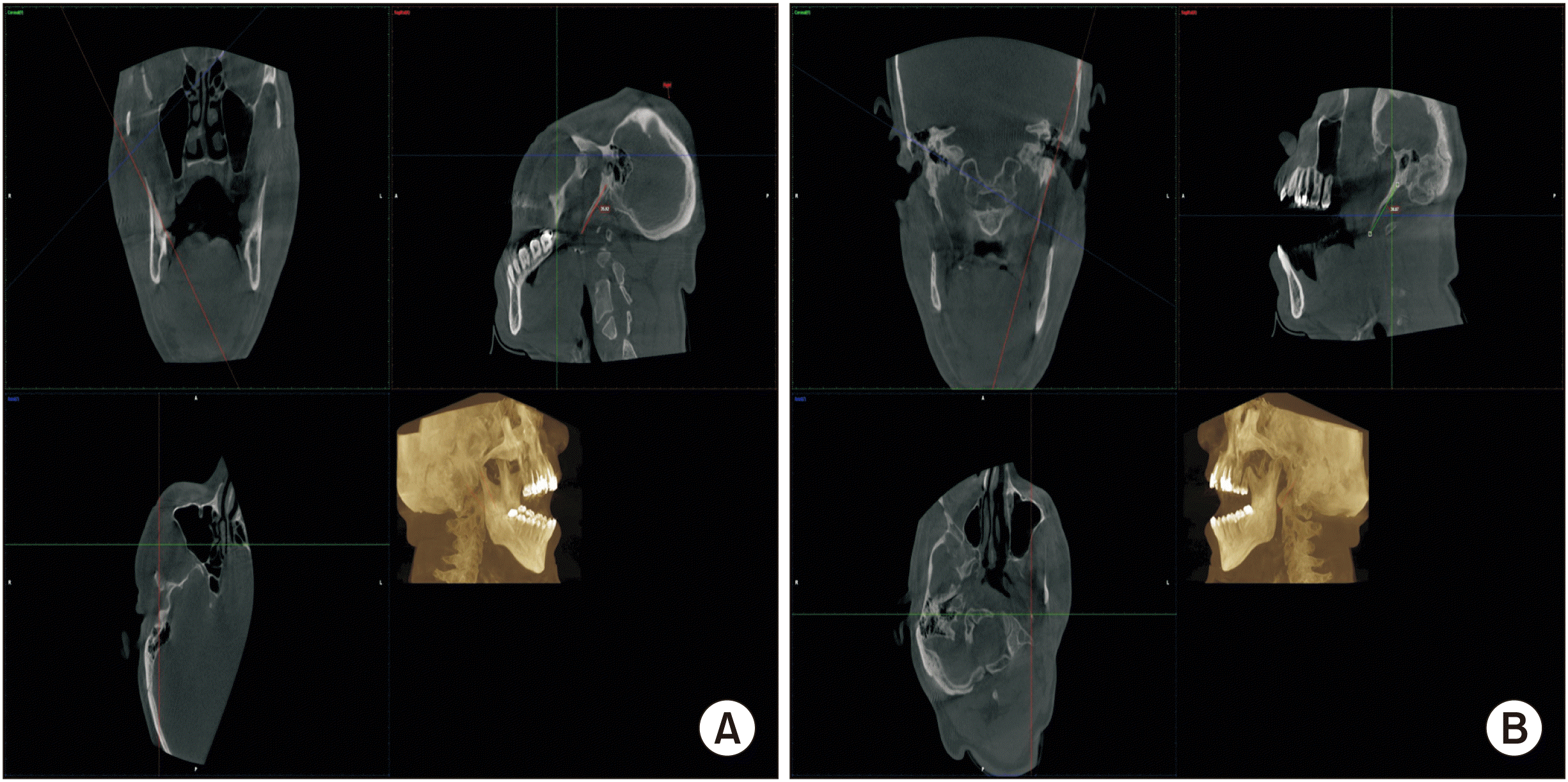
CBCT imaging of the TMJ indicated arthritic changes with flattening of the head of the right mandibular condyle. A bilateral elongated SP 35.92 mm in length was noted on the right side and 38.87 mm on the left.(Fig. 2) Both were morphologically interpreted as type 1 based on Langlais’ classification4. The correlation between patient presentation and clinical and imaging findings was consistent with Eagle’s syndrome. The patient was informed of his diagnosis, the symptoms, possible complications, and surgical management approaches. The patient declined further treatment and preferred to remain under observation.
The patient was referred to the otolaryngology clinic of our hospital for further assessment of foreign body sensation and progressive hearing loss to exclude concurrent pharyngeal inflammatory or neoplastic conditions. Fiberoptic endoscopic evaluation of swallowing indicated that both vocal folds were freely mobile with complete closure during phonation and safe oropharyngeal swallowing physiology with all tested volumes and consistencies. Both vocal folds moved freely with complete closure during phonation. Imaging for the internal auditory canal and labyrinth did not detect any abnormalities, and no auditory pathology or hemorrhage was noted with the vestibulocochlear nerve. Screening for auditory disorders was inconclusive. The 2-, 6-, and 12-month follow-up reviews at oral medicine clinics indicated no new symptoms or discomfort with fewer episodic sounds and avoidance of severe head turning. The patient agreed to continue to attend the scheduled appointments with both clinics and to consider surgical management if there were any unanticipated symptoms.
Go to :
III. Discussion
It is not unusual for individuals with ESS to be initially diagnosed and managed as having TMJD owing to myriad similar craniomandibular manifestations between both diagnoses1,7,14. Additionally, factors such as the degree of elongation, ossification, and involvement of vital anatomical structures can contribute to the diverse clinical presentations of ESS and deter its early recognition15.
A concurrent diagnosis of myalgia or TMJD was not considered despite the concurrent unilateral flattening morphological condylar changes and tenderness on palpation of the temporalis and masseter muscles. This was due to the lack of DC/TMD criteria for myalgia and intra-articular TMJD, including previous or present orofacial pain reports or jaw sounds during the examination9. Regardless of the possible coexistence of TMJD and ESS or SP fracture15, imaging-based studies of patients with TMJD indicated no correlation between TMJD symptoms and the degree of SP elongation14 or angulation10.
In contrast, radiographic interpretation of 50 individuals with TMJD indicated a 76% incidence rate of unilateral or bilateral elongated SP12. The previous study also noted a possible but not statistically significant correlation between elongation and pain intensity, headache, and tinnitus in the same patient population12. In line with these findings, Bardhoshi et al.16 noted an increased styloid length among patients with TMJD compared to controls. Furthermore, conservative management of TMJD seems to reduce the orofacial symptoms of ESS1,7. The evidence supporting the synchronous and overlapping TMJD and ESS due to muscular disturbance14 was observed in case reports of individuals with both diagnoses. For instance, a 37-year-old female initially reported headaches, orofacial pain, and foreign body sensations upon swallowing, and she was managed with an occlusal appliance, which significantly improved her symptoms7. Similarly, a 55-year-old female presented with a five-year history of episodic preauricular pain with ipsilateral radiation to the temporal area and was managed with occlusal appliances, non-steroidal anti-inflammatory drugs, and benzodiazepines11. The persistent foreign body sensation, recurrence, and progression of symptoms in these cases led to further assessments, and a diagnosis of ESS was made, as was for the previous patients7,11.
Other differential diagnoses that have been reported to be relevant to ESS include atypical facial and cervicofacial pain, odontogenic pain, referred otalgia, trigeminal neuralgia, Sluder’s syndrome, hyoid bursitis, headache, Ernest’s syndrome, glossitis, carotidynia, vertebral arteritis, vertigo, salivary gland sialolithiasis, as well as sialadenitis, oesophageal diverticula, head and neck tumors, and psychosomatic disorders3,11,13.
Although imaging in a dental care setting, such as orthopantomography and periapical films, can be used to diagnose long SP7, clinicians commonly attribute the varied clinical findings to TMJD or other attributes that have been initially reported as TMJD (e.g., primary or metastatic neoplasms, infections, and neurological disorders)15,16. Furthermore, ultrasonography can be a cost-effective alternative with relatively comparative accuracy to CT when determining SP length and anatomical displacement17. However, SP might seem longer on this imaging modality and sometimes appears internally deflected owing to its low resolution17. To diagnose the carotid and jugular variants of ESS, transoral carotid and intra-vascular ultrasound imaging can detail the SP and its relevance to cervical vessels18. Further investigation is needed, however, to determine the feasibility for pre- and postoperative evaluations in patients with ESS.
Assessing SP pathology on MRI can be challenging due to positioning errors and compression/displacement of vital cervical structures17. Thus, CBCT is often used to confirm the diagnosis and determine the operability of elongated processes when the clinical presentation suggests an ESS diagnosis11,14. The management of ESS can include conservative approaches using pain medications (e.g., nonsteroidal anti-inflammatory drugs, acetaminophen, and tramadol), local anesthetics (e.g., triamcinolone and mepivacaine), anticonvulsants (e.g., carbamazepine, gabapentin, and pregabalin), antidepressants (e.g., amitriptyline and tianeptine), corticosteroids (e.g., dexamethasone), and physical manipulation1,11,15. However, evidence-based guidelines have yet to confirm the long-term effectiveness and safety of these medications in managing ESS1,19.
Surgical resection of the elongated SP (styloidectomy) using transoral or extraoral techniques remains a definitive treatment modality of long-lasting efficacy1,19. The advantages of the transoral approach include shorter operative procedures, lower pre and postoperative complications, and its possible use with local anesthetics19. However, the risk of perioperative tonsillar bleeding, bacterial infection, and neurovascular lesions have been reported with this approach13,19. Alternatively, the extraoral (transcutaneous cervical) approach can be used with a higher cure rate, better access and vascular control to dissect the SP, and a lower risk of infection than the intraoral approach19. Endoscopy also decreases the operative duration and improves esthetics19.
Determining the surgical criteria for the elongated SP for either approach, especially for patients with neurological deficits, can be challenging, as most published records are of retrospective methods and often lack longitudinal assessments of efficacy or complications19. Upon shared clinical decision-making, consideration should be made for symptomatic improvement, length of stay, and post-surgical complications (e.g., uncontrolled oral bleeding with or without carotid artery injury, airway edema, scarring, and facial nerve injury)1,11,19.
The present study was limited by the absence of surgical management of pharyngeal symptoms based on patient preference, as reported in a similar case report7. Thus, examining the mineralized process and assessing the elongation anatomically and patient-reported surgical outcomes was not possible. Evidence recommending surgical and non-surgical approaches to achieve favorable outcomes for patients with ESS remains lacking, with the available systematic reviews focusing on stylohyoid syndrome and not necessarily ESS or surgical methods19. Therefore, further longitudinal and systematic appraisal studies are needed to assess the clinician-reported and patient-reported outcomes (e.g., health-related quality of life and physical functioning) with these approaches. There is also a need to include the less frequent variant of ESS (stylo-carotid-artery syndrome) in the recognized diagnostic criteria because of its lack of recognition in the current version of the International Classification of Headache Disorders (ICHD-3)20.
In conclusion, clinicians should consider ESS and its less frequent variant, stylo-carotid-artery syndrome, in their differential diagnosis of complaints about jaw sounds. As styloid elongation may not be evident or may be under-detected in two-dimensional imaging, clinicians should consider CBCT for patients with TMJD who exhibit oropharyngeal symptoms of unclear etiology. They are also encouraged to develop a consensus on evidence-based conservative and surgical management of patients with ESS.
Go to :
 XML Download