

 PDF
PDF Citation
 Print
Print


I. Introduction
Plexiform neurofibromas are rare benign tumors and a special subtype of neurofibromatosis type 1 (NF-1), which is a slow-growing benign tumor1,2. Neurofibromatosis type 1 (NF-1), or Von Recklinghausen disease, is an autosomal dominant disease that affects about 1 in 3,000 people and may involves the bones, skin, eyes, nerves, and vascular system3. Plexiform neurofibroma is a benign peripheral nerve tumor seen in nearly 27%-44% of NF-1 patients and most often occurs in the craniomaxillofacial region4. The World Health Organization has subdivided neurofibromas into dermal and plexiform5. Dermal neurofibroma occurs in a single peripheral nerve, whereas plexiform neurofibroma involves nerve bundle5. Neurofibromas may present as solitary lesions or as generalized syndromes of neurofibromatosis or Von Recklinghausen disease6,7. Neurofibromas are immuno-positive to the S-100 protein in 80%-100% of cases, suggesting that they are neural origin6. There is no specific treatment for NF-1, and clinical manifestations are treated individually8. Surgical intervention is a desirable treatment for plexiform neurofibroma; however, significant vascularization of the tumor and consequent significant bleeding during surgery complicate the procedure8. Hematoma of the scalp, chest, and limbs, which occurs spontaneously or after minimal trauma as a rare complication of neurofibromatosis, has been mentioned but is very unusual in the maxillofacial area9.
We present a case of hematoma resulting from minor trauma in a patient with a history of surgical removal of neurofibromas is the right lower facial region.
II. Case Report
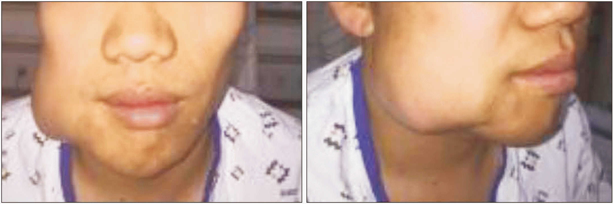
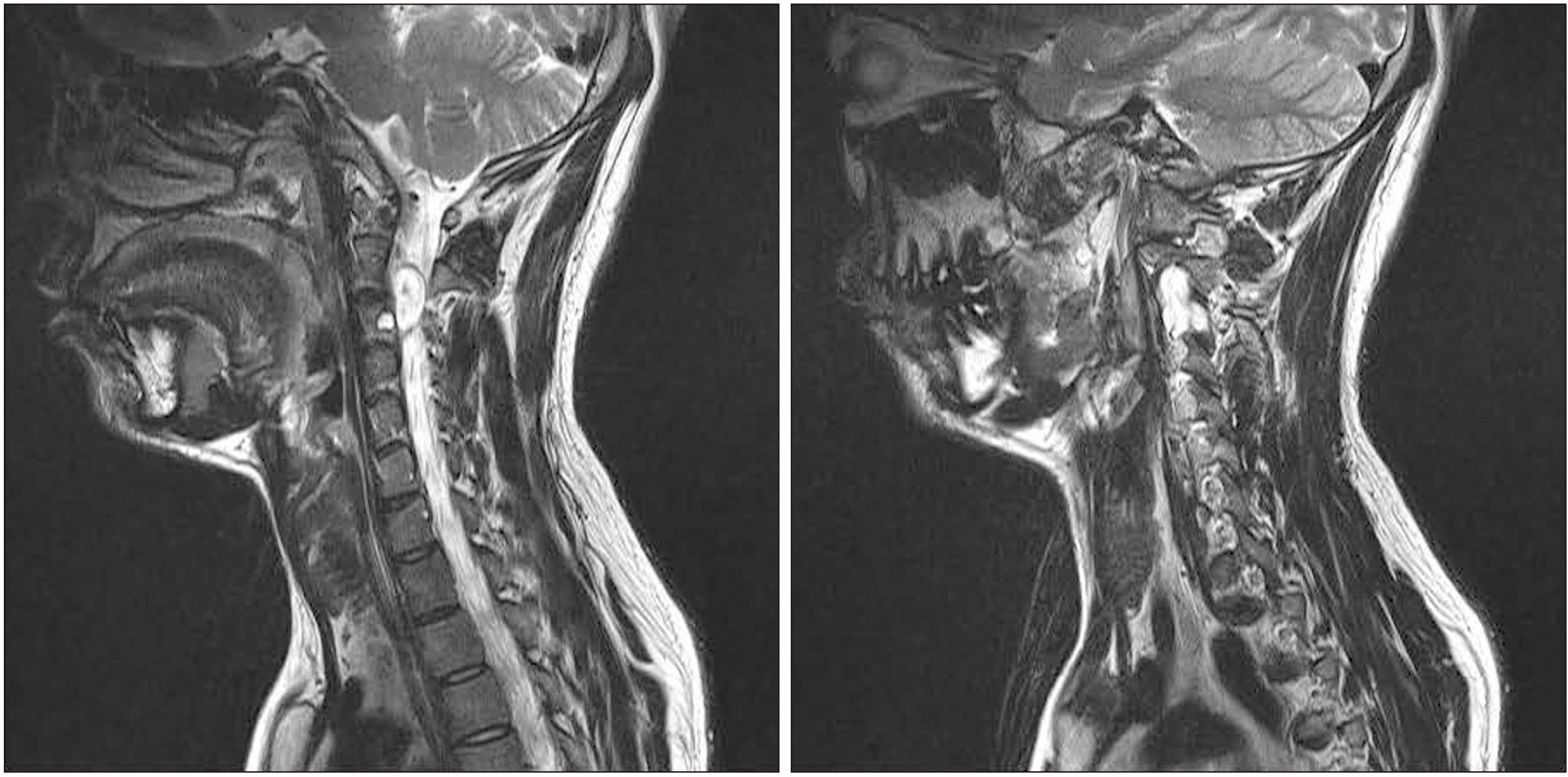
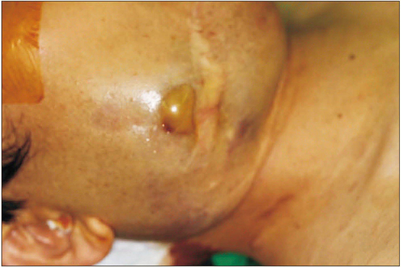
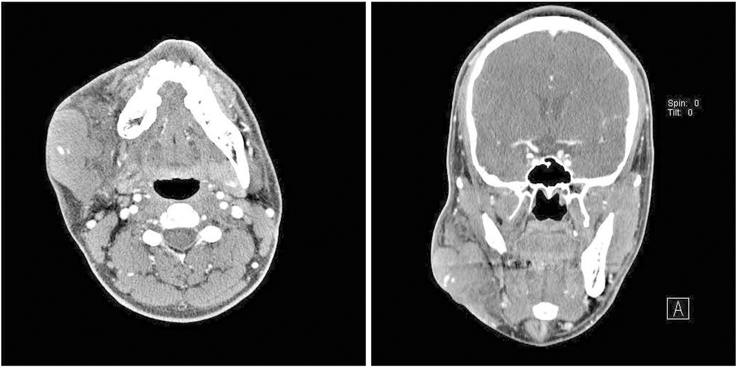
A 31-year-old male patient visited the emergency room with rapidly increasing, painful swelling of the lower right side of the face after blunt trauma of hitting his face against a door two hours prior.(Fig. 1) He had undergone two previous surgeries on the right lower face to remove neurofibromas, at 22 years (nine years prior) and 19 years (11 years prior). When the neurofibroma was removed, biopsy revealed plexiform neurofibroma. Magnetic resonance imaging (MRI) showed that the neurogenic tumor was located at the C2, C3 disc levels, spanned the central spinal canal and left neural foramen, and severely compressed the spinal cord to the right.(Fig. 2) Upon clinical examination, the overlying skin on the right lower face was bluish and taut.(Fig. 3) Vital signs were stable, hematocrit was 42.3%, and hemoglobin was 12.7 g/dL. Hemodynamic parameters were within normal limits. Upon angiography of the right external carotid artery, vascular lesions were not observed. A computed tomography (CT) scan revealed a large hematoma collection sized about 5 cm in diameter in the buccal and submandibular space, and a vascular structure was confirmed in the contrast-enhanced CT image.(Fig. 4) A facial approach was performed to remove the hematoma. After muscle dissection, the hematoma remained relatively stable. The mean arterial pressure (MAP) during the operation was maintained in the range of 55-60 mmHg. After careful removal of the hematoma, several slow bleeds from the muscle were controlled by electrocautery. Since there was no pulsatile active bleeding, layered sutures were performed, and the operation was completed. A compression bandage was applied and an ice pack was recommended to reduce pain and swelling. However, several hours later, the patient complained of persistent swelling at the surgical site. Upon clinical examination, the bandage was wet with leaked blood. The patient was transferred to the operating room again, and the bleeding site was closely observed. Another hematoma was removed and the buccal artery was prophylactically ligated. A hemostatic matrix composed of thrombin and gelatin was applied to the surgical site. Such a hemostatic matrix requires careful use because it may cause extensive coagulation when administered intravascularly. While it should not be used as a substitute for vascular ligation, it is effective for oozing or spurting bleeding. Layered sutures and compression dressing were applied. After surgery, ice pack compression and transfusion of four units of packed red blood cells and four units of fresh frozen plasma were prescribed. An ice pack was applied for 48 hours, and the compression dressing was removed on the third day of surgery. Hemoglobin was 13.4 g/dL, and hematocrit was 38.8%. One week after surgery, the patient was discharged from the hospital after all facial stitches were removed. Hemoglobin was 12.8 g/dL, and hematocrit was 38.5%. Biopsy revealed diffuse neurofibroma. The patient was followed two weeks after surgery on an outpatient basis, and no complication such as hematoma, seroma, swelling, infection, and wound dehiscence were observed.
In the present study, to find reports of facial hematomas due to spontaneity or minimal trauma, the following search strategy was implemented. Two authors (S.M.L. and D.H.L.) independently searched PubMed for articles of any year and country using the terms ((facial hematoma) OR (facial bleeding)) AND (neurofibromatosis). Initially, 86 related articles were identified.
The articles in English-language literature, case reports, and case series were included. Non-English-language literature and genetic studies were excluded.
The following data were independently extracted from each eligible article: first author, year of publication, age, gender, facial area of hematoma, vector, previous diagnostic history of neurofibromatosis, methods of hemostasis, and postoperative blood transfusion.
All discrepancies related to the qualification criteria were resolved though discussion with team members. Among the initial 86 articles, those with the following traits were excluded: non-English language (n=9), genetic studies (n=7), cochlear hematoma (n=2), and irrelevant (n=63). Finally, five related articles were selected.
Six patients described in five articles were five males and one female, ranging from 18 to 60 years, with a mean age of 40.17 years. The vectors of facial hematoma were as follows: spontaneous4,10, blunt trauma10, minimal trauma9, fall11, and jaw thrust maneuvers performed during recovery from general anesthesia3. There were four patients previously diagnosed with neurofibromatosis4,9-11. In two cases, embolization was performed prior to open surgery4,11. One patient underwent open surgery immediately, and the other patient underwent open surgery five days after embolization to remove the mass and hematoma. As a result, regardless of embolization, all six patients underwent open surgery to remove the hematoma. The mentioned methods of hemostasis were vascular ligation in five patients and hypotensive anesthesia in two patients, as shown in Table 1. In studies using hypotensive anesthesia, MAP was maintained at 45-50 mmHg. In our case, angiography was performed but was not preceded by embolization. Under hypotensive anesthesia, the hematoma site was surgically opened, and vascular ligation was performed. The target MAP was less than 60 mmHg and was maintained at approximately 55 mmHg. After hematoma removal and hemostasis confirmation, prophylactic Floseal (Baxter) was applied. There were four cases of postoperative blood transfusion, including in our case.
III. Discussion
Vascular manifestations associated with NF-1 are occlusion, stenosis, aneurysm, and ectasia. The incidence of vascular involvement of NF-1, mainly stenosis and aneurysm, is approximately 3.6%12. Arterial rupture, fistulas, and occlusion are rare and are presumably caused by arterial fragility secondary to arterial dysplasia or vascular invasion by the neurofibroma13. Hemorrhage caused by neurofibromatosis can be severe, and the most common sites of bleeding are intrathoracic, intraluminal, and subcutaneous areas. Cerebral, organic, and retroperitoneal hemorrhages are less common14.
The pathophysiological mechanism of neurofibroma and hemorrhage is as follows. Neurofibromin, which is identified in endothelial and smooth muscle cells of the heart and blood vessels, in encoded by NF-1. Angiopathy associated with NF-1 may result from alterations in neurofibromin function in these cells15. Decreased NF-1 gene function leads to proliferation of vascular endothelial and smooth muscle cells, thickening the vessel wall and functionally narrowing the lumen11. Decreased neurofibromin may also decrease the bone ability to respond to functional demands, and the skeleton is more prone to intraosseous defects16. Bone mineral density impairment in adult patients with NF-1 presents an established complication. In childhood, bone density impairment becomes more evident with development and puberty17.
For hemorrhage in plexiform fibroma after spontaneous or minor trauma, NF-1-related arterial dysplasia and increased vascular fragility due to neurofibromatous venous involvement have been proposed18. The tissue of the neurofibroma itself has an abnormal vascular structure with thin-walled ecstatic blood vessels lying in a loose neural stroma that replaces the normal adipose tissue19.
Lin and Chen20 suggested three key points for reducing bleeding to a minimum level for treatment of neurofibromatosis complications: (1) hypotensive anesthesia, (2) preliminary sutures around the lesion, and (3) ligation of the limited numbers of feeding vessels in the vascular malformation of the neurofibroma.
In conclusion, bleeding that occurs during surgical removal of neurofibroma is a predictable complication. However, in NF-1 patients, hematoma can occur spontaneously or with minor trauma due to vascular abnormality. In some cases, neurofibromatosis has already been diagnosed or there is history of neurofibroma removal at the site. In other cases, NF-1 has been diagnosed after formation of a hematoma. According to the review of the reported literature, ligation is essential for hemostasis, and hypotensive anesthesia and embolization were used depending on the situation.
 XML Download