Abstract
Bimaxillary transverse width discrepancies are commonly encountered among patients with dentofacial deformities. Skeletal discrepancies should be diagnosed and managed appropriately with possible surgical corrections. Transverse width deficiencies can present in varieties of combinations involving the maxilla and mandible. We observed that in a significant proportion of cases, the maxilla is normal, and the mandible showed deficiency in the transverse dimension after pre-surgical orthodontics. We designed novel osteotomy techniques to enhance mandibular transverse width correction, as well as simultaneous genioplasty. Chin repositioning along any plane is applicable concomitant with mandibular midline arch widening. When there is a requirement for larger widening, gonial angle reduction may be necessary. This technical note focuses on key points in management of patients with transversely deficient mandible and the factors affecting the outcome and stability. Further research on the maximum amount of stable widening will be conducted. We believe that developing evidence-based additional modifications to existing conventional surgical procedures can aid precise correction of complex dentofacial deformities.
Identification and management of maxillomandibular transverse width discrepancies constitute an important part of treating dentofacial deformities. Previous literature shows extensive reports of managing maxillary transverse width discrepancies that are widely accounted for. The surgical methods employed to correct a transverse width deficiency of maxilla consists of rapid maxillary expansion and its variations1, depending on the amount of discrepancy and direction of the bony corrections. In severe cases, multiple maxillary osteotomies2 at Le Fort I level3,4 may be required. These are, however, technique-dependent procedures and carry the risk of many potential complications, such as relapse5, periodontal defects, prolonged intraoperative time, alterations in gingival contour, loss of tooth vitality, postoperative mobile segments, and massive hemorrhage6. It is feasible to adjust minor bimaxillary transverse deformities by orthodontic camouflage without surgery. Nonetheless, moderate to severe cases still need some form of surgical correction procedure to re-instate the ideal maxillary transverse width.
Though historically not a new technique, mandibular midline osteotomies have been employed to correct excess and discrepancies in the transverse dimension. When patients present with a normal maxilla and mandibular width excess, expansion of the maxilla is mostly preferred7. Hara et al.7 performed a mandibular width reduction by performing osteotomies at symphyseal and posterior mandibular regions in a bimaxillary discrepancy patient, having mandibular excess. When the maxilla presents adequate transverse width or a mild width deficit that can be corrected with orthodontics, isolated mandibular surgery may be useful in adjustment of bimaxillary problems.
In bimaxillary discrepancies wherein a normal maxilla and an abnormal mandible are observed, the standard management approach is either a mandibular surgery or a multi-segmented maxillary transverse width variation7,8. In any case, the normal jaw with adequate dimensions should be preserved as such and any surgical correction must involve the affected jaw.
While performing the diagnosis, extra concern should be exerted to identify the presence of transverse width problems in the dentoalveolar level as well as in the skeletal level. When the transverse discrepancy is present at both levels, osteotomies for bimaxillary problems including the chin should be well-planned. The presence of other deformities and the requisite for their concomitant correction, and post-treatment stability should be considered. Here, we introduce a novel technique for the correction of bimaxillary transverse width discrepancy wherein the maxilla had normal transverse width and the mandible showed deficiency in the transverse dimension following pre-surgical orthodontic treatment.
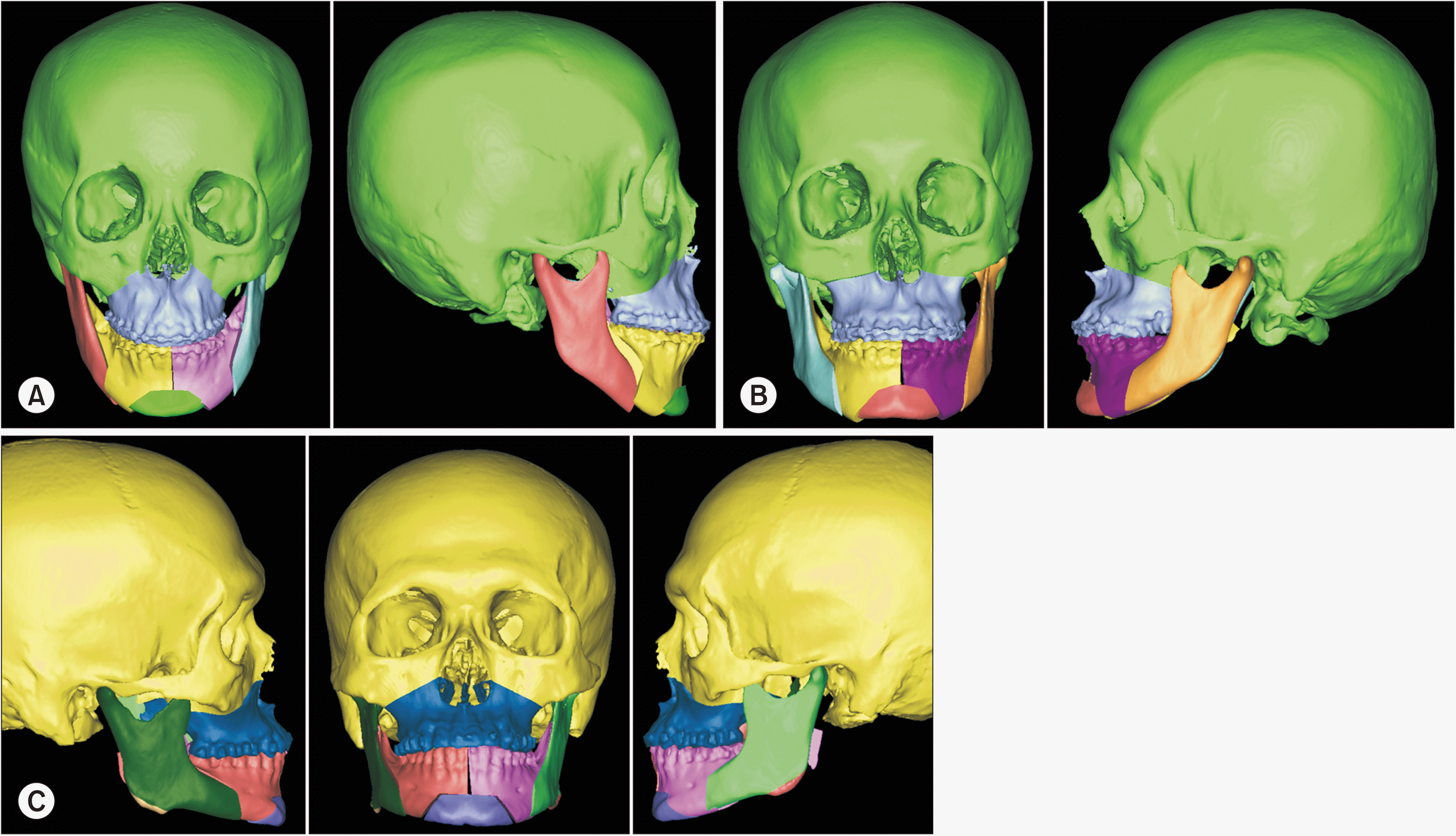
All patients reported to the outpatient clinic at the Department of Oral and Maxillofacial Surgery, Shimane University Faculty of Medicine and underwent pre-surgical orthodontic treatment for approximately 6 to 12 months; orthodontic decompensation was achieved. Virtual surgical planning was done using the ProPlan CMF software (Materialise) (Fig. 1) and interocclusal splints were fabricated for intraoperative use. Informed consent was obtained before surgery. The same single surgical team performed all the surgeries.
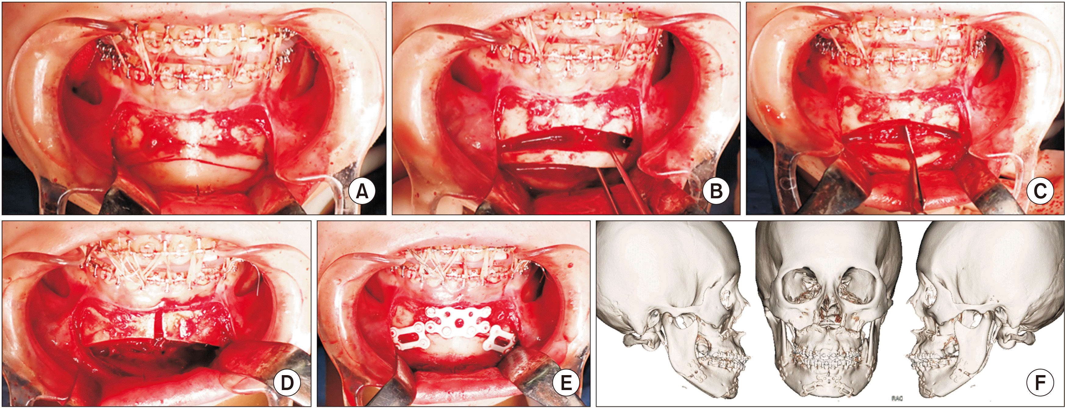
After completion of Le Fort I osteotomy bilateral sagittal split ramus osteotomy (BSSRO) and genioplasty were carried out to build the appropriate skeletal form and soft tissue profile in the mental region using preoperative computer simulation. Local anaesthetic solution of 2% lignocaine with adrenaline was infiltrated at the surgical site. Soft tissue marking was done with sutures. Vestibular incision was made, and a healthy cuff of attached gingiva was retained. The anterior mandible was exposed up to the lower border; the soft tissues were reflected carefully to expose the osteotomy site. Mental nerves at both sides were identified and preserved. We employed the sliding osteotomy design for genial correction. The osteotomy was performed using saw. Once the cut was completed, the genial fragment was down-fractured. Then, the mandibular midline osteotomy was followed. A straight saw was used to cut in between the roots of central incisors, with precaution to avoid periodontal ligament (PDL) damage. The split mandibular segments were mobilized and brought into position using the final interocclusal splint and stabilized with elastics and wires. The chin was repositioned accordingly (advancement, reduction, and asymmetry correction) and fixation was carried out; first at the chin segment (2 stepped – four-hole three-dimensional [3D] plates on either side), then followed by the midline (eight-hole 3D plate with gap). We were able to perform various combinations of genioplasties customized for the patient’s specific deformity as anterior advancement (Fig. 2) and vertical height reduction.(Fig. 3)
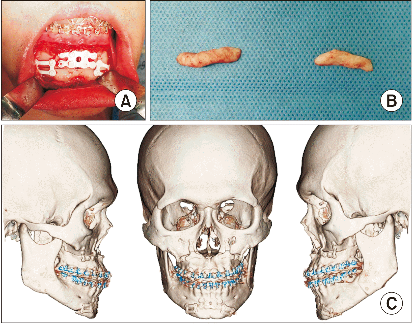
After Le Fort I osteotomy and BSSRO were completed, local anaesthetic solution was infiltrated at the anterior mandibular region with 2% Lignocaine with adrenaline. A vestibular incision was made, and the anterior mandible was exposed. Periosteal and muscular attachments were retained on the lingual side of the lower border to avoid compromise in blood supply. We first performed a sliding type of osteotomy cut and the chin segment was mobilized. A straight saw was then used to perform the midline osteotomy. The osteotomy was initiated at the upper part of the genial osteotomy and ended between the roots of the central incisors. Care was taken to prevent PDL injury to the lower central incisors. After the bony fragments were separated, an interocclusal splint was positioned with elastics and wires to prevent midline diastema. The bony segments were then fixed at the desired position with bioresorbable osteosynthesis. Thus, we were able to retain the ideal genial morphology by performing the midline osteotomy and fixing the mobilized chin at the same position.(Fig. 4)
Mandibular midline osteotomies can be combined with genioplasty when an alteration of the genial form and position is required. When symphyseal expansion is necessary with posterior region widening, plain midline osteotomy while conserving the existing ideal genial morphology can be a good choice. Using our technique, widening of 2-4 mm can be easily achieved at the first molar region, as planned by preoperative simulation. If widening of more than 4 mm is needed, then ostectomy at the gonial angle should be carried out to prevent a broad lower facial appearance.(Fig. 4. B)
Osteosynthesis across all osteotomised sites was achieved using SuperFixorb MX (Teijin)—the third generation bioactive bioresorbable 3D plates and screws. Intraoperative passive occlusion was checked, and the planned occlusion was well seen for all patients. Wound closure was done using 3-0 Vicryl resorbable sutures. Recovery was uneventful. From postoperative day 2, liquid and semi-solid diet was resumed. The final interocclusal splint was used for 4 weeks postoperatively at only night time to gain occlusal stabilization. In addition, we used soft training elastics for 4 weeks to guide proper occlusion. The patients were discharged 7-10 days post-surgery and regular follow-up was done at the outpatient clinic. Post-surgical comparison imaging showed good outcomes in all patients. We have applied our technique to more than 20 patients and no perioperative complications were observed so far.
To establish a stable functional occlusion, adequate correction of bimaxillary transverse discrepancies is required. Concurrent vertical and sagittal discrepancies often hide the presence of an underlying transverse problem. Common combinations seen in maxillomandibular discrepancy is: (a) narrow maxilla and normal mandible, (b) normal maxilla and wide mandible, (c) narrow maxilla and wide mandible9, (d) narrow maxilla and narrow mandible8, and (e) normal maxilla and narrow mandible1. Studies have also evidently shown that underdeveloped maxillae often are accompanied with smaller mandibles with increased articular angle and overjet10. The surgical options include maxillary constriction/widening surgery and mandibular constriction/widening surgery or distraction osteogenesis (DO). In this study, the maxilla with adequate transverse width and constricted mandible having arch width discrepancy was indication for a surgical correction as described here. Although literature shows evidence that favours maxillary expansion via surgery or DO, it can prove to be more stressful for the patient than when bi-jaw surgery is performed7. As mentioned earlier, multi-piece segmental maxillary osteotomies carry the risk of a myriad of complications. There have been concerns regarding the stability after multi-piece segmental osteotomies, mainly because of soft tissue surrounding the maxilla. A recent publication noted a relapse rate of 26.3% after segmental Le Fort I osteotomy after one-year follow-up duration11. Though traditional orthodontic correction/appliance expansion can be employed, neglecting the underlying skeletal issue leads to relapse and rates12 of about 20%-30% have been noted13.
Vertical midline symphyseal cuts with or without augmentation are associated with complications such as large relapse, mucosal issues, and necessity for rigid fixation13. Modified osteotomy cuts suited to correct midline defects have also been described. Surgical midline widening is portrayed as an underused technique in orthognathic surgery due to (1) relapse concerns, (2) TMJ effects, (3) procedural unfamiliarity, and (4) no adequate address of the skeletal problem14. Besides the above-mentioned, impending complications such as (1) condylar torquing, (2) midline diastema, (3) bi-angle width control, (4) lingual flaring, and (5) vascularity compromise in multiple osteotomies often make surgical midline widening an uncommon procedure15. None of the potential complications were observed in any of our cases. Intraoperatively, when the mandible was in multiple segments, condylar torquing/positioning15, control of bi-angle width and lingual flaring were anticipated and prevented appropriately.
Osteotomy designs similar to our study have been described before; however, it was designed mainly for narrowing of the mandibular arch16 and combined with tooth-borne distraction osteogenesis17. Moreover, the combined procedure of BSSRO, genioplasty and midline symphyseal split to correct anterior mandibular asymmetry was advocated by Anghinoni et al.18. Though numerous multi-segmental mandibular osteotomies involving the midline have been described, this study is the first report of a single stage mandibular widening procedure with or without genial form alterations for bimaxillary transverse discrepancy. We did not use any rigid fixation or distraction, but the third generation bioactive bioresorbable 3D plate and screw fixation was used.
The presence of healthy periodontium surrounding teeth at the osteotomy and minimum 1 mm bone surrounding the tooth root has been followed19. Even if there is no prior divergence between the central incisor roots orthodontically, the roots should at least be parallel to each other and perpendicular to the mandibular occlusal plane14. Some authors have not noted any kind of periodontal issues if the osteotomy line runs through the PDL of one incisor root, with proper postoperative maintenance14. Palpation of the lingual side during osteotomy aids to assess saw position and depth during osteotomy, thereby preventing soft tissue injury17. Our pre-surgical orthodontic plan was set to retain sufficient space for osteotomy and posed no problem intra- and postoperatively.
We found that our technique can be successfully applied and resulted in widening of about 2-4 mm at first molar region without any stability or relapse complications. We observed gonial flaring in expansions of more than 4 mm, which requires simultaneous angle ostectomy. Previous study mentioned that widening more than 7 mm requires distraction to maintain stability13. As such, less stability after conventional mandibular widening have been reported, the maximum amount of mandibular widening and the degree of stability that can be achieved without distraction remains a debatable topic.
Since the posterior mandibular width decreases when the mandible is widened anteriorly, in case of large widening movements, transverse crossbite can be presented and progressively corrected postoperatively19. It has been noticed that midline widening is more difficult than narrowing mainly due to tissue resistance. Therefore, rigid fixation may be necessary in the anterior mandible to prevent torquing movement of the bony segments by muscle pull20. It was recommended to use rigid fixation using bi-cortical screws or rigid osteosynthesis for some mandibular widening cases due to potential instability, however, it was not required in our study. We prefer to use bioactive/bioresorbable osteosynthesis for the patients, because titanium osteosynthesis mandates a second removal surgery and moreover, the bioresorbable systems have consistently proved to be effective21. The bioactive/bioresorbable 3D plating system has bio-conductive properties, thus enabling faster bone regeneration. Furthermore, application of 3D plate helped us to resist torquing of the osteotomized segments, which was especially advantageous in this study22.
We conclude that, in patients exhibiting normal maxillary width with transversely small mandibles, mandibular width expansion surgery is beneficial in correcting the existing discrepancy. Our novel techniques showed good postoperative outcomes and are highly useful to correct bimaxillary transverse width deficits. It consumes short intraoperative time, is relatively less invasive, and simple to perform.
Notes
Authors’ Contributions
M.R. participated in treatment, data collection and manuscript writing. R.S.O. participated in treatment and manuscript writing. Y.S. participated in treatment and data collection. T.O. participated in treatment and data collection. T.K. participated in treatment planning, treatment, conceptualization of the article and manuscript editing. All authors read and approved the final manuscript.
References
1. Andrucioli MCD, Matsumoto MAN. 2020; Transverse maxillary deficiency: treatment alternatives in face of early skeletal maturation. Dental Press J Orthod. 25:70–9. https://doi.org/10.1590/2177-6709.25.1.070-079.bbo. DOI: 10.1590/2177-6709.25.1.070-079.bbo. PMID: 32215481. PMCID: PMC7077945.

2. Starch-Jensen T, Blæhr TL. 2016; Transverse expansion and stability after segmental Le Fort I osteotomy versus surgically assisted rapid maxillary expansion: a systematic review. J Oral Maxillofac Res. 7:e1. https://doi.org/10.5037/jomr.2016.7401. DOI: 10.5037/jomr.2016.7401. PMID: 28154745. PMCID: PMC5279767.

3. Corega C, Corega M, Băciuţ M, Vaida L, Wangerin K, Bran S, et al. 2010; Bimaxillary distraction osteogenesis--an effective approach for the transverse jaw discrepancies in adults. Chirurgia (Bucur). 105:571–5. PMID: 20941985.
4. Aktop P, Biren S, Aktop S, Motro M, Delilbasi C, Gurler G, et al. 2018; Evaluation of two different rapid maxillary expansion surgical techniques and their effects on the malar complex based on 3d cone-beam computed tomography. Niger J Clin Pract. 21:13–21. https://doi.org/10.4103/1119-3077.224794. DOI: 10.4103/1119-3077.224794. PMID: 29411717.

5. Lee HW, Kim SJ, Kwon YD. 2015; Salvage rapid maxillary expansion for the relapse of maxillary transverse expansion after Le Fort I with parasagittal osteotomy. J Korean Assoc Oral Maxillofac Surg. 41:97–101. https://doi.org/10.5125/jkaoms.2015.41.2.97. DOI: 10.5125/jkaoms.2015.41.2.97. PMID: 25922822. PMCID: PMC4411735.

6. Morgan TA, Fridrich KL. 2001; Effects of the multiple-piece maxillary osteotomy on the periodontium. Int J Adult Orthodon Orthognath Surg. 16:255–65. PMID: 12390003.
7. Hara S, Mitsugi M, Hirose H, Tatemoto Y. 2015; Combination of mandibular constriction and intraoral vertical ramus osteotomies for a transverse jaw discrepancy. Plast Reconstr Surg Glob Open. 3:e521. https://doi.org/10.1097/gox.0000000000000505. DOI: 10.1097/GOX.0000000000000505. PMID: 26495234. PMCID: PMC4596446.

8. Chaudhary DC, Sharma R, Bagga DS, Sharma V. 2015; Management of bimaxillary transverse discrepancy with vertical excess. J Indian Orthod Soc. 49:213–9. https://doi.org/10.4103/0301-5742.171315. DOI: 10.4103/0301-5742.171315.

9. Betts NJ, Sturtz DH, Aldrich DA. 2004; Treatment of transverse (width) discrepancies in patients who require isolated mandibular surgery: the case for maxillary expansion. J Oral Maxillofac Surg. 62:361–4. https://doi.org/10.1016/j.joms.2003.08.018. DOI: 10.1016/j.joms.2003.08.018. PMID: 15015171.

10. García Menéndez M, Perdomo Gutiérrez L, Valdés Massó D. 2021; Negative maxillary transverse discrepancy and cephalometric lateral differences according to the width of upper arch. Rev Cubana Estomatol. 58:e3627.
11. Kim H, Cha KS. 2018; Evaluation of the stability of maxillary expansion using cone-beam computed tomography after segmental Le Fort I osteotomy in adult patients with skeletal Class III malocclusion. Korean J Orthod. 48:63–70. https://doi.org/10.4041/kjod.2018.48.1.63. DOI: 10.4041/kjod.2018.48.1.63. PMID: 29423378. PMCID: PMC5799308.

12. Housley JA, Nanda RS, Currier GF, McCune DE. 2003; Stability of transverse expansion in the mandibular arch. Am J Orthod Dentofacial Orthop. 124:288–93. https://doi.org/10.1016/s0889-5406(03)00450-5. DOI: 10.1016/S0889-5406(03)00450-5. PMID: 12970662.

13. Rougier G, Diner PA, Rachwalski M, Galliani E, Tomat C, Picard A, et al. 2019; Mandibular symphyseal distraction osteogenesis: 20 years of experience treating transverse deficiencies with an internal hybrid device. J Craniomaxillofac Surg. 47:586–91. https://doi.org/10.1016/j.jcms.2019.01.001. DOI: 10.1016/j.jcms.2019.01.001. PMID: 30718215.

14. Bloomquist D, Joondeph D. Naini FB, Gill DS, editors. 2017. Mandibular midline osteotomy. Orthognathic surgery: principles, planning and practice. Wiley-Blackwell;p. 630–4. DOI: 10.1002/9781119004370.ch37.

15. Bloomquist DS. 2007; Anterior segmental mandibular osteotomies for the correction of facial-skeletal deformities. Oral Maxillofac Surg Clin North Am. 19:369–79. vihttps://doi.org/10.1016/j.coms.2007.04.005. DOI: 10.1016/j.coms.2007.04.005. PMID: 18088891.

16. Mommaerts MY, Voisin C, Joshi Otero J, Loomans NAJ. 2019; Mandibular feminization osteotomy-preliminary results. Int J Oral Maxillofac Surg. 48:597–600. https://doi.org/10.1016/j.ijom.2018.10.003. DOI: 10.1016/j.ijom.2018.10.003. PMID: 30342756.

17. Guerrero CA, Bell WH, Contasti GI, Rodriguez AM. 1997; Mandibular widening by intraoral distraction osteogenesis. Br J Oral Maxillofac Surg. 35:383–92. https://doi.org/10.1016/s0266-4356(97)90712-9. DOI: 10.1016/S0266-4356(97)90712-9. PMID: 9486441.

18. Anghinoni ML, Magri AS, Di Blasio A, Toma L, Sesenna E. 2009; Midline mandibular osteotomy in an asymmetric patient. Angle Orthod. 79:1008–14. https://doi.org/10.2319/102908-550.1. DOI: 10.2319/102908-550.1. PMID: 19705948.

19. Guerrero CA, Bell WH, Mujica EV, Gonzalez M, Dominguez E. Fonseca RJ, editor. 2018. Intraoral distraction osteogenesis. Oral and maxillofacial surgery. 3rd ed. Elsevier;p. 196–221. DOI: 10.1016/s0278-2391(00)80016-6.
20. Mani V. Bonanthaya K, Panneerselvam E, Manuel S, Kumar VV, Rai A, editors. 2021. Orthognathic surgery for mandible. Oral and maxillofacial surgery for the clinician. Springer;Singapore: p. 1477–512. DOI: 10.1007/978-981-15-1346-6_68.

21. Kanno T, Sukegawa S, Furuki Y, Nariai Y, Sekine J. 2018; Overview of innovative advances in bioresorbable plate systems for oral and maxillofacial surgery. Jpn Dent Sci Rev. 54:127–38. https://doi.org/10.1016/j.jdsr.2018.03.003. DOI: 10.1016/j.jdsr.2018.03.003. PMID: 30128060. PMCID: PMC6094489.

22. Budhraja NJ, Shenoi RS, Badjate SJ, Bang KO, Ingole PD, Kolte VS. 2018; Three-dimensional locking plate and conventional miniplates in the treatment of mandibular anterior fractures. Ann Maxillofac Surg. 8:73–7. https://doi.org/10.4103/ams.ams_175_17. DOI: 10.4103/ams.ams_175_17. PMID: 29963428. PMCID: PMC6018293.

Fig. 1
Preoperative virtual planning with Proplan CMF software (Materialise). A. Mandibular midline widening with advancement genioplasty, frontal and right lateral profile views. B. Mandibular midline widening with reduction genioplasty, frontal and left lateral profile views. C. Mandibular midline widening preserving genial morphology, frontal and lateral profile views.

Fig. 2
Fig.2.
Intra- and postoperative photos of mandibular midline widening with advancement genioplasty 46-year-old female patient with apertognathia and asymmetry. Our treatment plan consisted of Le Fort I osteotomy with impaction (4 mm), bilateral sagittal split ramus osteotomy setback (7 mm) with advancement genioplasty (4 mm) and 3 mm midline mandibular widening at first molar. A. Positioning of saw to perform the midline cut. B. Completion of osteotomy cut. C, D. Separation and mobilization of osteotomized segments. E. Stabilizing the fragments with interocclusal splint. F. Bioresorbable osteosynthesis placement. G. Postoperative three-dimensional computed tomography demonstrating skeletal changes, frontal and profile views.

Fig. 3
Intra- and postoperative photos of mandibular midline widening with reduction genioplasty 17-year-old female patient with Class III dentofacial profile and asymmetry. Our treatment protocol consisted of Le Fort I osteotomy with advancement (4 mm), bilateral sagittal split ramus osteotomy setback (right 3 mm and left 6 mm) with reduction genioplasty (4 mm) and 2.5 mm midline mandibular widening at first molar. A. Osteotomy for reduction genioplasty. B. Removal of intervening bone. C. Completed midline osteotomy cut. D. Separation and fragment mobilization with subsequent splint placement. E. Bioresorbable osteosynthesis placement. F. Postoperative three-dimensional computed tomography demonstrating skeletal changes, frontal and profile views.

Fig. 4
Intra- and postoperative photos of mandibular midline widening with preserving genial morphology 19-year-old male patient with Class III malocclusion and dentofacial deformity. Our treatment plan was Le Fort I osteotomy with advancement (3 mm), bilateral sagittal split ramus osteotomy setback (right 4 mm and left 6 mm) with genial osteotomy and 4.5 mm midline mandibular widening at first molar. A. Completed osteosynthesis with bioresorbable plates and screws. B. Ostectomy of gonial angles. C. Postoperative three-dimensional computed tomography demonstrating skeletal changes, frontal and profile views.


 PDF
PDF Citation
 Print
Print


 XML Download