Abstract
Hand involvement in sarcoidosis is rare and it presents as tenosynovitis, dactylitis, nodules and osteoarticular bony destruction. We describe an unusual presentation of progressive intrinsic muscle contracture of both hands in a 42-year-old woman with sarcoid myopathy who presented with painful swelling and weakness of all four extremities. Her systemic symptoms improved with oral corticosteroids, but the hand muscle contracture remained after resolution of myositis. Serial soft tissue releases of intrinsic muscle contracture improved hand function markedly. This case highlights that surgery is a viable option to treat intrinsic muscle contracture in patients with chronic sarcoid myopathy complicated with severe muscle contracture.
Sarcoidosis or Besnier-Boeck-Schaumann syndrome, is an idiopathic multisystem inflammatory disease characterized by non-caseating granulomas primarily involving the lungs and mediastinal lymph nodes. Musculoskeletal involvements such as sarcoid arthropathy and myopathy were reported in up to 10% of patients [1]. It is associated with chronic, prolonged disease course. Hand manifestations have been reported in 0.2% of patients [2]. The presentation of sarcoidosis in the hand can vary from digital swelling, tenosynovitis, soft tissue nodular masses or cutaneous lesions to osteoarticular lytic destruction [3]. Muscle involvement in sarcoidosis or sarcoid myopathy is common but only 0.5% to 2.5% of them are symptomatic [4]. Sarcoid myopathy can result in muscle contracture with significant impairment of joint mobility. We hereby describe a rare case of intrinsic muscle contracture in bilateral hands which was successfully corrected with surgical soft tissue release procedures. The patient was informed that data concerning the case would be submitted for publication, and she consented.
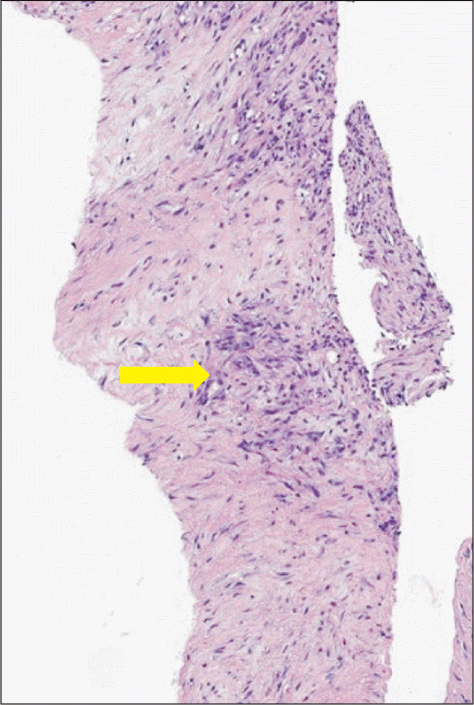
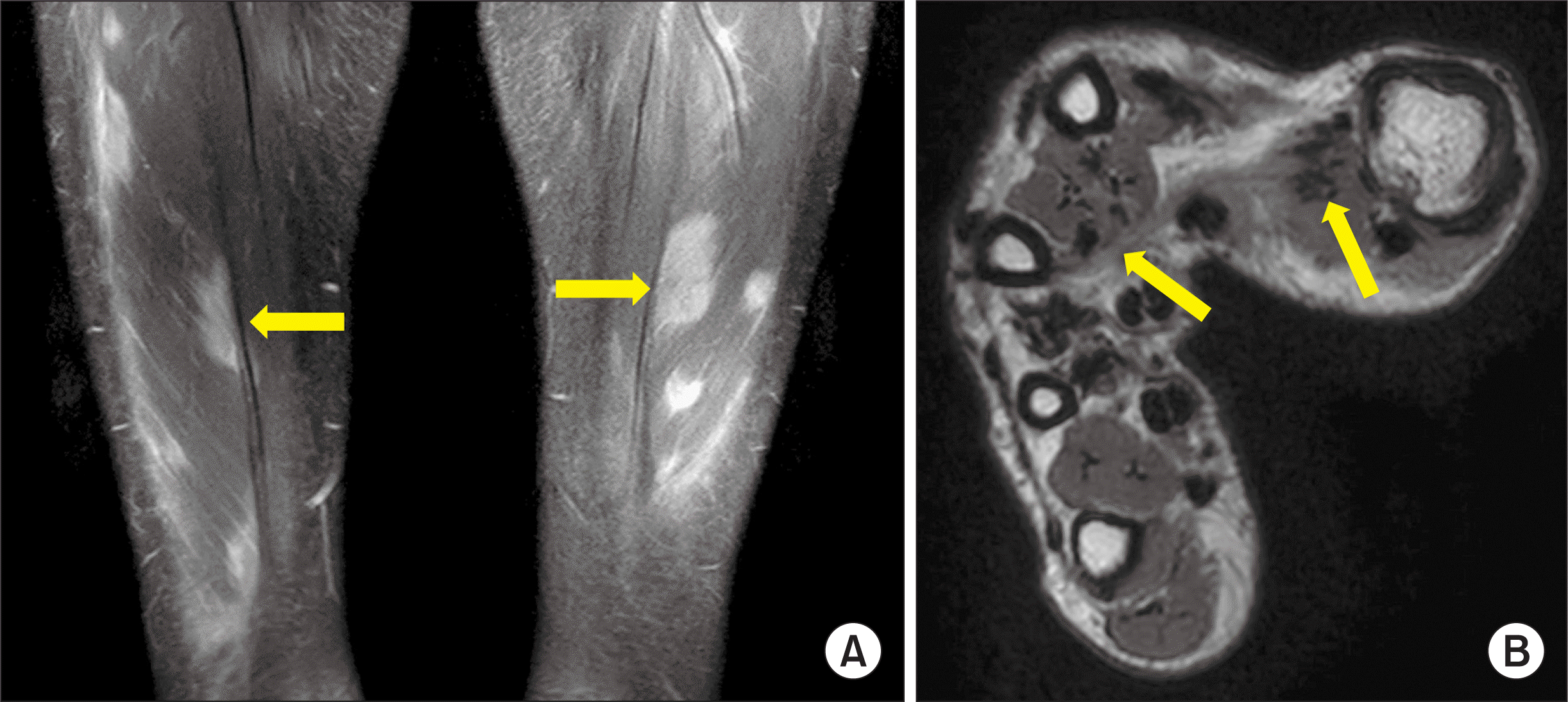
A 42-year-old right-handed woman presented to a rheumatology clinic with weakness in both upper and lower extremities. She developed edema in both forearms distal to the elbows with inability to make of a full grip due to finger muscle contracture. Laboratory results showed: white blood cell count 6.1×10⁹/L, hemoglobin 11.3 g/dL, platelet 241×109/L. C-reactive protein was 0.05 mg/dL and erythrocyte sedimentation rate was 11 mm/h. Calcium level was 9.1 mg/dL. Creatinine kinase and aldolase levels were 45 IU/L and 5.0 IU/L, respectively. Angiotensin converting enzyme level was 27.9 IU/L (normal: 7.5~53 IU/L). Anti-nuclear antibody was absent. Computed tomography (CT) of her chest revealed a 3.9 cm enhancing mass in thymus. Lung parenchyma was normal with no evidence of hilar lymphadenopathy. A Duplex Doppler scan of the lower extremities revealed deep vein thrombosis (DVT) in her left popliteal, posterior tibial and peroneal vein. Magnetic resonance imaging (MRI) showed extensive intramuscular T2 hyperintense nodular lesions with increased perfascial signal intensities involving multiple muscles of the upper and lower extremities (Figure 1). Electromyography showed a myopathic pattern. Muscle biopsy of right gastrocnemius muscle revealed inflammation with non-caseating granulomas, consistent with sarcoidosis (Figure 2).
A partial thymectomy was performed and biopsy was consistent with a thymoma. Prednisolone 40mg daily and apixaban 5 mg twice daily and corticosteroid were started for sarcoid myopathy and DVT. Bilateral upper limbs pain and edema improved markedly, but the hands contracture became more prominent, limiting her activities of daily living. Prednisolone dose was tapered down to 5 mg/day over 4 months. Azathioprine was added with no further improvement. The patient was referred to the orthopedic hand clinic 7 months after symptom onset after the hand function did not respond to medical treatment.
The range of motion of her both wrists were 60° flexion and 30° extension; bilateral second and third metacarpophalangeal joints (MCPJs) had flexion contracture at 30°. There were bilateral first and second webspace contracture and left flexor pollicis longus (FPL) and carpometacarpal joint contracture (Figure 3). A follow-up MRI of her bilateral hands showed multifocal T2 high signal changes with enhancement in hand muscles, consistent with non-specific myopathy. Multifocal band-like T2 low signal intensity lesions in both hands involving the hand intrinsic muscles indicated post-inflammatory sequelae.
Surgical contracture release was performed to her left and then right hand. For the right hand, first webspace (thumb) scar excision, adductor pollicis and thenar muscles release with Z-plasty, first, second and third palmar and dorsal interosseous release from the metacarpal origin were performed. Similar surgery was performed to the left hand with additional FPL fractional lengthening at the forearm. Intra-operative biopsy of tendon showed nonspecific inflammation with lymphocytic infiltration. She recovered uneventfully and achieved good hand functions with rehabilitation physiotherapy (Figure 4). No myositis relapse or recurrent contracture were observed up to 2 years of follow-up. The patient expressed high satisfaction with the surgical results. Prednisolone was further tapered down to 2.5 mg/day and Azathioprine was stopped.
Sarcoid myopathy is symptomatic in only 0.5% to 2.5% of patients and it often involves the lower extremities [4]. It can present as acute myositis, chronic myopathy or nodular myopathy [5]. The acute myositis form accounts approximately for 10% of sarcoid myopathy. It typically occurs in patients less than 40 years of age, manifesting as diffuse swelling and pain of affected muscles. Disease can progress into contracture with resulting limitation in activity of daily living. The acute sarcoid myositis can be accompanied by other symptoms including fatigue, fever, joint pain, and erythema nodosum. Chronic myopathy is the most common form (86%), commonly affecting women between 50~60 years of age. It is characterized by insidious, slowly progressive symmetrical proximal muscle weakness. The nodular form of sarcoid myopathy is the least common type (3%); it presents with tender, palpable nodules in single or multiple muscles, commonly in the lower limbs. The nodular form is not accompanied by weakness or limitation of movement.
In this report, the patient presented with acute myositic sarcoidosis of her both arms and legs. MRI of upper and lower extremities showed diffuse myositis, which is typical of acute myositis. The diagnosis was confirmed by the presence of non-caseating granulomas on the muscle biopsy. The classical treatment for sarcoidosis is corticosteroids, which is usually effective in acute myositis and nodular myopathy. Relapses have been reported around 16%~74% when corticosteroids are tapered down or stopped [6]. Other medical treatment includes chloroquine, azathioprine, methotrexate, thalidomide, and tumor necrosis factor inhibitor such as infliximab [7]. The patient in this report responded well to corticosteroids with resolution of pain and swelling but the muscle fibrosis with intrinsic muscle contracture persisted with marked impairment of hand function. This is unusual since in acute myositis of sarcoidosis, proximal muscles are affected, while the intrinsic hand muscles are usually spared. To our best of our knowledge, there were only seven cases on finger flexion contracture associated with sarcoid myopathy reported in English literature [8]. All of them presented with little and ring fingers flexion contracture of flexor muscles at the forearm (extrinsic muscles).
Diagnosis of sarcoid myopathy in early disease course can be challenging because a wide range of disease include inflammatory myopathies, and fibrosing diseases such as Dupuytren’s contracture, complicated trigger finger, flexor tenosynovitis, Volkmann’s contracture can mimic it.
Contractures of intrinsic muscles disrupt the complex balance between extrinsic and intrinsic muscles. Interossei and lumbrical contractures causes MCPJ into flexion and proximal interphalangeal joint into extension, creating an “intrinsic plus” hand. In severe cases, MCPJs are fixed in adducted or abducted position and fingers in swan-neck deformity due to secondary contracture of the MCP volar plate and laxity of the PIP volar plate [9]. Surgical management is dictated by its etiology, severity, duration of symptoms and joints involved [10]. The procedures include but not limited to distal intrinsic release, radial lateral band resection, intrinsic tenodesis, lateral band translocation and mobilization, cross intrinsic transfer, proximal interosseous muscle slide, proximal intrinsic release, interosseous tendon fractional lengthening and ulnar neurectomy. We performed proximal interosseous muscle slide for this case for first, second and third interosseous muscles, Z-plasty with adductor pollicis and thenar muscles release for first webspace contracture and FPL fractional lengthening for thumb IPJ contracture. The role of surgery is particularly important late in the sarcoidosis course, when causing mechanical symptoms or neurovascular compression due to chronic changes or fibrosis do not respond to medical treatment.
Here, we report a rare case of sarcoid myopathy complicated by contracture of intrinsic hand muscles with joint contracture and severe functional limitation which was successfully corrected with soft tissue release surgery. A surgical management should be considered in treatment-refractory chronic sarcoid myopathy with muscle contracture.
Notes
REFERENCES
1. Alemdaroğlu E, Ertürk A, Eroğlu AG. 2013; A sarcoidosis patient with hand involvement and large pulmonary lymph nodes: results of 1-year treatment with methotrexate. Clin Rheumatol. 32 Suppl 1:S71–3. DOI: 10.1007/s10067-010-1487-2. PMID: 20577892.

2. Motomiya M, Iwasaki N, Kawamura D. 2013; Finger flexion contracture due to muscular involvement of sarcoidosis. Hand Surg. 18:85–7. DOI: 10.1142/S0218810413720015. PMID: 23413857.

3. Namas R, Meysami A. 2016; Sarcoidosis of hands. J Clin Rheumatol. 22:278–9. DOI: 10.1097/RHU.0000000000000389. PMID: 27464776.

4. Fayad F, Lioté F, Berenbaum F, Orcel P, Bardin T. 2006; Muscle involvement in sarcoidosis: a retrospective and followup studies. J Rheumatol. 33:98–103.
5. Crawford B, Badlissi F, Lozano Calderón SA. 2018; Orthopaedic considerations in the management of skeletal sarcoidosis. J Am Acad Orthop Surg. 26:197–203. DOI: 10.5435/JAAOS-D-16-00252. PMID: 29443705.

6. Aptel S, Lecocq-Teixeira S, Olivier P, Regent D, Teixeira PG, Blum A. 2016; Multimodality evaluation of musculoskeletal sarcoidosis: imaging findings and literature review. Diagn Interv Imaging. 97:5–18. DOI: 10.1016/j.diii.2014.11.038. PMID: 25883076.

7. Walter MC, Lochmüller H, Schlotter-Weigel B, Meindl T, Müller-Felber W. 2003; Successful treatment of muscle sarcoidosis with thalidomide. Acta Myol. 22:22–5.
8. Tada K, Ikeda K, Tomita K. 2009; Flexion contracture of the fingers caused by sarcoidosis: an 11-year follow-up. Scand J Plast Reconstr Surg Hand Surg. 43:171–3. DOI: 10.1080/02844310701510306. PMID: 19401937.

9. Paksima N, Besh BR. 2012; Intrinsic contractures of the hand. Hand Clin. 28:81–6. DOI: 10.1016/j.hcl.2011.10.001. PMID: 22117926.

10. Tosti R, Thoder JJ, Ilyas AM. 2013; Intrinsic contracture of the hand: diagnosis and management. J Am Acad Orthop Surg. 21:581–91. DOI: 10.5435/JAAOS-21-10-581. PMID: 24084432.

Fig. 1
(A) Magnetic resonance imaging (MRI) of bilateral thighs. Coronal T2 MRI showed hyperintense lesions involving multiple bilateral thigh muscles (arrows). (B) MRI of right hand. Trans axial T2-weighted low signal intensity lesions seen on adductor pollicis and intrinsic muscles of the hand indicating fibrotic bands (arrows).

Fig. 2
Right gastrocnemius muscle biopsy showed hallmark of non-caseating granuloma of sarcoidosis. Central area of multinucleated giant cells, epithelioid cells and macrophages surrounded by lymphocytes and fibroblasts (arrow). Gastrocnemius biopsy; hematoxylin–eosin (H&E) stain; original magnification, 40×.


 PDF
PDF Citation
 Print
Print


 XML Download