Journal List > Korean J Physiol Pharmacol > v.27(4) > 1516083039

Lim and Kim: Roles of non-coding RNAs in intercellular crosstalk in cardiovascular diseases

Abstract

Complex diseases including cardiovascular disease are caused by a combination of the alternation of many genes and the influence of environments. Recently, non-coding RNAs (ncRNAs) have been shown to be involved in diverse diseases, and the functions of various ncRNAs have been reported. Many researchers have elucidated the mechanisms of action of these ncRNAs at the cellular level prior to in vivo and clinical studies of the diseases. Due to the characteristics of complex diseases involving intercellular crosstalk, it is important to study communication between multiple cells. However, there is a lack of literature summarizing and discussing studies of ncRNAs involved in intercellular crosstalk in cardiovascular diseases. Therefore, this review summarizes recent discoveries in the functional mechanisms of intercellular crosstalk involving ncRNAs, including microRNAs, long non-coding RNAs, and circular RNAs. In addition, the pathophysiological role of ncRNAs in this communication is extensively discussed in various cardiovascular diseases.

INTRODUCTION

Atherosclerosis

For decades, various genetic and environmental factors have been studied to elucidate the onset, progression, and causes of diseases [1-4]. Cardiovascular disease (CVD) as a representative disease with high mortality rates progresses through complex pathophysiological processes through communication between the many involved cells [5,6]. Atherosclerosis, a representative CVD, has traditionally focused on evaluating immune cells, especially macrophages that phagocytose lipids to become foam cells and secrete various inflammatory substances [7-9]. Recently, many studies have been published showing that endothelial cells (ECs) and smooth muscle cells (SMCs) constituting the intima and media of arteries play major roles in the formation of atherosclerotic lesions [10-12]. Blood factors such as cholesterol and low-density lipoprotein (LDL) cause endothelial dysfunction in the vascular intima and allow primary immune cells such as monocytes and macrophages to infiltrate the intima and media [13,14]. These macrophages are transformed into foam cells and inflammatory phenotypes that secrete inflammatory substances to stimulate vascular smooth muscle cells (VSMCs). VSMCs, which are capable of phenotypic switching, are converted to proliferative and migratory phenotypes by inflammatory stimuli and play a major role in plaque formation [15-17]. Interestingly, new cell types with characteristic transcriptome patterns within atherosclerotic lesions have been discovered with the recently developed single-cell RNA sequencing technology suggesting that there are still unknown cell types involved in atherosclerosis [18-21]. Therefore, evaluation of the crosstalk between the cell types involved in the lesion is important for understanding these diseases.

Non-coding RNAs

Studies on the pathophysiological mechanisms of CVDs have been conducted for numerous genes [22-25]. Furthermore, several regulatory non-coding RNAs (ncRNAs) involved in this mechanism have been discovered [26]. Based on 200 nucleotides, it can be divided into small ncRNAs and long ncRNAs (lncRNAs) [27]. First, microRNA (miRNA), a typical regulatory small ncRNA, forms a complex with proteins including argonaute (AGO), and binds to the 3ʹ-untranslated region (3ʹ-UTR) of the target messenger RNA (mRNA) in a specific base pair, thereby interfering with mRNA translation [27]. Many lncRNAs have also been identified, and they regulate the expression of various genes [28]. The types and roles of lncRNAs are further subdivided according to their location on the genome (intronic, intergenic, bidirectional), the direction of transcription relative to the nearby protein-coding gene (sense and antisense), and the regulation manner for other genes (cis and trans) [27]. Finally, circular RNA (circRNA), the most recently identified regulatory ncRNA, is being actively studied for its biogenesis and function [29]. Early on, it was discovered in eukaryotes that circRNAs have closed and single-stranded structures made from the back-splicing of host mRNAs [30,31]. The analysis from high-throughput RNA sequencing has confirmed that many circRNAs are widely expressed in cells and tissues [29,32].

Co-culture systems

Although many researchers are conducting experiments using animal models and analyzing patient-derived clinical samples to elucidate the molecular mechanisms of disease, there are some limitations [33]. In vivo experiments using animal models such as mice are of great help in predicting disease mechanisms but have limitations in that the molecular mechanisms do not completely match those of humans [34]. In addition, research using clinical samples is difficult to expand to molecular study and the supply of samples is also limited. Therefore, a plan must be designed to reduce the excessive time and cost of in vivo experiments and to efficiently utilize clinical samples after confirming the precise mechanisms in cells through in vitro experiments. Elucidation of the action mechanism of a specific gene in a single cell type has continuously progressed. However, to further explore the complex process in a particular disease, it is also important to examine the intercellular crosstalk response according to gene expression changes by co-cultivating two or more cells. Furthermore, cell co-culture systems have been well established, and they are often used in in vitro experiments in disease mechanism studies [17].
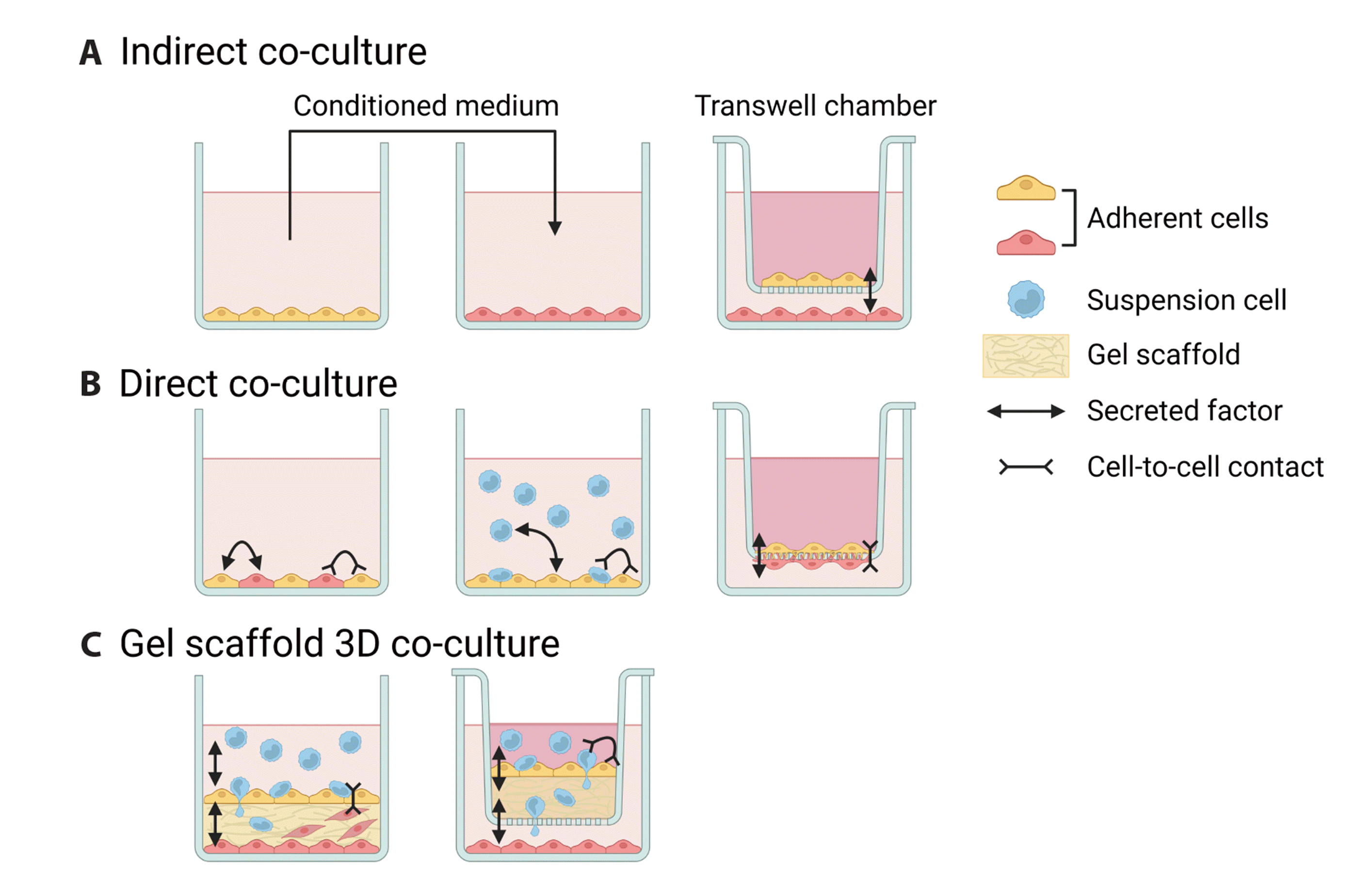
For co-culture, donor cells that generate signals upon stimulation by external factors are required, as in diseases [35-37]. A recipient cell is also needed to receive these signals. There are several co-culture techniques for CVD research [38]. First, indirect co-culture confirms the effect only through secreted factors (cytokines, growth factors, and extracellular vesicles [EVs]) produced by donor cells [38]. Indirect co-culture is divided into a method that involves direct treatment of the donor cell-cultured medium and a method that involves exchanging only secreted factors in a separate space through a transwell chamber (Fig. 1A). Conversely, direct co-culture induces cell-to-cell contact by placing two cells in a culture dish or attaching each cell to the opposite sides of a transwell chamber (Fig. 1B) [38,39]. In addition to secreted factors, the effects of adhesion molecules such as intercellular adhesion molecule 1 (ICAM-1) and vascular cell adhesion molecule 1 (VCAM-1) can also be tested [38]. Finally, there is another method that involves creating a three-dimensional environment such as tissue using two or more types of cells and a gel scaffold platform (Fig. 1C) [38]. This is a typical atherosclerosis-simulating co-culture model in which the intima (ECs), media (VSMCs) with the extracellular matrix (gel scaffold), and immune cells (monocyte/macrophage) infiltrating these layers of the blood vessel model are all structured together [38].
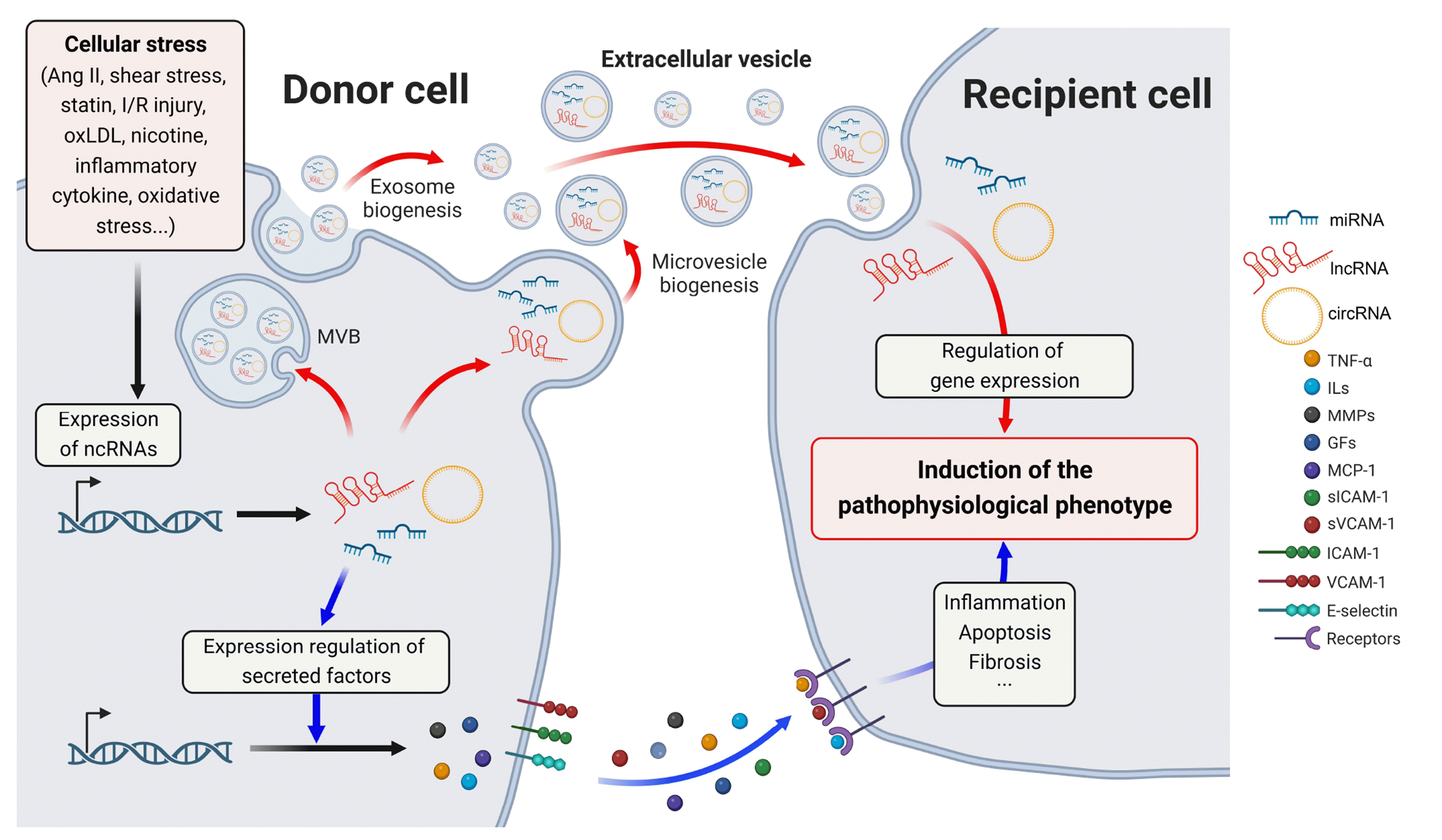
Intercellular crosstalk studies that utilized the abovementioned co-culture systems to create various disease-mimic environments have been performed widely [17,40,41]. However, intercellular crosstalk studies of ncRNAs are relatively scarce, and few reviews have summarized the utilization of co-culture systems. Therefore, this review summarizes intercellular crosstalk studies of various ncRNAs and the co-culture systems used therein. In particular, among complex diseases, research on CVDs including atherosclerosis is intensively discussed herein. The studies involving the direct delivery of ncRNAs through EVs and those evaluating the indirect influence of ncRNAs on other cells through the regulation of secreted factors and cell adhesion molecules are mainly described (Fig. 2).

REGULATION OF INTERCELLULAR CROSSTALK OF NCRNAS

Direct delivery of ncRNAs via EVs

EV-derived miRNAs: The pathophysiological role and mechanism of action of ncRNAs identified in each disease have been extensively studied and summarized in other reviews [28,42]. Several EV-derived ncRNAs that play important roles in CVDs, including atherosclerosis, are reviewed here. In studies of ncRNAs delivered directly through EVs (exosomes and microvesicles), many miRNAs, which are relatively small in their size compared with lncRNAs and circRNAs, have been discovered and studied [43,44].
Representatively, the miRNA produced from the miR-143/145 cluster is well known as an EV-derived miRNA that regulates the phenotype of VSMCs [45-47]. Krüppel-like factor 2 (KLF2), a transcription factor associated with various diseases including atherosclerosis, induces upregulation of the miR-143/145 cluster in ECs [43]. The miR-143/145 cluster is enriched in the EVs released from human umbilical vein ECs (HUVECs) in which KLF2 is overexpressed or stimulated by shear stress. These are delivered to human aortic SMCs (HASMCs) and reduce the expression of miR-143/145 targets to prevent dedifferentiation of VSMCs [43]. This result was confirmed by the isolation of EVs and the co-culture of these two cell types. In this study, when the phospholipid membrane of the EV was disrupted by phospholipase (Triton X-100 or cyclodextrin), miR-143/145 in the EV was mostly degraded by RNase [43]. This suggests that EVs such as exosomes and microvesicles play an important role in stably delivering ncRNAs to a target.
MiR-155 is another EV-derived miRNA that is highly expressed in human carotid plaque samples [48]. It plays an important role in vascular inflammation and atherogenesis by suppressing the expression of B-cell lymphoma 6 (BCL6), a transcription factor that attenuates the pro-inflammatory nuclear factor-κB (NF-κB) signal. In addition, miR-155, which is strongly expressed in VSMCs by overexpression of KLF5, is transferred to ECs through exosomes [49]. This transferred miR-155 facilitates endothelial dysfunction and atherosclerosis progression, suggesting that exosomal miR-155 could be a target for atherosclerosis treatment.
Exosomal miR-155 has not only been studied in atherosclerosis but also in acute myocardial infarction (AMI) [50]. Mouse macrophages in the heart injured by AMI were found to secrete a large amount of miR-155 through exosomes. When mouse cardiac fibroblasts were treated with the injured macrophage-conditioned medium, the expression of son of sevenless 1 (SOS1) and suppressor of cytokine signaling 1 (SOCS1) was suppressed, and inflammation of cardiac fibroblasts was induced [50].
EV-derived lncRNAs: EV-mediated functions of lncRNAs and circRNAs are also being elucidated [28,51]. Metastasis-associated lung adenocarcinoma transcript 1 (MALAT1) is a representative multifunctional lncRNA that regulates various diseases and molecular mechanisms, and many studies have been conducted on CVDs [52-55]. MALAT1 expression is greatly increased in ECs by hypoxia, and silencing of MALAT1 enhances the migratory response of ECs and suppresses their proliferation [56]. Interestingly, plaque size and CD45+ cell infiltration were found to be increased in a MALAT1-deficient atherosclerosis mouse model (ApoE-/- Malat1-/-), and significantly lower expression of MALAT1 was observed in human plaques than in normal arteries [52]. These lines of evidence indicate that reduced levels of MALAT1 are associated with atherosclerotic lesion formation in mice and humans.
In addition, it was recently verified that a large amount of MALAT1 is distributed in the exosomes of HUVEC treated with oxidized LDL (oxLDL), a key risk factor for CVDs [57]. When monocytes were co-cultured with oxLDL-treated HUVECs, exosomal MALAT1 reduced the expression of pro-inflammatory markers (IL-12) and increased the expression of anti-inflammatory markers (CD206, Arg-1, IL-10) in macrophages. This was demonstrated by the reversal of the progression of M2 macrophage polarization in MALAT1-inhibited monocytes [57]. In summary, MALAT1 is a lncRNA that mediates the communication among several related vascular cells through exosomes in the pathophysiology of atherosclerosis.
Another lncRNA, growth arrest-specific 5 (GAS5), is also involved in intercellular mechanisms in vascular cells [58]. GAS5 inhibits SMC differentiation by competitively binding with SMAD3 protein and preventing SMAD3 from binding to the Smad-binding elements of the transforming growth factor (TGF)-β-responsive contractile gene promoter [58]. Interestingly, the expression of GAS5 was greatly increased in plaques of human patients with atherosclerosis and animal models, and its inhibition reduced apoptosis of oxLDL-treated THP-1 cells [59]. GAS5 was also found in the exosomes of oxLDL-treated THP-1 cells. When these exosomes were isolated and directly treated with HUVECs, apoptosis was enhanced [59]. These findings suggest that GAS5 regulates the apoptosis of macrophages and ECs through exosomes and can be an effective therapeutic target for atherosclerosis.
EV-derived circRNAs: Among the aforementioned regulatory ncRNAs, circRNAs have been identified most recently, and their functions are less known than those of miRNAs and lncRNAs. One of the well-known roles of circRNAs is as competing endogenous RNAs [60]. In particular, many circRNAs act as sponges for miRNAs [61,62]. CircNPHP4, a circRNA that is produced from the nephronophthisis 4 (NPHP4) gene locus and abundant in small extracellular vesicles (SEVs) of monocyte, is significantly upregulated in patients with coronary heart atherosclerotic disease [63]. Treatment of human coronary artery endothelial cells (HCAECs) with these monocyte-derived SEVs significantly enhanced heterogeneous adhesion between monocytes and HCAECs. CircNPHP4 acts as a sponge for miR-1231, which suppresses the protein expression of epidermal growth factor receptor in HCAECs [63].
Another circRNA, circUbe3a, has been studied in myocardial fibrosis caused by AMI [64]. CircUbe3a was increased and loaded into SEVs during increased M2 macrophage infiltration after AMI. This circRNA sponges miR-138-5p and then regulates the translation of the Ras homolog gene family member C (RhoC), the target of miR-138-5p [64]. Co-culture with M2 macrophages differentiated from bone marrow-derived macrophages through transwell chambers increased fibrosis markers (collagen I, collagen III, and α-SMA) and proliferation and migration of mouse primary cardiac fibroblasts [64].
A recent study revealed the role and mechanism of circHIPK3 in the regulation of cardiac senescence [65]. This study showed an interesting mechanism in which circHIPK3 regulates p21 expression by serving as a scaffold for HuR and E3 ubiquitin ligase β-TrCP. In circHIPK3-knockout mice, cardiac function is poor, and the telomere length in the heart is shortened [65]. However, aging and weakening of heart function were recovered in cardiomyocytes (mouse primary cardiomyocytes and H9C2 cells) and mice injected with exosomes derived from human umbilical cord mesenchymal stem cells, which have a high amount of circHIPK3 [65]. Another report has shown that circR-284 (another name for circHIPK3) contains a binding site for miR-221 and that the ratio of circHIPK3 to miR-221 in serum can be a diagnostic biomarker for carotid plaque rupture and stroke [66,67].
The studies of EV-derived ncRNAs associated with the CVDs described here and others that have been further investigated are summarized in Table 1. In the section below, several representative studies of the indirect effects of ncRNAs through the regulation of secreted factors are reviewed (summarized in Table 2).

Indirect functions of ncRNAs via secreted factors

It has been reported that the synthetic SMCs converted in the stagnant state of the blood flow caused by atherosclerotic lesions lead to abnormal miRNA profiles of ECs [68]. Anti-inflammatory miRNAs (miR-146a, miR-708, miR-451, and miR-98) derived by shear stresses target NF-κB-related genes (IRAK1, IKBKG, IL6R, and IKBKA) in EC, which results in the relieve of inflammation of adjacent synthetic SMCs [68]. In this study, the co-culture of human aortic ECs and SMCs was established by plating each cell on the opposite side of a porous membrane and incorporating it into a parallel-plate flow chamber [39,68]. Furthermore, treatment with GW4869, a neutral sphingomyelinase inhibitor that suppresses the secretion of exosomal miRNAs, demonstrated that miRNAs in ECs were not directly delivered to regulate SMC inflammation [68,69].
Activated immune cells, including monocytes, macrophages, dendritic cells (DCs), and T cells, which actively produce factors such as cytokines, are the main cause of atherosclerotic inflammation [20]. Interestingly, the miRNA let-7c was increased in oxLDL-treated DCs, and its suppression reversed the oxLDL-induced increase in pro-inflammatory cytokines (TNF-α, IL-1β, and IL-6) and decrease in anti-inflammatory cytokines (TGF-β and IL-10) [70]. In addition, inhibition of let-7c limited the activation of T cells co-cultured with mature DCs and promoted the induction of T regulatory cells, which was confirmed by flow cytometry following the co-culture of these two cells [70].
A few years ago, a large-scale meta-analysis using the genome-wide association studies datasets showed the possibility that the lncRNA nexilin antisense RNA 1 (NEXN-AS1) may contribute to lung cancer susceptibility [71]. However, its detailed mechanism of action was reported for the first time in a study on arteriosclerosis [72]. NEXN-AS1 was decreased in human atherosclerotic plaques, and its increase in ECs inhibited Toll-like receptor 4 oligomerization and NF-κB activity and downregulated the expression of adhesion molecules (ICAM-1 and VCAM-1), inflammatory cytokines (MCP-1, TNF-α, and IL-6), and extracellular matrix-degrading enzymes (MMP-1 and MMP-9) [72]. These findings were confirmed by the co-culture of ECs and monocytes. Interestingly, NEXN-AS1 interacts with chromatin remodeler BAZ1A (bromodomain adjacent to zinc finger domain protein 1A) at the promoter region of NEXN to upregulate its expression [72]. This function of regulating the expression of nearby protein-coding genes on the same chromatin is a typical cis-acting mode of action of lncRNAs [28].
LINC00305 was identified as an atherosclerosis-related lncRNA from the genome-wide association studies database (GWASdb) [73]. Indeed, LINC00305 was more abundant in human peripheral blood mononuclear cell (PBMC)-derived CD14+ cells, THP-1 cells, and atherosclerotic plaques than in HASMCs and HUVECs [73]. Its overexpression promotes the expression of inflammatory genes (IL1B, IL8, TNF, MMP9, and CD14) in THP-1 cells and reduces the expression of contractile markers (CNN1, MYH11, and SMTN) in co-cultured HASMCs [73]. In this study, lipocalin-interacting membrane receptor (LIMR), a binding partner of LINC00305, was identified, and the mechanism of inflammation regulation by LIMR was reported for the first time [73,74]. LINC00305-LIMR binding enhances the interaction between LIMR and aryl hydrocarbon receptor repressor (AHRR), which cooperates with NF-κB in inflammation, promoting protein expression and nuclear localization of AHRR [73,75,76].
Another lncRNA, vascular inflammation and atherosclerosis lncRNA (VINAS), that modulates vascular inflammation was discovered through RNA sequencing profiling of atherosclerotic intimal lesions [77]. Interestingly, VINAS knockdown inhibited vascular wall inflammation and reduced atherosclerotic lesion formation by more than half in LDL receptor-deficient (LDLR-/-) mice. This lncRNA regulated the expression of key inflammatory markers (MCP-1, TNF-α, IL-1β, and COX-2) and adhesion molecules (VCAM-1, ICAM-1, and E-selectin) by regulating NF-κB and MAPK signaling in ECs [77]. This effect was confirmed through the co-culture of mouse ECs (b.End.3 cells) and PBMC-derived monocytes, and this was also verified for a conserved human ortholog of VINAS, DEP domain containing 4 (DEPDC4), in the experiment using HUVECs and THP-1 cells [77]. The group that conducted this study recently discovered a new endothelial lncRNA called lncRNA-MAP3K4 that regulates vascular inflammation, which has similar functions to VINAS [78]. In addition, lncRNA-MAP3K4 shares a bidirectional promoter with the neighboring protein-coding gene MAP3K4 and, similar to NEXN-AS1, cis-regulates the expression of this neighboring gene. Interestingly, the three abovementioned lncRNAs NEXN-AS1, VINAS, and lncRNA-MAP3K4 are expressed not only in ECs but also in other atherosclerosis-related cells (SMCs and macrophages) and have some regulatory functions in inflammation, indicating their broad role in CVDs [72,77,78].
In the human specimen of abdominal aortic aneurysms (AAAs), a common CVD, the circRNA hsa_circ_0087352, produced from ubiquilin 1 (UBQLN1) gene locus, is highly expressed [79]. The progression of AAA is significantly influenced by the focal distribution of macrophages to the arterial wall. Recently, hsa_circ_0087352 has been found to be upregulated in LPS-induced THP-1 inflammatory macrophages [79]. Hsa_circ_0087352 sponges miR-149-5p promoting IL6 mRNA expression and increasing the secretion of inflammatory cytokine. In addition, overexpression of hsa_circ_0087352 in macrophages was found to induce apoptosis of human aortic-VSMCs in transwell co-cultures through induction of ERK/NF-κB signaling and release of apoptotic inflammatory cytokines (IL-6 and TNF-α) [79].

CONCLUSION

Recently, the diverse roles of many regulatory ncRNAs in CVDs have been extensively studied [26,80]. However, compared with studies of direct delivery through EVs, there are relatively fewer reports on indirect intercellular crosstalk mediated by ncRNAs [81,82]. miRNAs and circRNAs are found in large numbers in serum and secreted vesicles due to their structural stability [67,83]. This point shows the potential of extracellular ncRNAs as a predictive and diagnostic biomarker for various diseases. Measurement of ncRNA expression in blood, which is easier to obtain than lesional tissue samples, can enhance clinical accessibility limited by ethical issues. In addition, the regulation of intercellular crosstalk of ncRNAs demonstrates their broad role in vascular disease. Along with the ncRNAs discussed in detail above, we briefly describe additional ncRNAs in Tables 1 and 2, which we hope will be helpful to researchers [84-88]. The study of the pathophysiological mechanisms of ncRNA utilizing co-culture systems discussed in this review will play an important role in obtaining reliable results in future experiments using animal models and clinical samples.

ACKNOWLEDGEMENTS

Figs. 1 and 2 were created using BioRender (https://biorender.com/).

Notes

CONFLICTS OF INTEREST

The authors declare no conflicts of interest.

REFERENCES

1. Hawa MI, Beyan H, Buckley LR, Leslie RD. 2002; Impact of genetic and non-genetic factors in type 1 diabetes. Am J Med Genet. 115:8–17. DOI: 10.1002/ajmg.10339. PMID: 12116172.
crossref
2. Pociot F, Lernmark Å. 2016; Genetic risk factors for type 1 diabetes. Lancet. 387:2331–2339. DOI: 10.1016/S0140-6736(16)30582-7. PMID: 27302272.
crossref
3. Hersi M, Irvine B, Gupta P, Gomes J, Birkett N, Krewski D. 2017; Risk factors associated with the onset and progression of Alzheimer's disease: a systematic review of the evidence. Neurotoxicology. 61:143–187. DOI: 10.1016/j.neuro.2017.03.006. PMID: 28363508.
crossref
4. Recillas-Targa F. 2022; Cancer epigenetics: an overview. Arch Med Res. 53:732–740. DOI: 10.1016/j.arcmed.2022.11.003. PMID: 36411173.
crossref
5. Araújo F, Gouvinhas C, Fontes F, La Vecchia C, Azevedo A, Lunet N. 2014; Trends in cardiovascular diseases and cancer mortality in 45 countries from five continents (1980-2010). Eur J Prev Cardiol. 21:1004–1017. DOI: 10.1177/2047487313497864. PMID: 23884980.
crossref
6. Pearson-Stuttard J, Buckley J, Cicek M, Gregg EW. 2021; The changing nature of mortality and morbidity in patients with diabetes. Endocrinol Metab Clin North Am. 50:357–368. DOI: 10.1016/j.ecl.2021.05.001. PMID: 34399950.
crossref
7. Colin S, Chinetti-Gbaguidi G, Staels B. 2014; Macrophage phenotypes in atherosclerosis. Immunol Rev. 262:153–166. DOI: 10.1111/imr.12218. PMID: 25319333.
crossref
8. Moore KJ, Sheedy FJ, Fisher EA. 2013; Macrophages in atherosclerosis: a dynamic balance. Nat Rev Immunol. 13:709–721. DOI: 10.1038/nri3520. PMID: 23995626. PMCID: PMC4357520.
crossref
9. Park SH. 2021; Regulation of macrophage activation and differentiation in atherosclerosis. J Lipid Atheroscler. 10:251–267. DOI: 10.12997/jla.2021.10.3.251. PMID: 34621697. PMCID: PMC8473962.
crossref
10. Pan H, Xue C, Auerbach BJ, Fan J, Bashore AC, Cui J, Yang DY, Trignano SB, Liu W, Shi J, Ihuegbu CO, Bush EC, Worley J, Vlahos L, Laise P, Solomon RA, Connolly ES, Califano A, Sims PA, Zhang H, et al. 2020; Single-cell genomics reveals a novel cell state during smooth muscle cell phenotypic switching and potential therapeutic targets for atherosclerosis in mouse and human. Circulation. 142:2060–2075. DOI: 10.1161/CIRCULATIONAHA.120.048378. PMID: 32962412. PMCID: PMC8104264.
crossref
11. Sorokin V, Vickneson K, Kofidis T, Woo CC, Lin XY, Foo R, Shanahan CM. 2020; Role of vascular smooth muscle cell plasticity and interactions in vessel wall inflammation. Front Immunol. 11:599415. DOI: 10.3389/fimmu.2020.599415. PMID: 33324416. PMCID: PMC7726011. PMID: e2a7ec5af1c6407892d3c44b55dd91a4.
crossref
12. Depuydt MAC, Prange KHM, Slenders L, Örd T, Elbersen D, Boltjes A, de Jager SCA, Asselbergs FW, de Borst GJ, Aavik E, Lönnberg T, Lutgens E, Glass CK, den Ruijter HM, Kaikkonen MU, Bot I, Slütter B, van der Laan SW, Yla-Herttuala S, Mokry M, et al. 2020; Microanatomy of the human atherosclerotic plaque by single-cell transcriptomics. Circ Res. 127:1437–1455. DOI: 10.1161/CIRCRESAHA.120.316770. PMID: 32981416. PMCID: PMC7641189.
crossref
13. Mehta JL, Basnakian AG. 2014; Interaction of carbamylated LDL with LOX-1 in the induction of endothelial dysfunction and atherosclerosis. Eur Heart J. 35:2996–2997. DOI: 10.1093/eurheartj/ehu122. PMID: 24694664.
crossref
14. Jiang H, Zhou Y, Nabavi SM, Sahebkar A, Little PJ, Xu S, Weng J, Ge J. 2022; Mechanisms of oxidized LDL-mediated endothelial dysfunction and its consequences for the development of atherosclerosis. Front Cardiovasc Med. 9:925923. DOI: 10.3389/fcvm.2022.925923. PMID: 35722128. PMCID: PMC9199460. PMID: 3258c06963e64f5c953c8bf0dfcbdf34.
crossref
15. Gabunia K, Jain S, England RN, Autieri MV. 2011; Anti-inflammatory cytokine interleukin-19 inhibits smooth muscle cell migration and activation of cytoskeletal regulators of VSMC motility. Am J Physiol Cell Physiol. 300:C896–906. DOI: 10.1152/ajpcell.00439.2010. PMID: 21209363. PMCID: PMC3074623.
crossref
16. Biros E, Reznik JE, Moran CS. 2022; Role of inflammatory cytokines in genesis and treatment of atherosclerosis. Trends Cardiovasc Med. 32:138–142. DOI: 10.1016/j.tcm.2021.02.001. PMID: 33571665.
crossref
17. Battiston KG, Ouyang B, Labow RS, Simmons CA, Santerre JP. 2014; Monocyte/macrophage cytokine activity regulates vascular smooth muscle cell function within a degradable polyurethane scaffold. Acta Biomater. 10:1146–1155. DOI: 10.1016/j.actbio.2013.12.022. PMID: 24361424.
crossref
18. Hajkarim MC, Won KJ. 2019; Single cell RNA-sequencing for the study of atherosclerosis. J Lipid Atheroscler. 8:152–161. DOI: 10.12997/jla.2019.8.2.152. PMID: 32821705. PMCID: PMC7379113.
crossref
19. Woo SH, Kyung D, Lee SH, Park KS, Kim M, Kim K, Kwon HJ, Won YS, Choi I, Park YJ, Go DM, Oh JS, Yoon WK, Paik SS, Kim JH, Kim YH, Choi JH, Kim DY. 2023; TXNIP suppresses the osteochondrogenic switch of vascular smooth muscle cells in atherosclerosis. Circ Res. 132:52–71. DOI: 10.1161/CIRCRESAHA.122.321538. PMID: 36448450. PMCID: PMC9829043.
crossref
20. Vallejo J, Cochain C, Zernecke A, Ley K. 2021; Heterogeneity of immune cells in human atherosclerosis revealed by scRNA-Seq. Cardiovasc Res. 117:2537–2543. DOI: 10.1093/cvr/cvab260. PMID: 34343272. PMCID: PMC8921647.
crossref
21. Williams JW, Winkels H, Durant CP, Zaitsev K, Ghosheh Y, Ley K. 2020; Single cell RNA sequencing in atherosclerosis research. Circ Res. 126:1112–1126. DOI: 10.1161/CIRCRESAHA.119.315940. PMID: 32324494. PMCID: PMC7185048.
crossref
22. Lim Y, Jeong A, Kwon DH, Lee YU, Kim YK, Ahn Y, Kook T, Park WJ, Kook H. 2021; P300/CBP-associated factor activates cardiac fibroblasts by SMAD2 acetylation. Int J Mol Sci. 22:9944. DOI: 10.3390/ijms22189944. PMID: 34576109. PMCID: PMC8472677. PMID: 597fbda4eb6c40bb8a9c9e55305fa574.
crossref
23. Cho DI, Ahn MJ, Cho HH, Cho M, Jun JH, Kang BG, Lim SY, Yoo SJ, Kim MR, Kim HS, Lee SJ, Dat LT, Lee C, Kim YS, Ahn Y. 2023; ANGPTL4 stabilizes atherosclerotic plaques and modulates the phenotypic transition of vascular smooth muscle cells through KLF4 downregulation. Exp Mol Med. 55:426–442. DOI: 10.1038/s12276-023-00937-x. PMID: 36782020. PMCID: PMC9981608. PMID: 39e89f9d113c4639b20e48c9acdb3920.
crossref
24. Cho DI, Kang HJ, Jeon JH, Eom GH, Cho HH, Kim MR, Cho M, Jeong HY, Cho HC, Hong MH, Kim YS, Ahn Y. 2019; Antiinflammatory activity of ANGPTL4 facilitates macrophage polarization to induce cardiac repair. JCI Insight. 4:e125437. DOI: 10.1172/jci.insight.125437. PMID: 31434807. PMCID: PMC6777833.
crossref
25. Cao Y, Zhang X, Wang L, Yang Q, Ma Q, Xu J, Wang J, Kovacs L, Ayon RJ, Liu Z, Zhang M, Zhou Y, Zeng X, Xu Y, Wang Y, Fulton DJ, Weintraub NL, Lucas R, Dong Z, Yuan JX, et al. 2019; PFKFB3-mediated endothelial glycolysis promotes pulmonary hypertension. Proc Natl Acad Sci U S A. 116:13394–13403. DOI: 10.1073/pnas.1821401116. PMID: 31213542. PMCID: PMC6613097.
crossref
26. Fasolo F, Di Gregoli K, Maegdefessel L, Johnson JL. 2019; Non-coding RNAs in cardiovascular cell biology and atherosclerosis. Cardiovasc Res. 115:1732–1756. DOI: 10.1093/cvr/cvz203. PMID: 31389987. PMCID: PMC7967706.
crossref
27. Hombach S, Kretz M. 2016; Non-coding RNAs: classification, biology and functioning. Adv Exp Med Biol. 937:3–17. DOI: 10.1007/978-3-319-42059-2_1. PMID: 27573892.
crossref
28. Uchida S, Dimmeler S. 2015; Long noncoding RNAs in cardiovascular diseases. Circ Res. 116:737–750. DOI: 10.1161/CIRCRESAHA.116.302521. PMID: 25677520.
crossref
29. Liu CX, Chen LL. 2022; Circular RNAs: characterization, cellular roles, and applications. Cell. 185:2016–2034. Erratum in: Cell. 2022;185:2390. DOI: 10.1016/j.cell.2022.04.021. PMID: 35584701.
crossref
30. Sanger HL, Klotz G, Riesner D, Gross HJ, Kleinschmidt AK. 1976; Viroids are single-stranded covalently closed circular RNA molecules existing as highly base-paired rod-like structures. Proc Natl Acad Sci U S A. 73:3852–3856. DOI: 10.1073/pnas.73.11.3852. PMID: 1069269. PMCID: PMC431239.
crossref
31. Hsu MT, Coca-Prados M. 1979; Electron microscopic evidence for the circular form of RNA in the cytoplasm of eukaryotic cells. Nature. 280:339–340. DOI: 10.1038/280339a0. PMID: 460409.
32. Jeck WR, Sharpless NE. 2014; Detecting and characterizing circular RNAs. Nat Biotechnol. 32:453–461. DOI: 10.1038/nbt.2890. PMID: 24811520. PMCID: PMC4121655.
crossref
33. Emini Veseli B, Perrotta P, De Meyer GRA, Roth L, Van der Donckt C, Martinet W, De Meyer GRY. 2017; Animal models of atherosclerosis. Eur J Pharmacol. 816:3–13. DOI: 10.1016/j.ejphar.2017.05.010. PMID: 28483459.
crossref
34. Getz GS, Reardon CA. 2012; Animal models of atherosclerosis. Arterioscler Thromb Vasc Biol. 32:1104–1115. DOI: 10.1161/ATVBAHA.111.237693. PMID: 22383700. PMCID: PMC3331926.
crossref
35. Kook YM, Jeong Y, Lee K, Koh WG. 2017; Design of biomimetic cellular scaffolds for co-culture system and their application. J Tissue Eng. 8:2041731417724640. DOI: 10.1177/2041731417724640. PMID: 29081966. PMCID: PMC5564857. PMID: 8cd6381b4d754d0a9bf116732288e2cd.
crossref
36. Donhauser N, Heym S, Thoma-Kress AK. 2018; Quantitating the transfer of the HTLV-1 p8 protein between T-cells by flow cytometry. Front Microbiol. 9:400. DOI: 10.3389/fmicb.2018.00400. PMID: 29563906. PMCID: PMC5850991. PMID: bd58bb64bfde49289ef8b0d955c7866f.
crossref
37. Borsci G, Barbieri S, Guardamagna I, Lonati L, Ottolenghi A, Ivaldi GB, Liotta M, Tabarelli de Fatis P, Baiocco G, Savio M. 2020; Immunophenotyping reveals no significant perturbation to PBMC subsets when co-cultured with colorectal adenocarcinoma Caco-2 cells exposed to X-rays. Front Immunol. 11:1077. DOI: 10.3389/fimmu.2020.01077. PMID: 32655551. PMCID: PMC7326036. PMID: aaf81347079c4241986f5a3ae2ecccca.
crossref
38. Zuniga MC, White SL, Zhou W. 2014; Design and utilization of macrophage and vascular smooth muscle cell co-culture systems in atherosclerotic cardiovascular disease investigation. Vasc Med. 19:394–406. DOI: 10.1177/1358863X14550542. PMID: 25204605.
crossref
39. Chiu JJ, Chen LJ, Lee PL, Lee CI, Lo LW, Usami S, Chien S. 2003; Shear stress inhibits adhesion molecule expression in vascular endothelial cells induced by coculture with smooth muscle cells. Blood. 101:2667–2674. DOI: 10.1182/blood-2002-08-2560. PMID: 12468429.
crossref
40. Mosaad E, Chambers K, Futrega K, Clements J, Doran MR. 2018; Using high throughput microtissue culture to study the difference in prostate cancer cell behavior and drug response in 2D and 3D co-cultures. BMC Cancer. 18:592. DOI: 10.1186/s12885-018-4473-8. PMID: 29793440. PMCID: PMC5968610. PMID: e8d35d12755849368460a9a63103d2ff.
crossref
41. Cattaneo CM, Dijkstra KK, Fanchi LF, Kelderman S, Kaing S, van Rooij N, van den Brink S, Schumacher TN, Voest EE. 2020; Tumor organoid-T-cell coculture systems. Nat Protoc. 15:15–39. DOI: 10.1038/s41596-019-0232-9. PMID: 31853056. PMCID: PMC7610702.
crossref
42. Long Q, Lv B, Jiang S, Lin J. 2023; The landscape of circular RNAs in cardiovascular diseases. Int J Mol Sci. 24:4571. DOI: 10.3390/ijms24054571. PMID: 36902000. PMCID: PMC10003248. PMID: 5a5ea986f851490985de75fb80dfba40.
crossref
43. Hergenreider E, Heydt S, Tréguer K, Boettger T, Horrevoets AJ, Zeiher AM, Scheffer MP, Frangakis AS, Yin X, Mayr M, Braun T, Urbich C, Boon RA, Dimmeler S. 2012; Atheroprotective communication between endothelial cells and smooth muscle cells through miRNAs. Nat Cell Biol. 14:249–256. DOI: 10.1038/ncb2441. PMID: 22327366.
44. Marques-Rocha JL, Samblas M, Milagro FI, Bressan J, Martínez JA, Marti A. 2015; Noncoding RNAs, cytokines, and inflammation-related diseases. FASEB J. 29:3595–3611. DOI: 10.1096/fj.14-260323. PMID: 26065857.
crossref
45. Quintavalle M, Elia L, Condorelli G, Courtneidge SA. 2010; MicroRNA control of podosome formation in vascular smooth muscle cells in vivo and in vitro. J Cell Biol. 189:13–22. DOI: 10.1083/jcb.200912096. PMID: 20351064. PMCID: PMC2854384.
crossref
46. Riches K, Alshanwani AR, Warburton P, O'Regan DJ, Ball SG, Wood IC, Turner NA, Porter KE. 2014; Elevated expression levels of miR-143/5 in saphenous vein smooth muscle cells from patients with Type 2 diabetes drive persistent changes in phenotype and function. J Mol Cell Cardiol. 74:240–250. DOI: 10.1016/j.yjmcc.2014.05.018. PMID: 24927876. PMCID: PMC4121534.
crossref
47. Sala F, Aranda JF, Rotllan N, Ramírez CM, Aryal B, Elia L, Condorelli G, Catapano AL, Fernández-Hernando C, Norata GD. 2014; MiR-143/145 deficiency attenuates the progression of atherosclerosis in Ldlr-/-mice. Thromb Haemost. 112:796–802. Erratum in: Thromb Haemost. 2015;114:210. DOI: 10.1160/TH15070001. PMID: 25008143. PMCID: PMC4180777.
crossref
48. Nazari-Jahantigh M, Wei Y, Noels H, Akhtar S, Zhou Z, Koenen RR, Heyll K, Gremse F, Kiessling F, Grommes J, Weber C, Schober A. 2012; MicroRNA-155 promotes atherosclerosis by repressing Bcl6 in macrophages. J Clin Invest. 122:4190–4202. DOI: 10.1172/JCI61716. PMID: 23041630. PMCID: PMC3484435.
crossref
49. Zheng B, Yin WN, Suzuki T, Zhang XH, Zhang Y, Song LL, Jin LS, Zhan H, Zhang H, Li JS, Wen JK. 2017; Exosome-mediated miR-155 transfer from smooth muscle cells to endothelial cells induces endothelial injury and promotes atherosclerosis. Mol Ther. 25:1279–1294. DOI: 10.1016/j.ymthe.2017.03.031. PMID: 28408180. PMCID: PMC5475247.
crossref
50. Wang C, Zhang C, Liu L, Chen B, Li Y, Du J. A X. 2017; Macrophage-derived mir-155-containing exosomes suppress fibroblast proliferation and promote fibroblast inflammation during cardiac injury. Mol Ther. 25:192–204. DOI: 10.1016/j.ymthe.2016.09.001. PMID: 28129114. PMCID: PMC5363311.
crossref
51. Werfel S, Nothjunge S, Schwarzmayr T, Strom TM, Meitinger T, Engelhardt S. 2016; Characterization of circular RNAs in human, mouse and rat hearts. J Mol Cell Cardiol. 98:103–107. DOI: 10.1016/j.yjmcc.2016.07.007. PMID: 27476877.
crossref
52. Cremer S, Michalik KM, Fischer A, Pfisterer L, Jaé N, Winter C, Boon RA, Muhly-Reinholz M, John D, Uchida S, Weber C, Poller W, Günther S, Braun T, Li DY, Maegdefessel L, Perisic Matic L, Hedin U, Soehnlein O, Zeiher A, et al. 2019; Hematopoietic deficiency of the long noncoding RNA MALAT1 promotes atherosclerosis and plaque inflammation. Circulation. 139:1320–1334. Erratum in: Circulation. 2019;140:e161. DOI: 10.1161/CIRCULATIONAHA.117.029015. PMID: 30586743.
crossref
53. Kim J, Piao HL, Kim BJ, Yao F, Han Z, Wang Y, Xiao Z, Siverly AN, Lawhon SE, Ton BN, Lee H, Zhou Z, Gan B, Nakagawa S, Ellis MJ, Liang H, Hung MC, You MJ, Sun Y, Ma L. 2018; Long noncoding RNA MALAT1 suppresses breast cancer metastasis. Nat Genet. 50:1705–1715. DOI: 10.1038/s41588-018-0252-3. PMID: 30349115. PMCID: PMC6265076.
crossref
54. Zhang X, Tang X, Liu K, Hamblin MH, Yin KJ. 2017; Long noncoding RNA Malat1 regulates cerebrovascular pathologies in ischemic stroke. J Neurosci. 37:1797–1806. DOI: 10.1523/JNEUROSCI.3389-16.2017. PMID: 28093478. PMCID: PMC5320610.
crossref
55. Zhang X, Hamblin MH, Yin KJ. 2017; The long noncoding RNA Malat1: its physiological and pathophysiological functions. RNA Biol. 14:1705–1714. DOI: 10.1080/15476286.2017.1358347. PMID: 28837398. PMCID: PMC5731810.
crossref
56. Michalik KM, You X, Manavski Y, Doddaballapur A, Zörnig M, Braun T, John D, Ponomareva Y, Chen W, Uchida S, Boon RA, Dimmeler S. 2014; Long noncoding RNA MALAT1 regulates endothelial cell function and vessel growth. Circ Res. 114:1389–1397. DOI: 10.1161/CIRCRESAHA.114.303265. PMID: 24602777.
crossref
57. Huang C, Han J, Wu Y, Li S, Wang Q, Lin W, Zhu J. 2018; Exosomal MALAT1 derived from oxidized low-density lipoprotein-treated endothelial cells promotes M2 macrophage polarization. Mol Med Rep. 18:509–515. DOI: 10.3892/mmr.2018.8982. PMID: 29750307.
crossref
58. Tang R, Zhang G, Wang YC, Mei X, Chen SY. 2017; The long non-coding RNA GAS5 regulates transforming growth factor β (TGF-β)-induced smooth muscle cell differentiation via RNA Smad-binding elements. J Biol Chem. 292:14270–14278. DOI: 10.1074/jbc.M117.790030. PMID: 28659340. PMCID: PMC5572929.
crossref
59. Chen L, Yang W, Guo Y, Chen W, Zheng P, Zeng J, Tong W. 2017; Exosomal lncRNA GAS5 regulates the apoptosis of macrophages and vascular endothelial cells in atherosclerosis. PLoS One. 12:e0185406. DOI: 10.1371/journal.pone.0185406. PMID: 28945793. PMCID: PMC5612752. PMID: 00ea6cf240874a76b85fcbe6d7d52a34.
crossref
60. Zhang S. 2021; The characteristics of circRNA as competing endogenous RNA in pathogenesis of acute myeloid leukemia. BMC Cancer. 21:277. DOI: 10.1186/s12885-021-08029-7. PMID: 33722210. PMCID: PMC7962291. PMID: 2d82e4e52e474dce933f68ff21fadfa9.
crossref
61. Hansen TB, Wiklund ED, Bramsen JB, Villadsen SB, Statham AL, Clark SJ, Kjems J. 2011; miRNA-dependent gene silencing involving Ago2-mediated cleavage of a circular antisense RNA. EMBO J. 30:4414–4422. DOI: 10.1038/emboj.2011.359. PMID: 21964070. PMCID: PMC3230379.
crossref
62. Hansen TB, Jensen TI, Clausen BH, Bramsen JB, Finsen B, Damgaard CK, Kjems J. 2013; Natural RNA circles function as efficient microRNA sponges. Nature. 495:384–388. DOI: 10.1038/nature11993. PMID: 23446346.
crossref
63. Xiong F, Mao R, Zhang L, Zhao R, Tan K, Liu C, Xu J, Du G, Zhang T. 2021; CircNPHP4 in monocyte-derived small extracellular vesicles controls heterogeneous adhesion in coronary heart atherosclerotic disease. Cell Death Dis. 12:948. DOI: 10.1038/s41419-021-04253-y. PMID: 34650036. PMCID: PMC8516978. PMID: 80afce83d7674017ae7f2865c8800876.
crossref
64. Wang Y, Li C, Zhao R, Qiu Z, Shen C, Wang Z, Liu W, Zhang W, Ge J, Shi B. 2021; CircUbe3a from M2 macrophage-derived small extracellular vesicles mediates myocardial fibrosis after acute myocardial infarction. Theranostics. 11:6315–6333. DOI: 10.7150/thno.52843. PMID: 33995660. PMCID: PMC8120198.
crossref
65. Ding F, Lu L, Wu C, Pan X, Liu B, Zhang Y, Wang Y, Wu W, Yan B, Zhang Y, Yu XY, Li Y. 2022; circHIPK3 prevents cardiac senescence by acting as a scaffold to recruit ubiquitin ligase to degrade HuR. Theranostics. 12:7550–7566. DOI: 10.7150/thno.77630. PMID: 36438474. PMCID: PMC9691369.
crossref
66. Bazan HA, Hatfield SA, Brug A, Brooks AJ, Lightell DJ Jr, Woods TC. 2017; Carotid plaque rupture is accompanied by an increase in the ratio of serum circR-284 to miR-221 levels. Circ Cardiovasc Genet. 10:e001720. DOI: 10.1161/CIRCGENETICS.117.001720. PMID: 28779016. PMCID: PMC5555615.
crossref
67. Kim YK. 2022; Circular RNAs as a promising biomarker for heart disease. Biomed Pharmacother. 156:113935. DOI: 10.1016/j.biopha.2022.113935. PMID: 36411622.
crossref
68. Chen LJ, Chuang L, Huang YH, Zhou J, Lim SH, Lee CI, Lin WW, Lin TE, Wang WL, Chen L, Chien S, Chiu JJ. 2015; MicroRNA mediation of endothelial inflammatory response to smooth muscle cells and its inhibition by atheroprotective shear stress. Circ Res. 116:1157–1169. DOI: 10.1161/CIRCRESAHA.116.305987. PMID: 25623956. PMCID: PMC4380766.
crossref
69. Zhou J, Li YS, Nguyen P, Wang KC, Weiss A, Kuo YC, Chiu JJ, Shyy JY, Chien S. 2013; Regulation of vascular smooth muscle cell turnover by endothelial cell-secreted microRNA-126: role of shear stress. Circ Res. 113:40–51. DOI: 10.1161/CIRCRESAHA.113.280883. PMID: 23603512. PMCID: PMC3772783.
crossref
70. Frostegård J, Zhang Y, Sun J, Yan K, Liu A. 2016; Oxidized low-density lipoprotein (OxLDL)-treated dendritic cells promote activation of T cells in human atherosclerotic plaque and blood, which is repressed by statins: microRNA let-7c is integral to the effect. J Am Heart Assoc. 5:e003976. DOI: 10.1161/JAHA.116.003976. PMID: 27650878. PMCID: PMC5079044. PMID: 596656a061f04b0eb38e57a011919879.
crossref
71. Yuan H, Liu H, Liu Z, Owzar K, Han Y, Su L, Wei Y, Hung RJ, McLaughlin J, Brhane Y, Brennan P, Bickeboeller H, Rosenberger A, Houlston RS, Caporaso N, Landi MT, Heinrich J, Risch A, Christiani DC, Gümüş ZH, et al. 2016; A novel genetic variant in long non-coding RNA gene NEXN-AS1 is associated with risk of lung cancer. Sci Rep. 6:34234. DOI: 10.1038/srep34234. PMID: 27713484. PMCID: PMC5054367.
crossref
72. Hu YW, Guo FX, Xu YJ, Li P, Lu ZF, McVey DG, Zheng L, Wang Q, Ye JH, Kang CM, Wu SG, Zhao JJ, Ma X, Yang Z, Fang FC, Qiu YR, Xu BM, Xiao L, Wu Q, Wu LM, et al. 2019; Long noncoding RNA NEXN-AS1 mitigates atherosclerosis by regulating the actin-binding protein NEXN. J Clin Invest. 129:1115–1128. DOI: 10.1172/JCI98230. PMID: 30589415. PMCID: PMC6391138.
crossref
73. Zhang DD, Wang WT, Xiong J, Xie XM, Cui SS, Zhao ZG, Li MJ, Zhang ZQ, Hao DL, Zhao X, Li YJ, Wang J, Chen HZ, Lv X, Liu DP. 2017; Long noncoding RNA LINC00305 promotes inflammation by activating the AHRR-NF-κB pathway in human monocytes. Sci Rep. 7:46204. DOI: 10.1038/srep46204. PMID: 28393844. PMCID: PMC5385552.
crossref
74. Hesselink RW, Findlay JB. 2013; Expression, characterization and ligand specificity of lipocalin-1 interacting membrane receptor (LIMR). Mol Membr Biol. 30:327–337. DOI: 10.3109/09687688.2013.823018. PMID: 23964685.
crossref
75. Evans BR, Karchner SI, Allan LL, Pollenz RS, Tanguay RL, Jenny MJ, Sherr DH, Hahn ME. 2008; Repression of aryl hydrocarbon receptor (AHR) signaling by AHR repressor: role of DNA binding and competition for AHR nuclear translocator. Mol Pharmacol. 73:387–398. DOI: 10.1124/mol.107.040204. PMID: 18000031. PMCID: PMC3263532.
crossref
76. Tian Y, Rabson AB, Gallo MA. 2002; Ah receptor and NF-kappaB interactions: mechanisms and physiological implications. Chem Biol Interact. 141:97–115. DOI: 10.1016/S0009-2797(02)00068-6. PMID: 12213387.
crossref
77. Simion V, Zhou H, Pierce JB, Yang D, Haemmig S, Tesmenitsky Y, Sukhova G, Stone PH, Libby P, Feinberg MW. 2020; LncRNA VINAS regulates atherosclerosis by modulating NF-κB and MAPK signaling. JCI Insight. 5:e140627. DOI: 10.1172/jci.insight.140627. PMID: 33021969. PMCID: PMC7710319.
crossref
78. Zhou H, Simion V, Pierce JB, Haemmig S, Chen AF, Feinberg MW. 2021; LncRNA-MAP3K4 regulates vascular inflammation through the p38 MAPK signaling pathway and cis-modulation of MAP3K4. FASEB J. 35:e21133. DOI: 10.1096/fj.202001654RR. PMID: 33184917. PMCID: PMC8443149.
crossref
79. Ma X, Xu J, Lu Q, Feng X, Liu J, Cui C, Song C. 2022; Hsa_circ_0087352 promotes the inflammatory response of macrophages in abdominal aortic aneurysm by adsorbing hsa-miR-149-5p. Int Immunopharmacol. 107:108691. DOI: 10.1016/j.intimp.2022.108691. PMID: 35286916.
crossref
80. Ooi JYY, Bernardo BC. 2020; Translational potential of non-coding RNAs for cardiovascular disease. Adv Exp Med Biol. 1229:343–354. DOI: 10.1007/978-981-15-1671-9_21. PMID: 32285423.
crossref
81. Zheng D, Huo M, Li B, Wang W, Piao H, Wang Y, Zhu Z, Li D, Wang T, Liu K. 2021; The role of exosomes and exosomal MicroRNA in cardiovascular disease. Front Cell Dev Biol. 8:616161. DOI: 10.3389/fcell.2020.616161. PMID: 33511124. PMCID: PMC7835482. PMID: ca86a76e45c94682a14020f9faf2347b.
crossref
82. Li C, Ni YQ, Xu H, Xiang QY, Zhao Y, Zhan JK, He JY, Li S, Liu YS. 2021; Roles and mechanisms of exosomal non-coding RNAs in human health and diseases. Signal Transduct Target Ther. 6:383. DOI: 10.1038/s41392-021-00779-x. PMID: 34753929. PMCID: PMC8578673. PMID: d78d97473e6e486fbcce13f6b6709a08.
crossref
83. Jiapaer Z, Li C, Yang X, Sun L, Chatterjee E, Zhang L, Lei J, Li G. 2023; Extracellular non-coding RNAs in cardiovascular diseases. Pharmaceutics. 15:155. DOI: 10.3390/pharmaceutics15010155. PMID: 36678784. PMCID: PMC9865796. PMID: ef2244091e394132aa1cf71776c3bf2c.
crossref
84. Lee TL, Lai TC, Lin SR, Lin SW, Chen YC, Pu CM, Lee IT, Tsai JS, Lee CW, Chen YL. 2021; Conditioned medium from adipose-derived stem cells attenuates ischemia/reperfusion-induced cardiac injury through the microRNA-221/222/PUMA/ETS-1 pathway. Theranostics. 11:3131–3149. DOI: 10.7150/thno.52677. PMID: 33537078. PMCID: PMC7847683.
crossref
85. Lai TC, Lee TL, Chang YC, Chen YC, Lin SR, Lin SW, Pu CM, Tsai JS, Chen YL. 2020; MicroRNA-221/222 mediates ADSC-exosome-induced cardioprotection against ischemia/reperfusion by targeting PUMA and ETS-1. Front Cell Dev Biol. 8:569150. DOI: 10.3389/fcell.2020.569150. PMID: 33344446. PMCID: PMC7744807. PMID: 3e58461d0de748459506825f92edb311.
crossref
86. Zhu J, Liu B, Wang Z, Wang D, Ni H, Zhang L, Wang Y. 2019; Exosomes from nicotine-stimulated macrophages accelerate atherosclerosis through miR-21-3p/PTEN-mediated VSMC migration and proliferation. Theranostics. 9:6901–6919. DOI: 10.7150/thno.37357. PMID: 31660076. PMCID: PMC6815950.
crossref
87. Liu Y, Zhang WL, Gu JJ, Sun YQ, Cui HZ, Bu JQ, Chen ZY. 2020; Exosome-mediated miR-106a-3p derived from ox-LDL exposed macrophages accelerated cell proliferation and repressed cell apoptosis of human vascular smooth muscle cells. Eur Rev Med Pharmacol Sci. 24:7039–7050. DOI: 10.26355/eurrev_202006_21697. PMID: 32633398.
88. Zhou Z, Chen Y, Zhang D, Wu S, Liu T, Cai G, Qin S. 2019; MicroRNA-30-3p suppresses inflammatory factor-induced endothelial cell injury by targeting TCF21. Mediators Inflamm. 2019:1342190. Erratum in: Mediators Inflamm. 2021;2021:9816785. DOI: 10.1155/2019/1342190. PMID: 31354385. PMCID: PMC6636441. PMID: 05b93e6c23024dde9e5e5c25cb854cfe.
crossref

Fig. 1

Co-culture systems.

(A) Indirect co-culture model. Intercellular crosstalk is mediated through factors secreted from cells. In a model, each cell type is cultured separately, and the supernatant cultivated with one cell type (conditioned medium) is applied to the other cell type. Otherwise, each cell type is cultured separative at the same culture well through the transwell chamber and the secreted factors (cytokines, chemokines, growth factors, and extracellular vesicles) are exchanged. (B) Direct co-culture model. Both cells are cultured together in the same culture well for interaction through secreted factors and adhesion molecules (ICAM-1, VCAM-1, E-selectin, and receptors) allowing cell-to-cell contact. Two different types of adherent cells (vascular smooth muscle cells or endothelial cells), or both adherent and suspension cells (immune cells), are cultured together. Otherwise, each cell type attached to the opposite sides of the permeable membrane of the transwell chamber is allowed to interact through contact and to exchange secreted factor. (C) Gel scaffold 3D co-culture model. An extracellular matrix (ECM) is used to construct a gel scaffold in a culture well or transwell chamber. Through the ECM layer, compartments are divided between different cell types, and intercellular crosstalk and cell migration can be observed through this layer. ICAM-1, intercellular adhesion molecule 1; VCAM-1, vascular cell adhesion molecule 1; 3D, three-dimensional.
kjpp-27-4-289-f1.tif
Fig. 2

The intercellular crosstalk and non-coding RNAs (ncRNAs) in diseases.

ncRNAs are differentially expressed by various cellular stresses that cause disease, and they transmit signals to other cells in two major ways. First, ncRNAs are directly delivered from donor cells to recipient cells by extracellular vesicles such as exosomes and microvesicles (red arrow). These delivered ncRNAs interfere with the protein translation of target mRNAs (miRNA) or regulate the expression of other genes by binding to RNA-binding proteins or miRNAs (lncRNA and circRNA). In another way, ncRNAs indirectly affect recipient cells by regulating the expression of secreted factors (cytokines, chemokines, and growth factors) or cell adhesion molecules (ICAM-1, VCAM-1, E-selectin, and receptors) in donor cells (blue arrow). miRNA, microRNA; lncRNA, long non-coding RNA; circRNA, circular RNA; MVB, multivesicular body; Ang II, angiotensin II; I/R injury, ischemia-reperfusion injury; oxLDL, oxidized low density lipoprotein; TNF-α, tumor necrosis factor-α; ILs, interleukins; MMPs, matrix metalloproteinases; GFs, growth factors; sICAM-1, soluble intercellular adhesion molecule-1; sVCAM-1, soluble vascular cell adhesion molecule-1.
kjpp-27-4-289-f2.tif
Table 1
Direct delivery of ncRNAs via extracellular vesicles
ncRNA Disease Donor cell Recipient cell Co-culture system Target in the recipient cell Reference
miRNA
miR-143/145 AS HUVEC HASMC Transwell co-culture ELK1, KLF4, CAMK2d, CFL1, PHACTR4, SSH2, MMP3 [43]
miR-155 AS HASMC HCMEC Transwell co-culture, exosome treatment ZO-1 [49]
AMI Mouse BMDM Mouse CF Conditioned medium, exosome treatment Sos1, Socs1 [50]
miR-221/222 AMI ADSC H9C2 cell Conditioned medium PUMA, ETS-1 [84]
Exosome treatment [85]
miR-21-3p AS RAW264.7 cell Mouse VSMC Transwell co-culture, exosome treatment PTEN [86]
miR-106a-3p AS THP-1 cell Human VSMC Transwell co-culture, exosome treatment CASP9 [87]
lncRNA
MALAT1 AS HUVEC
/TERT2
THP-1 cell Exosome treatment Unknown [57]
GAS5 AS THP-1 cell HUVEC Exosome treatment Unknown [59]
circRNA
circNPHP4 CAD PBMC-derived
monocyte
HCAEC Co-culture (monocyte adhesion assay), conditioned media, SEV treatment miR-1231/EGFR [63]
circUbe3a AMI Mouse BMDM Mouse CF Transwell co-culture, SEV treatment miR-138-5p/RhoC [64]
circHIPK3 Cardiac
senescence
UMSC Mouse CM,
H9C2 cell
Exosome treatment β-TrCP, HuR [65,66]

ncRNA, non-coding RNA; miRNA, microRNA; lncRNA, long ncRNA; circRNA, circular RNA; AS, atherosclerosis; AMI, acute myocardial infarction; CAD, coronary heart atherosclerotic disease; HUVEC, human umbilical vein endothelial cell; BMDM, bone marrow-derived macrophage; ADSC, adipose-derived stem cell; HUVEC/TERT2, hTERT-immortalized HUVEC; PBMC, peripheral blood mononuclear cell; UMSC, umbilical cord mesenchymal stem cell; HASMC, human aortic smooth muscle cell; HCMEC, human cardiac microvascular endothelial cell; CF, cardiac fibroblast; VSMC, vascular smooth muscle cell; HCAEC, human coronary artery endothelial cell; CM, cardiomyocyte; SEV, small extracellular vesicle; ZO-1, zonula occludens-1; Sos1, son of sevenless homolog 1; Socs1, suppressor of cytokine signaling 1; PUMA, p53 upregulated modulator of apoptosis; ETS-1, ETS proto-oncogene 1; PTEN, phosphatase and tension homologue; CASP9, caspase-9; EGFR, epidermal growth factor receptor; RhoC, ras homolog gene family member C; β-TrCP, E3 ubiquitin ligase; HuR, human antigen R.

Table 2
Indirect functions of ncRNAs via secreted factors
ncRNA Disease Donor
cell
Recipient
cell
Co-culture system Target in the donor cell Secreted factors Reference
miRNA
miR-146a,
-708, -451,-98
AS HAEC HASMC Transwell co-culture (flow chamber) IRAK1, IKBKG,
IL6R, IKBKA
NF-κB-related genes [68]
let-7c AS Monocyte-derived
DC
CD4+ T cell Co-culture(96-well plate, FACS) Unknown TNF-α, IL-1β/6/10, TGF-β [70]
miR-30-5p AS THP-1 cell HUVEC Transwell co-culture TCF21 TNF-α [88]
lncRNA
NEXN-AS1 AS HUVEC THP-1 cell Co-culture (monocyte adhesion assay) BAZ1A/
NEXN
MCP-1, ICAM-1, VCAM-1, TNF-α, IL-6, MMP-1/9 [72]
LINC00305 AS THP-1 cell HASMC Co-culture LIMR/
AHRR
IL-1β/8, TNF-α, MMP-9, CD14 [73]
VINAS (DEPDC4
in human)
AS b.End.3 cell,
HUVEC
Mouse PBMCs,
THP-1 cell
Co-culture (monocyte adhesion assay) Unknown MCP-1, TNF-α, IL-1β, COX-2, VCAM-1, ICAM-1, E-selectin [77]
lncRNA-MAP3K4 AS b.End.3 cell Mouse PBMCs Co-culture (monocyte adhesion assay) MAP3K4 MCP-1, TNF-α, IL-1β, COX-2, ICAM-1, E-selectin [78]
circRNA
Hsa_circ_
0087352
AAA THP-1 cell HA-VSMC Transwell co-culture miR-149-5p/IL6 IL-6/1B, TNFa [79]

ncRNA, non-coding RNA; miRNA, microRNA; lncRNA, long ncRNA; circRNA, circular RNA; AS, atherosclerosis; AAA, abdominal aortic aneurysms; HAEC, human aortic endothelial cell; DC, dendritic cell; HUVEC, human umbilical vein endothelial cell; HASMC, human aortic smooth muscle cell; PBMC, peripheral blood mononuclear cell; HA-VSMC, human aortic-vascular smooth muscle cell; IRAK1, IL-1 receptor-associated kinase; IKBKG, NF-κB kinase subunit-γ; IL6R, IL-6 receptor; IKBKA, NF-κB kinase subunit-α; TCF21, transcription factor 21; BAZ1A, bromodomain adjacent to zinc finger domain protein 1A; NEXN, nexilin F-actin-binding protein; LIMR, lipocalin-1 interacting membrane receptor; AHRR, aryl-hydrocarbon receptor repressor; MAP3K4, mitogen-activated protein kinase kinase kinase 4; IL-6, interleukin-6; NF-κB, nuclear factor-κB; TNF-α, tumor necrosis factor-α; TGF-β, transforming growth factor-β; MCP-1, monocyte chemoattractant protein 1; ICAM-1, intercellular adhesion molecule 1; VCAM-1, vascular cell adhesion molecule 1; MMP-1, matrix metalloproteinase-1; COX-2, cyclooxygenase-2.

TOOLS
Similar articles