Abstract
Background/Aims
Gastrointestinal (GI) bezoars are relatively rare diseases with clinical characteristics and treatment modalities that depend on the location of the bezoars. This study evaluated the clinical characteristics and treatment outcomes in patients with GI bezoars.
Methods
Seventy-five patients diagnosed with GI bezoars were enrolled in this study. Data were collected on the demographic and clinical characteristics and the characteristics of the bezoars, such as type, size, location, treatment modality, and clinical outcomes.
Results
Among the 75 patients (mean age 71.2 years, 38 males), 32 (42.6%) had a history of intra-abdominal surgery. Hypertension (43%) and diabetes (30%) were common morbidities. The common location of the bezoars was the stomach in 33 (44%) and the small intestine in 33 (44%). Non-surgical management, including adequate hydration, chemical dissolution, and endoscopic removal, was successful in 2/2 patients with esophageal bezoars, 26/33 patients with gastric bezoars, 7/9 patients with duodenal bezoars, and 20/33 patients with small intestinal bezoars. The remaining patients had undergone surgical management.
A bezoar is a collection or concretion of indigestible food or foreign materials in the gastrointestinal (GI) tract. The prevalence is low and varies depending on the anatomical location. Previous studies reported gastric bezoars in less than 0.5% of cases during endoscopic procedures1,2 and bezoars in the small intestine in 0.4-4.8% of cases presenting with ileus.3,4 Although bezoars are the most commonly reported in the stomach, they can move distally and lead to small bowel obstruction.4
GI bezoars can cause various symptoms depending on the location, including gastric outlet obstruction, ileus, ulceration caused by pressure necrosis, and subsequent GI bleeding or perforation.5 The treatment options determined by the location and nature of the bezoar include the dissolution of the bezoar using Coca-Cola® or proteolytic enzymes and endoscopic or surgical treatments.5,6 Currently, with the development of endoscopic techniques or devices, endoscopic treatment may be considered even when previously surgical treatment would have been required.7,8
This study examined the clinical characteristics and the treatment outcomes in patients with GI bezoars and the factors affecting the decision on treatment modalities.
A computerized search of the authors’ clinical database was made for patients with suspected bezoars diagnosed by an endoscopic examination or CT between January 2010 and December 2021. First, 116 patients with suspected bezoars were identified. Patients whose bezoar was not visible in the first image examination or subsequent follow-up examinations were excluded. Therefore, 26 patients without a bezoar on the initial endoscopic image and 15 without a bezoar on the initial abdomen CT were excluded. Finally, 75 patients were enrolled in this study. Data were collected on the demographic and clinical characteristics, and characteristics of the bezoars, such as type, size, location, treatment modality, and clinical outcomes. This study was conducted according to the ethical guidelines of the Declaration of Helsinki. This study was approved by the institutional review board of the Chonnam National University Hospital (IRB No. CNUH-2022-260).
Conservative treatments, including fluid replacement and nutritional support, were initiated since the patients were admitted. The goal of fluid replacement was to correct abnormalities in the volume status or electrolytes, such as hypernatremia or hypokalemia combined with hypokalemia. The rate of fluid administration was 50 to 100 mL/hour. The fluid composition was dependent on the concurrent electrolyte imbalance or the presence of metabolic acidosis. In some patients, drinking or a nasogastric lavage of Coca-Cola® and administering proteolytic enzyme (Pancreatin Enteric Un-Coated Tablet, Norzyme cap®) 457.7 mg were used to dissolve bezoars. Endoscopic removal was performed under mild to moderate sedation with an intravenous dose of midazolam (0.05 mg/kg) and pethidine (25 mg). The fragmentation of bezoars was performed using a single-channel endoscope (290 series Olympus duodenoscopes, Olympus®) with equipment, such as a rat-tooth or alligator-jaw grasping forceps (FG-47L-1; Olympus, Tokyo, Japan), snare (φ25 mm, SnareMaster; Olympus®), Argon plasma coagulation (APC; ESG-300; Olympus®), knife (KD-650Q; Olympus®), basket (FG-24SX-1; Olympus®), and retrieval net (Roth Net; Olympus®). Surgical removal with an open ileostomy or enterostomy with a primary repair or interrupt closure was chosen according to the location of the bezoar or underlying patient’s condition. Surgery was performed under general anesthesia.
The success of non-surgical treatment was defined as the absence of the bezoar in the follow-up examination through endoscopic removal and chemical dissolution. Endoscopic treatment failure was defined as a case in which surgical treatment was performed because of the incomplete removal of the bezoar through endoscopic removal or chemical dissolution. The success of surgical treatment was defined as the complete removal of the bezoar through surgical management or chemical dissolution.
Table 1 lists the baseline demographic characteristics of the 75 patients (mean age 71.2 years, 38 males). There were 61 (81.3%) patients aged 65 years or older and 40 (53.3%) patients aged 75 years or older. There were 32 (42.6%) patients with gastric bezoars, 10 (13%) patients with duodenal bezoars, and 33 (44%) patients with bezoars in the small intestine. The common underlying disorders were hypertension (43%) and diabetes (30%). There were 7 (9.1%) patients with chronic analgesic use. The most common type of bezoar was a phytobezoar in 72 (96%) patients. Thirty-two (41.6%) patients had undergone intraabdominal surgery.
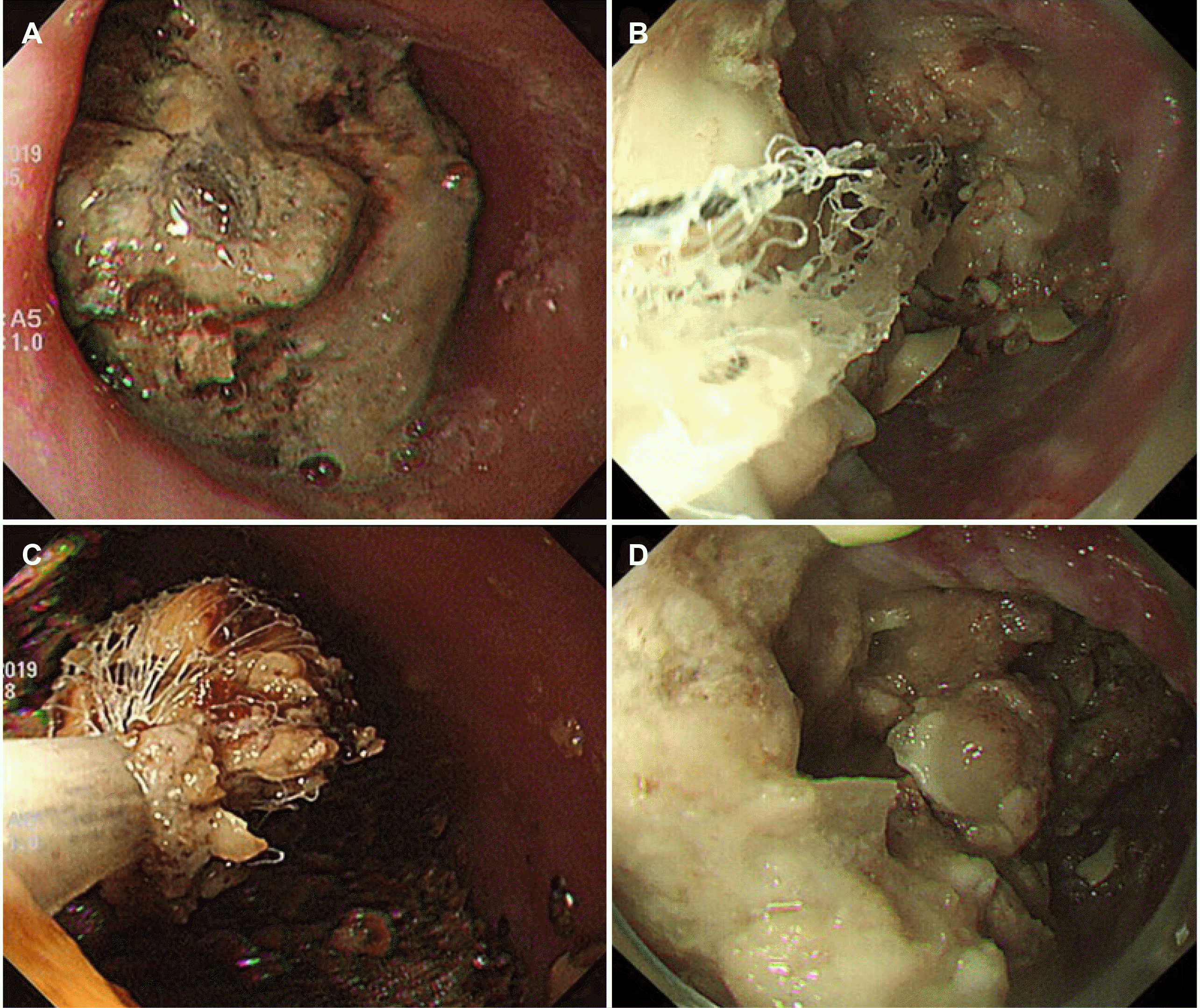
Two patients were diagnosed with esophageal bezoars. One of them (a 72-year-old man) had undergone a total gastrectomy with esophagojejunostomy to manage gastric cancer eight years earlier. The patient complained of epigastric pain 10 days before the visit. He was instructed to go on a Coca-Cola® diet because he could swallow liquids. The follow- up duodenoscopy after two days revealed no remnant bezoar. Another patient (an 85-year-old woman) with a previous history of cerebrovascular infarction presented with recent aggravated dysphagia and odynophagia. Duodenoscopy revealed a bezoar in the distal esophagus, which was partially fragmented and removed using a basket and retrieval net and disappeared in the follow-up endoscopy after two days (Fig. 1). On the other hand, the patient presented with similar symptoms after six months and was diagnosed with a recurrent bezoar.
Thirty-three patients (20 males) were diagnosed with gastric bezoar. A bezoar in the small intestine (ileum) was noted simultaneously in two patients. The median age was 72.7 years (47-98 years). All of them had a phytobezoar. Ten (31.2%) patients had hypertension, and five (15.6%) had diabetes mellitus. Seven (21.2%) out of the 33 patients had a history of previous gastric surgery. Twenty-one (63.6%) patients had a gastric ulcer. The most common location of the gastric ulcer was the antrum (14/21, 66.7%), followed by the angle (3/21, 15%). There were three patients with small bowel ileus
There were 30 patients without small bowel ileus. In six patients, gastric bezoars disappeared spontaneously after a median of two days (1-8 days) after conservative treatment, including Nil per os (NPO) and adequate hydration. Three patients were treated only by chemical dissolution. Endoscopic removal was performed in 18 patients, and the bezoars were successfully removed in 88.9% (16/18) of patients in a single session (n=12), two sessions (n=2), three sessions (n=1), and seven sessions (n=1). Among the 16 patients with successful endoscopic removal, 12 (75 %) patients were treated with the coadministration of Coca-Cola®, pancreatic enzymes, or both.
Surgical removal was performed in five patients: two with endoscopic treatment failure, one with a previous history of endoscopic treatment failure, one with a history of surgical bezoar, and one with their own choice.
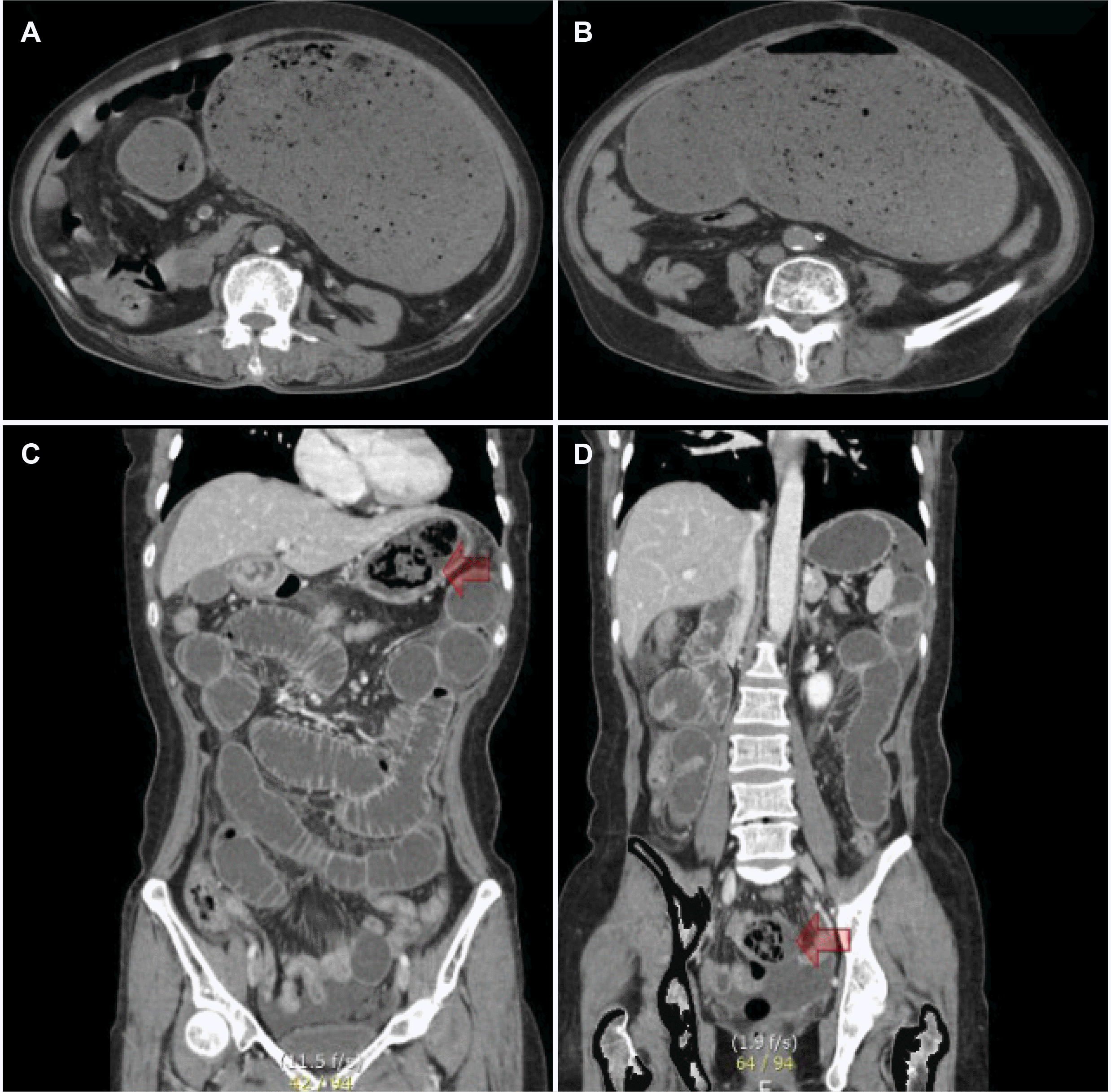
Three patients had a concomitant small bowel ileus. One was a 79-year-old female with long-standing diabetes in acute gastric dilatation as a symptom of an outlet obstruction, followed by metabolic acidosis with a blood pH of 7.2, leading to small bowel ileus without a transition zone. This patient improved through conservative management, including the correction of metabolic acidosis and chemical dissolution of the bezoar (Fig. 2A, B). Two patients were diagnosed with small bowel obstruction due to a concomitant bezoar in the small intestine. They had undergone surgical removal to manage obstructive ileus (Fig. 2C, D).
There were nine patients (median age 69.2 years [range, 52-90 years], seven males) with duodenal bezoars. There were four patients with duodenal ulcers. The most common location for bezoars was the second portion (n=5). Two patients with a mechanical small bowel ileus had undergone surgical removal. Six patients underwent successful endoscopic removal with a median of two sessions (range, 1-5), and one patient underwent conservative management with chemical dissolution.
Thirty-three patients (11 males) were diagnosed with bezoars in the small intestine. The median age was 69.8 years (range, 18-90 years). Twenty-nine (87.9%) patients had an ileus at presentation. Fourteen (42.4%) patients had a history of diabetes with a mean duration of 12.8 years, and 17 (51.5%) patients had a history of previous abdominal surgery. Twenty (60.6%) patients underwent surgical management, and 13 (39.4%) underwent conservative management.
This reported the clinical outcomes of patients with bezoars in different locations of the GI tract in a single tertiary center over 12 years. The bezoar was most commonly observed in the stomach and small intestine. Previous histories of abdominal surgery, especially gastric surgery, hypertension, and diabetes, were common predisposing factors for bezoar formation, consistent with the predisposing factors suggested in previous reports.9-11
Two patients with esophageal bezoars did not have dysphagia before bezoar formation in the esophagus. Bezoars in the esophagus rarely occur in patients with esophageal structural or motor disorders.12-14 Endoscopic approach with fragmentation and evacuation of the bezoar is the primary treatment for esophageal bezoar. Chemical dissolution by mouth can be considered for patients who tolerate a liquid diet. In this study, one patient with esophagojejunostomy was prescribed Coca-Cola® because he could swallow liquids, and no mucosal injuries, such as erosions or ulcers, were found on duodenoscopy. His symptoms improved, and the bezoar disappeared on follow-up endoscopy in two days. Although this outcome was successful, it is necessary to consider the risk of aspiration or esophageal perforation before initiating chemical dissolution to manage esophageal bezoar. Chemical dissolution must be determined cautiously, considering the availability of a liquid diet, esophageal mucosal injury, and the risk of aspiration during chemical dissolution.
This study showed that 63.6% of patients with gastric bezoars had simultaneous gastric ulcers, of which the most frequently affected area was the antrum. Gastric ulcers associated with gastric bezoars may be due to pressure necrosis and may be accompanied by complications, such as bleeding or perforation.5,7 The probability of concomitant gastric bezoar and small bowl bezoar was 17.5-21.0%.15,16 In this study, three patients had a small bowel ileus: two patients with concomitant small bowel bezoar and one patient with acute gastric dilatation due to gastric outlet obstruction. Currently, the main treatment options for gastric bezoars include chemical dissolution by Coca-Cola® or enzymatic treatment, endoscopic removal, or surgical removal. In this study, the gastric bezoar disappeared spontaneously in 20% of patients with only adequate fluid and electrolyte replacement to manage the imbalance due to recurrent vomiting and poor oral intake. Previous studies also showed that the rate of spontaneous resolution was 10-29.4%.4,8 Managing dehydration and electrolyte imbalance in patients with bezoars is essential before considering removal methods.11 Endoscopic removal is an effective treatment method with good accessibility and various available types of endoscopic devices.4,7,11 The reported success rate of endoscopic removal for gastric bezoar was 71.5-100%.9,17 The present study reported an 89% success rate for the endoscopic removal in patients with only gastric bezoar. In cases of multiple endoscopic sessions or chemical dissolution, crushed gastric bezoars may migrate downward, leading to a small bowel obstruction. Therefore, even though endoscopic management showed a high success rate for the removal of gastric bezoars, surgical management may need to be considered in cases with endoscopic treatment failure, new onset of small bowel ileus during multiple sessions of endoscopic removal, or concomitant gastric and small bowel bezoars or small bowel ileus during treatment.18
Patients with a bezoar in the small bowel present with ileus, intestinal ischemia, or perforation, in which surgical procedures are inevitable.11,18 In this study, 88% of patients with a small bowel bezoar presented with ileus, and surgical management was necessary for 61% of patients. A minimally invasive surgical procedure is a preferred method.3 Nevertheless, segmental resections with anastomosis or stoma are considered in cases of ischemia or perforation.10
This study has some limitations, including a small sample size because of the low incidence of bezoar and the fact that this is a retrospective study, where there may be some selection bias of treatment modalities and incomplete medical records. On the other hand, the authors’ experience provides valuable clinical information on GI bezoars regarding the clinical course and the necessity of surgical intervention.
In conclusion, managing gastrointestinal bezoars requires multidisciplinary approaches, including correcting fluid and electrolyte imbalance, chemical dissolution, endoscopic treatment, and surgical treatment.
REFERENCES
1. Ahn YH, Maturu P, Steinheber FU, Goldman JM. 1987; Association of diabetes mellitus with gastric bezoar formation. Arch Intern Med. 147:527–528. DOI: 10.1001/archinte.1987.00370030131025. PMID: 3827430.

2. Mihai C, Mihai B, Drug V, Cijevschi Prelipcean C. 2013; Gastric bezoars--diagnostic and therapeutic challenges. J Gastrointestin Liver Dis. 224:111.
3. Ghosheh B, Salameh JR. 2007; Laparoscopic approach to acute small bowel obstruction: review of 1061 cases. Surg Endosc. 21:1945–1949. DOI: 10.1007/s00464-007-9575-3. PMID: 17879114.

4. Park SE, Ahn JY, Jung HY, et al. 2014; Clinical outcomes associated with treatment modalities for gastrointestinal bezoars. Gut Liver. 8:400–407. DOI: 10.5009/gnl.2014.8.4.400. PMID: 25071905. PMCID: PMC4113045.

5. Iwamuro M, Okada H, Matsueda K, et al. 2015; Review of the diagnosis and management of gastrointestinal bezoars. World J Gastrointest Endosc. 7:336–345. DOI: 10.4253/wjge.v7.i4.336. PMID: 25901212. PMCID: PMC4400622.

6. Koulas SG, Zikos N, Charalampous C, Christodoulou K, Sakkas L, Katsamakis N. 2008; Management of gastrointestinal bezoars: an analysis of 23 cases. Int Surg. 93:95–98.
7. Gökbulut V, Kaplan M, Kaçar S, Akdoğan Kayhan M, Coşkun O, Kayaçetin E. 2020; Bezoar in upper gastrointestinal endoscopy: A single center experience. Turk J Gastroenterol. 31:85–90. DOI: 10.5152/tjg.2020.18890. PMID: 32141815. PMCID: PMC7062142.
8. Iwamuro M, Tanaka S, Shiode J, et al. 2014; Clinical characteristics and treatment outcomes of nineteen Japanese patients with gastrointestinal bezoars. Intern Med. 53:1099–1105. DOI: 10.2169/internalmedicine.53.2114. PMID: 24881731.

9. Kement M, Ozlem N, Colak E, Kesmer S, Gezen C, Vural S. 2012; Synergistic effect of multiple predisposing risk factors on the development of bezoars. World J Gastroenterol. 18:960–964. DOI: 10.3748/wjg.v18.i9.960. PMID: 22408356. PMCID: PMC3297056.

10. Paschos KA, Chatzigeorgiadis A. 2019; Pathophysiological and clinical aspects of the diagnosis and treatment of bezoars. Ann Gastroenterol. 32:224–232. DOI: 10.20524/aog.2019.0370.

11. Dikicier E, Altintoprak F, Ozkan OV, Yagmurkaya O, Uzunoglu MY. 2015; Intestinal obstruction due to phytobezoars: An update. World J Clin Cases. 3:721–726. DOI: 10.12998/wjcc.v3.i8.721. PMID: 26301232. PMCID: PMC4539411.

12. Qureshi SS. 2005; Esophageal bezoar in a patient with normal esophagus. Indian J Gastroenterol. 24:38.
13. Kim KH, Choi SC, Seo GS, Kim YS, Choi CS, Im CJ. 2010; Esophageal bezoar in a patient with achalasia: case report and literature review. Gut Liver. 4:106–109. DOI: 10.5009/gnl.2010.4.1.106. PMID: 20479921. PMCID: PMC2871618.

14. Yaqub S, Shafique M, Kjæstad E, et al. 2012; A safe treatment option for esophageal bezoars. Int J Surg Case Rep. 3:366–367. DOI: 10.1016/j.ijscr.2012.04.008. PMID: 22609703. PMCID: PMC3376689.

15. Wang PY, Skarsgard ED, Baker RJ. 1996; Carpet bezoar obstruction of the small intestine. J Pediatr Surg. 31:1691–1693. DOI: 10.1016/S0022-3468(96)90052-4. PMID: 8986991.

16. Escamilla C, Robles-Campos R, Parrilla-Paricio P, Lujan-Mompean J, Liron-Ruiz R, Torralba-Martinez JA. 1994; Intestinal obstruction and bezoars. J Am Coll Surg. 179:285–288.
17. Erzurumlu K, Malazgirt Z, Bektas A, et al. 2005; Gastrointestinal bezoars: a retrospective analysis of 34 cases. World J Gastroenterol. 11:1813–1817. DOI: 10.3748/wjg.v11.i12.1813. PMID: 15793871. PMCID: PMC4305881.

18. Yang S, Cho MJ. 2021; Clinical characteristics and treatment outcomes among patients with gastrointestinal phytobezoars: A single-institution retrospective cohort study in Korea. Front Surg. 8:691860. DOI: 10.3389/fsurg.2021.691860. PMID: 34250009. PMCID: PMC8263911.

Fig. 1
Esophagoscopy was performed on an 85-year-old female with acute onset of dysphagia. The esophageal bezoar is located at a gastroesophageal junction from the upper incisor 35-40 cm; (A). Endoscopic removal by retrieval net and lithotripsy basket; (B, C). The esophageal bezoar recurred six months later; (D).

Fig. 2
Abdomen CT is showing acute gastric dilatation and small bowel ileus in a 79-year-old female with gastric bezoar; (A, B). Abdomen CT is showing small bowel ileus in a 77-year-old female with concomitant gastric bezoar (C, arrow) and small bowel bezoar (D, arrow); (C, D).

Table 1
Demographic Factors of the 75 Patients with Gastrointestinal Bezoars


 PDF
PDF Citation
 Print
Print


 XML Download