Journal List > J Korean Foot Ankle Soc > v.27(2) > 1516082987

당뇨족으로 인한 비외상성 하퇴부 절단 환자에서 시행한 절단부 창상의 치유 기간과 Ischemia-Modified Albumin과의 관계

Abstract

Purpose

The present study examined the effectiveness of the preoperative ischemia-modified albumin (IMA) levels in predicting the healing period of amputation wounds in patients with diabetes mellitus following a non-traumatic below-knee amputation (BKA).

Materials and Methods

This study enrolled 41 diabetic foot ulcer patients who underwent BKA at the authors’ hospital diabetic foot center from April 2016 to April 2022. Among the 41 patients, 29 (70.7%) were male and 12 (29.3%) were female. Their mean age was 64.54±11.38 years (41~81 years). The mean follow-up period was 19.48±5.56 weeks (14~48 weeks) after BKA. The patients were divided into two groups (high IMA group and normal IMA group), which evaluated the healing period, wound dehiscence, and revision operation rate using a Fisher's exact test and Mann–Whitney U test. Three orthopedic surgeons performed stump wound evaluation, and they were evaluated as healing when all sutures were fused without oozing.

Results

Thirty patients (73.2%) (group A) showed a high level of IMA (median: 91.2 U/mL), and 11 (26.8%) patients (group B) showed a normal range of IMA (median: 82.7 U/mL). In group A, the median period for wound healing took 1.4 weeks longer, which was significant (p=0.001). No statistical relationship was observed between wound dehiscence, revision operation rate, and IMA value. There was no correlation between the other risk factors (estimated glomerular filtration rate, HbA1c) and the wound healing period.

Conclusion

Although there was a limitation in using IMA as the sole factor to predict the healing period of amputation wounds in patients after BKA, this study revealed a significant positive correlation between IMA and the period of stump healing after BKA. Therefore, the preoperative IMA levels may help predict the period of stump healing after BKA.

서 론

당뇨병성 족부 병변은 당뇨병의 합병증 중 하나로 당뇨 환자의 약 15%∼20% 정도에서 평생 한 번은 경험하는 것으로 알려져 있다.1) 진행된 족부 궤양은 치료에 잘 반응하지 않는 경우가 많으며 절단에 이르기도 한다. 미국 당뇨학회의 추산에 따르면 당뇨 환자에서의 절단은 매년 80,000건 이상 시행되고 있으며 전체 절단 원인의 60% 이상을 차지한다.2) 당뇨발 환자에서의 절단은 감염과 괴사된 조직의 광범위 제거라는 궁극적 치료 방법이다. 절단은 발가락 절단과 같은 소절단(minor amputation)에서부터 족관절 상부에서 시행하는 대절단(major amputation)까지 다양하게 시행되며, 수술 전 혈류 상태를 파악하기 위해 발목상완지수(ankle brachial index, ABI), 도플러 동맥압 측정(arterial Doppler sonography), 경피적 산소분압(transcutaneous oxygen pressure, TcPO2), 족지압(toe pressure) 측정 등이 시행되고 있다. 실제 절단술을 시행한 이후의 절단단의 창상 예후와 관련된 인자들은 술 전 시행한 경피적 산소분압, 도플러 동맥압 측정(arterial Doppler sonography)과 연관성이 있다고 알려져 있다.3,4) 그러나 절단술을 요하는 족부병변은 대부분 와그너 분류(Wagner classification) grade 4 혹은 grade 5에 해당하는 괴사를 동반된 병변이며 경피적 산소분압 혹은 도플러 동맥압 측정과 같은 방법들을 시행하기에 여러 제한점이 있다.
족부 궤양 발생 이후 절단술로까지의 이행 위험인자에는 당화혈색소(HbA1c), 말초혈관 질환, 신경병증, 기저 신기능 등 다양한 인자들이 보고되어 있으며,5) 이러한 절단단의 예후를 예측하는 데 있어 간편하게 시행 가능한 혈청검사는 소개된 바 없으나 다만, HbA1c와 신장합병증이 궤양 발생 이후 절단술까지 이어지는 위험인자로 보고되고 있다.6-9) 비교적 최근에 소개된 생화학적 검사 중 하나인 IMA (Ischemia-modified albumin)는 국소적인 심근 허혈 시에 세포에서 유리되는 copper에 의해 형성되는 oxygen free radical (03-)과 hydroxyl free radical (OH-)에 의해 손상받은 알부민으로 심근경색뿐만 아니라 다른 허혈 초기 상태를 찾아내는 능력이 우월한 생체표지자이다.10,11) 본 연구는 하퇴절단술 전 시행한 IMA 혈액검사와 평균 창상 치유 기간, 창상 불유합 및 재수술 여부와의 상관관계를 통해 하퇴절단술 이후 절단부 창상의 치유 기간을 예측하는 데 있어서 IMA 효용성을 분석하고자 한다.

대상 및 방법

1. 연구 대상

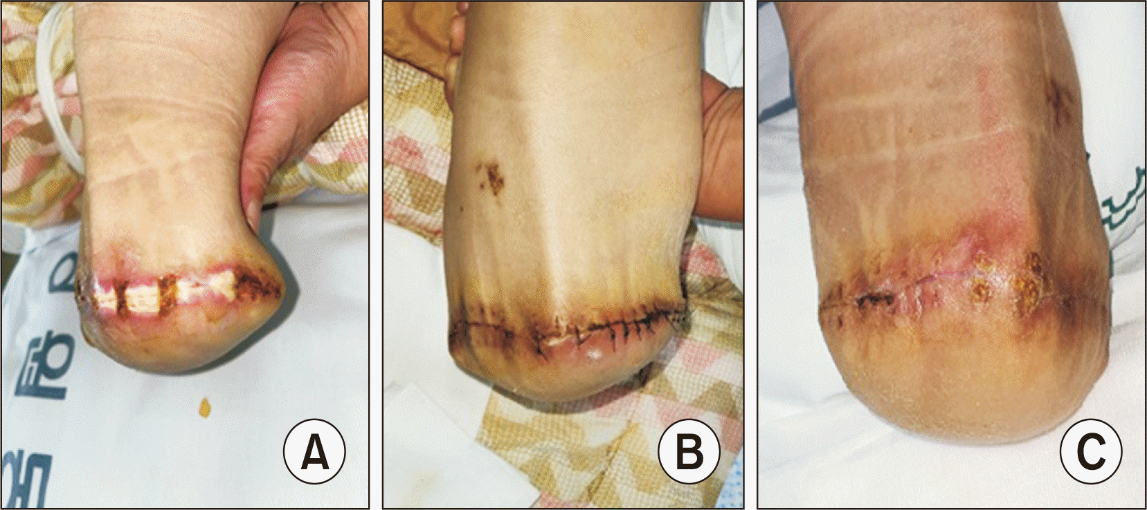
본 연구는 후향적 연구로 본원의 생명윤리위원회로부터 연구 승인을 받았다. 2016년 4월부터 2022년 4월 사이 본원에서 당뇨병성 족부 병변으로 하퇴절단술을 시행한 41예의 환자들을 대상으로 하였다. 최소 추시 경과 기간은 하퇴절단술을 시행한 후 4개월을 기준으로 하였으며, 4개월 내 사망 혹은 추적관찰이 되지 않았던 5예는 제외하였다. 의무기록상 당뇨병의 종류를 확인할 수 없는 경우가 많아 당뇨병의 종류를 따로 분류하지는 않았다. 연구 대상 간 절단술 시행 부위의 해부학적 특성, 수술 후 창상의 형태 및 범위의 차이를 최소화하기 위해 하퇴절단술을 시행한 환자들만을 대상으로 하였다. 모든 수술은 한 명의 정형외과 족부족관절 세부전문의에 의해 시행되었으며, 수술 후 창상 평가는 3명의 정형외과 의사에 의해 시행되었다. 각 증례에 대한 의견은 모두 동일하였다. 삼출물 없이 모든 하퇴절단 봉합부가 유합되었을 때를 창상유합으로 평가하였으며 삼출물 없이 봉합부가 벌어졌을 경우에는 지연 유합으로 판단하였다(Fig. 1).

2. 연구 방법

저자들은 환자의 나이, 성별, 흡연 여부에 대해 파악하였으며 말초혈관 질환에 대해서는 발목상완지수 및 도플러 동맥압 측정을 시행한 후 말초혈관 질환이 확인된 경우 컴퓨터단층촬영 혈관조영술을 시행하였다. 이후 순환기내과, 흉부외과 협진을 통해 하퇴절단술 전 15예의 환자에서 하지 혈관 경피적 풍선혈관성형술(percutaneous transluminal angioplasty, PTA)을 시행하였으며, 10예의 환자에서 혈관우회술(bypass surgery)을 시행하였다(Table 1). 궤양의 정도는 Wagner classification을 따랐으며,12) 모든 환자에서 Wagner classification grade 5 궤양으로 하퇴절단술을 시행하였다. 술 후 2주 뒤에 봉합사를 제거하였으며 최소 4개월간 창상 추적관찰을 시행하였다. 추적관찰 중 삼출물이 있거나 벌어짐이 보이면 1주 간격으로 경과관찰 하였으며 위의 스케줄은 모든 환자에서 동일하였다. 정상 IMA 수치 85 U/mL를 초과하는 군과 초과하지 않는 군, 정상 HbA1c 수치 6.5%를 초과하는 군과 초과하지 않는 군, 사구체여과율(estimated glomerular filtration rate, eGFR) 수치 60을 초과하는 군과 초과하지 않는 군으로 나누어 창상 유합 기간과 창상 불유합으로 인해 재수술을 시행한 경우를 조사하여 IMA, HbA1c 및 eGFR과의 통계적 유의성을 확인하였다. HbA1c 및 eGFR은 최초 외래 내원 시를 기준으로 하였으며, IMA는 경피적 풍선혈관성형술 혹은 혈관우회술을 이후, 하퇴절단술 이전 검사 결과를 기준으로 하였다. 수술 전 시행한 IMA 증가를 보이는 환자들은 트로포닌-I (Troponin-I), 심전도를 시행 후, 수술 전 순환기내과 협진을 통해 심근허혈 가능성을 배제하였다.

3. 통계 방법

통계처리는 IBM SPSS version 20.0 (IBM Corp., Armonk, NY, USA) 프로그램을 이용하였다. 작은 n수를 고려하여 빈도는 Fisher’s exact test, 평균은 Mann–Whitney U test를 실시하였다. 군 간 위험 인자들과의 상관관계를 알기 위해서 다중회귀분석을 실시하였으며 p값 0.05 이하인 경우 유의한 것으로 하였다.

결 과

평균 추시 기간은 19.48±5.56주(범위 14∼48주)였다. 남자 환자가 29명(70.7%)을 차지하였으며 평균 나이는 64.54±11.38세(41∼81세)였다. 41명의 환자 중 수술 전 혈액검사에서 정상 IMA 수치가 85 U/mL보다 높은 군은 30명(73.2%)이었다. 높은 IMA 수치를 보이는 군의 IMA 수치 중앙값은 91.2 U/mL (사분위 범위 89.0∼94.4 U/mL)로 정상 IMA군의 중앙값 82.7 U/mL (사분위 범위 80.2∼83.5 U/mL)보다 8.5 U/mL 높은 IMA 수치를 나타냈다. 창상 유합 기간 중앙값은 높은 IMA를 보이는 환자군에서 5.1주(사분위 범위 4.1∼6.0주)로 정상 IMA 환자군의 3.7주(사분위 범위 3.1∼4.1주)와 비교하여 1.4주(p=0.001) 더 길었다. IMA 수치와 창상 유합 기간은 양의 상관관계를 보였다(Fig. 2). 높은 IMA를 보이는 환자군에서의 창상 불유합으로 재수술을 시행한 경우는 13예(43.3%), 정상 IMA군에서는 5예(45.5%)였으나 IMA 수치와 통계적 유의성(p=0.716)은 없었다(Table 2).
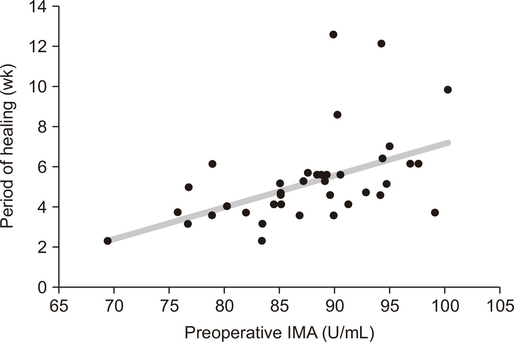
HbA1c와 평균 창상 유합 기간 및 재수술 여부는 통계적 유의성을 보이지 않았으나(Table 3) 높은 HbA1c를 보이는 군에서 창상 유합 기간의 중앙값은 0.9주 더 소요되었으며 재수술 증례도 정상 HbA1c군에서는 5예(35.7%)인데 반해 13예(48.1%)로 높았다. eGFR 60 미만의 신기능과도 통계적 유의성은 없었으나 eGFR 60 미만의 환자군에서 창상 유합 기간의 중앙값은 0.4주 더 높았으며, 재수술 증례 역시 12회(48.0%)로 정상 신기능군에서의 6예(37.5%)에 비해 6예 더 많았다(Table 4).
다중회귀분석 시행 결과, 다중회귀분석 모델은 F=4.832, p=0.013로 적합한 회귀모형이라고 할 수 있으며, HbA1c (p=0.950)와 eGFR (p=0.613)은 유의미한 결과를 보이지 않았으나 IMA (p=0.002)의 경우 창상 유합 기간과 유의미한 관계가 있었다(Table 5).

고 찰

IMA는 흉통을 호소하는 환자에서 급성심근경색의 회복 가능한 허혈 초기 상태의 빠른 진단검사를 위해 최초로 연구되었으며 그 임상적 의의를 인정받아 급성심근경색의 조기진단 및 예후 판정에 사용되고 있다.11,13) IMA는 뇌 허혈, 말기 신부전, 간질환, 악성 신생물, 염증반응 및 말초혈관 질환에서도 증가하며 운동 유발 골격근 허혈 시에도 증가하는 것으로 알려져 있다.14-16) 현재까지 알려진 바에 따르면, 혈장 알부민의 아미노 말단부는 정상적인 상태에서는 금속 이온에 대한 친화성을 갖고 있으며 허혈성 질환이 진행 중일 때 혈액 내의 알부민 말단 부분이 변형되게 된다.
IMA는 2형 당뇨병(type 2 diabetes mellitus, T2DM) 및 말초동맥질환(peripheral artery disease, PAD)과의 연관성에도 불구하고 급성심근경색, 장간막허혈, 뇌 허혈 등 체내 주요 장기와 관련된 조기진단 및 예후 판정과 관련하여 주로 연구되어 왔다. Ma 등17)의 연구에 따르면 T2DM 기저병력을 가진 환자에서 PAD를 조기진단하는데 IMA가 진단적 가치가 있으며 IMA의 증가 정도는 PAD의 중증도와도 연관이 있다고 하였다. 그러나 PAD 및 이로 인한 당뇨병성 족부 궤양의 예후인자, 절단술 이후 창상의 예후인자 등에 대해서는 많은 연구가 이루어지지 못하였다.
HbA1c와 T2DM은 합병증 발생빈도 및 그 중증도에 대해 밀접한 관련이 있는 것으로 여러 문헌을 통해 보고되었으며 Zhou 등6)의 연구에 따르면 HbA1c가 5%에서 9%로 증가한 경우 하지 절단술로 이행되는 경우가 급격히 증가하는 것으로 보고되었다. 그러나 당뇨병성 족부 궤양의 발생 및 치료에 대한 HbA1c의 영향에 대한 증거는 알려진 바가 없으며 절단으로의 이행을 줄이기 위해 혈당 조절이 필수적이라는 사실만을 주지시킬 뿐이다. 고혈당은 조직 내의 활성산소를 생성하는 다양한 산화 스트레스의 한 가지 원인일 뿐이며, HbA1c는 이러한 고혈당의 연속선상에 있는 지표인데 반해 IMA는 조직 내의 산화 스트레스를 직접 반영한다는 사실을18,19) 감안하여 보았을 때, IMA가 HbA1c보다 직접적으로 조직의 허혈 상태를 반영한다는 것을 알 수 있다.
만성 콩팥병 환자에서 역시 신기능과 하퇴절단술 이후 사망률 및 창상 불유합과 높은 연관성을 보인다는 많은 연구가 보고되었으나,18,19) 만성 콩팥병 역시 PAD의 위험인자로서20) 만성 콩팥병 단계가 증가함에 따라 함께 증가하는 산화 스트레스로 인한 결과이며,21) HbA1c와 마찬가지로 직접적인 조직의 허혈 상태를 반영하지는 못한다.
본 연구는 비록 41예의 작은 표본한계가 있으나 하퇴절단술 이후 절단단 평균 유합 기간과 IMA 간의 통계적 유의성을 확인할 수 있었으며 이는 저산소로 인한 산화스트레스가 창상 유합에 영향을 미치며 IMA를 통해 수치화되었다고 볼 수 있다.22) 그러나 이러한 유용성에도 불구하고 앞서 언급하였던 IMA를 증가시킬 수 있는 다양한 원인들에 대한 배제 진단이 우선시되어야 한다. IMA는 트레드밀을 통한 운동 유발 허혈 혹은 수술 중 토니켓(tourniquet) 사용으로도 증가될 수 있으며,23-25) PTA 혹은 혈관우회술 이후 재관류로 인한 영향에 대해 명확히 연구된 바가 없다는 한계점들이 있어 앞으로 추가적인 연구를 요할 것으로 생각한다.

결 론

IMA 수치는 다양한 장기들의 허혈성 상태에 따라 증가할 수 있으므로 타 장기에 대한 평가가 필수적이며 수술 전 시행한 IMA 검사 수치만으로 당뇨족으로 인한 비외상성 하퇴부 절단 환자의 술 후 절단부 창상의 치유 기간을 예측하는 데에는 임상적 한계가 있다. 그러나 본 연구에서 IMA 검사 수치와 절단부 창상의 치유 기간 사이에 통계적으로 의미 있는 양의 상관관계를 보였으므로 수술 전 시행한 IMA 검사 수치는 절단부 창상의 치유 기간을 예측하는 데에 도움이 될 수 있다.

Notes

Financial support

None.

Conflict of interest

None.

REFERENCES

1. Boulton AJ. 2013; The pathway to foot ulceration in diabetes. Med Clin North Am. 97:775–90. doi: 10.1016/j.mcna.2013.03.007. DOI: 10.1016/j.mcna.2013.03.007. PMID: 23992891.
crossref
2. Gregg EW, Sorlie P, Paulose-Ram R, Gu Q, Eberhardt MS, Wolz M, et al. 2004; Prevalence of lower-extremity disease in the US adult population ≥40 years of age with and without diabetes: 1999-2000 national health and nutrition examination survey. Diabetes Care. 27:1591–7. doi: 10.2337/diacare.27.7.1591. DOI: 10.2337/diacare.27.7.1591. PMID: 15220233.
3. Kram HB, Appel PL, Shoemaker WC. 1989; Prediction of below-knee amputation wound healing using noninvasive laser Doppler velocimetry. Am J Surg. 158:29–31. doi: 10.1016/0002-9610(89)90310-3. DOI: 10.1016/0002-9610(89)90310-3. PMID: 2662788.
crossref
4. Ratliff DA, Clyne CA, Chant AD, Webster JH. 1984; Prediction of amputation wound healing: the role of transcutaneous pO2 assessment. Br J Surg. 71:219–22. doi: 10.1002/bjs.1800710320. DOI: 10.1002/bjs.1800710320. PMID: 6697129.
crossref
5. Reiber GE, Pecoraro RE, Koepsell TD. 1992; Risk factors for amputation in patients with diabetes mellitus. A case-control study. Ann Intern Med. 117:97–105. doi: 10.7326/0003-4819-117-2-97. DOI: 10.7326/0003-4819-117-2-97. PMID: 1605439.
crossref
6. Zhou ZY, Liu YK, Chen HL, Yang HL, Liu F. 2015; HbA1c and lower extremity amputation risk in patients with diabetes: a meta-analysis. Int J Low Extrem Wounds. 14:168–77. doi: 10.1177/1534734615593190. DOI: 10.1177/1534734615593190. PMID: 26130760.
7. Zhao W, Katzmarzyk PT, Horswell R, Wang Y, Johnson J, Heymsfield SB, et al. 2013; HbA1c and lower-extremity amputation risk in low-income patients with diabetes. Diabetes Care. 36:3591–8. doi: 10.2337/dc13-0437. DOI: 10.2337/dc13-0437. PMID: 24062322. PMCID: PMC3816880.
crossref
8. Matsushita K, Ballew SH, Coresh J, Arima H, Ärnlöv J, Cirillo M, et al. 2017; Measures of chronic kidney disease and risk of incident peripheral artery disease: a collaborative meta-analysis of individual participant data. Lancet Diabetes Endocrinol. 5:718–28. doi: 10.1016/S2213-8587(17)30183-3. DOI: 10.1016/S2213-8587(17)30183-3. PMID: 28716631.
9. Kurniawati A, Ismiarto YD, Hsu IL. 2019; Prognostic factors for lower extremity amputation in diabetic foot ulcer patients. J Acute Med. 9:59–63. doi: 10.6705/j.jacme.201906_9(2).0003.
10. Shevtsova A, Gordiienko I, Tkachenko V, Ushakova G. 2021; Ischemia-modified albumin: origins and clinical implications. Dis Markers. 2021:9945424. doi: 10.1155/2021/9945424. DOI: 10.1155/2021/9945424. PMID: 34336009. PMCID: PMC8315882.
crossref
11. Sinha MK, Gaze DC, Tippins JR, Collinson PO, Kaski JC. 2003; Ischemia modified albumin is a sensitive marker of myocardial ischemia after percutaneous coronary intervention. Circulation. 107:2403–5. doi: 10.1161/01.CIR.0000072764.18315.6B. DOI: 10.1161/01.CIR.0000072764.18315.6B. PMID: 12742986.
crossref
12. Calhoun JH, Cantrell J, Cobos J, Lacy J, Valdez RR, Hokanson J, et al. 1988; Treatment of diabetic foot infections: Wagner classification, therapy, and outcome. Foot Ankle. 9:101–6. doi: 10.1177/107110078800900301. DOI: 10.1177/107110078800900301. PMID: 3229695.
crossref
13. Sbarouni E, Georgiadou P, Kremastinos DT, Voudris V. 2008; Ischemia modified albumin: is this marker of ischemia ready for prime time use? Hellenic J Cardiol. 49:260–6.
14. Apple FS, Wu AH, Mair J, Ravkilde J, Panteghini M, Tate J, et al. 2005; Future biomarkers for detection of ischemia and risk stratification in acute coronary syndrome. Clin Chem. 51:810–24. doi: 10.1373/clinchem.2004.046292. DOI: 10.1373/clinchem.2004.046292. PMID: 15774573.
crossref
15. Bourdon E, Loreau N, Blache D. 1999; Glucose and free radicals impair the antioxidant properties of serum albumin. FASEB J. 13:233–44. doi: 10.1096/fasebj.13.2.233. DOI: 10.1096/fasebj.13.2.233. PMID: 9973311.
crossref
16. Chan B, Dodsworth N, Woodrow J, Tucker A, Harris R. 1995; Site-specific N-terminal auto-degradation of human serum albumin. Eur J Biochem. 227:524–8. doi: 10.1111/j.1432-1033.1995.tb20419.x. DOI: 10.1111/j.1432-1033.1995.tb20419.x. PMID: 7851432.
crossref
17. Ma SG, Wei CL, Hong B, Yu WN. 2011; Ischemia-modified albumin in type 2 diabetic patients with and without peripheral arterial disease. Clinics (Sao Paulo). 66:1677–80. doi: 10.1590/s1807-59322011001000003.
18. Wukich DK, Ahn J, Raspovic KM, Gottschalk FA, La Fontaine J, Lavery LA. 2017; Comparison of transtibial amputations in diabetic patients with and without end-stage renal disease. Foot Ankle Int. 38:388–96. doi: 10.1177/1071100716688073. DOI: 10.1177/1071100716688073. PMID: 28103735.
crossref
19. Pollard J, Hamilton GA, Rush SM, Ford LA. 2006; Mortality and morbidity after transmetatarsal amputation: retrospective review of 101 cases. J Foot Ankle Surg. 45:91–7. doi: 10.1053/j.jfas.2005.12.011. DOI: 10.1053/j.jfas.2005.12.011. PMID: 16513503.
crossref
20. Chen J, Xie D, Shlipak MG, Townsend RR, Appel LJ, et al. Mohler ER 3rd. 2012; Risk factors for peripheral arterial disease among patients with chronic kidney disease. Am J Cardiol. 110:136–41. doi: 10.1016/j.amjcard.2012.02.061. DOI: 10.1016/j.amjcard.2012.02.061. PMID: 22465315. PMCID: PMC3586781.
crossref
21. Dounousi E, Papavasiliou E, Makedou A, Ioannou K, Katopodis KP, Tselepis A, et al. 2006; Oxidative stress is progressively enhanced with advancing stages of CKD. Am J Kidney Dis. 48:752–60. doi: 10.1053/j.ajkd.2006.08.015. DOI: 10.1053/j.ajkd.2006.08.015. PMID: 17059994.
crossref
22. Ruthenborg RJ, Ban JJ, Wazir A, Takeda N, Kim JW. 2014; Regulation of wound healing and fibrosis by hypoxia and hypoxia-inducible factor-1. Mol Cells. 37:637–43. doi: 10.14348/molcells.2014.0150. DOI: 10.14348/molcells.2014.0150. PMID: 24957212. PMCID: PMC4179131.
crossref
23. Refaai MA, Wright RW, Parvin CA, Gronowski AM, Scott MG, Eby CS. 2006; Ischemia-modified albumin increases after skeletal muscle ischemia during arthroscopic knee surgery. Clin Chim Acta. 366:264–8. doi: 10.1016/j.cca.2005.10.020. DOI: 10.1016/j.cca.2005.10.020. PMID: 16337614.
crossref
24. Zapico-Muñiz E, Santaló-Bel M, Mercé-Muntañola J, Montiel JA, Martínez-Rubio A, Ordóñez-Llanos J. 2004; Ischemia-modified albumin during skeletal muscle ischemia. Clin Chem. 50:1063–5. doi: 10.1373/clinchem.2003.027789. DOI: 10.1373/clinchem.2003.027789. PMID: 15161723.
crossref
25. Lee DH, Jeon HK, Park HJ, Shin WS, Lee SW, Youn HJ, et al. 2010; Change in ischemia-modified albumin and its clinical significance during exercise stress testing. Circ J. 74:484–9. doi: 10.1253/circj.cj-09-0581. DOI: 10.1253/circj.CJ-09-0581. PMID: 20057159.
crossref

Figure 1
Clinical photos show postoperative wound dehiscence (A) and after wound revision operation (B). Amputation evaluation was performed by 3 orthopedic surgeons, and when all sutures were fused without oozing (C), the healing state was assessed union.
jkfas-27-2-49-f1.tif
Figure 2
The graph represents the scatter plot of IMA levels and the periods of wound healing. It shows a positive correlation between IMA levels and the periods of wound healing. IMA: Ischemia-modified albumin.
jkfas-27-2-49-f2.tif
Table 1
Demographic and Clinical Patient Characteristics
Variable Value
Number 41
Age (yr) 64.54±11.38 (41~81)
Sex (male:female) 29:12
Smoking 22 (53.7)
Wagner classification
Grade 1 0 (0)
Grade 2 0 (0)
Grade 3 0 (0)
Grade 4 0 (0)
Grade 5 41 (100)
IMA (Ischemia modified albumin) 87.61±6.68
HbA1c 7.86±2.59
eGFR 51.88±40.28
ABPI (affected side) 21 (51.2)
0.88±0.29
Follow-up period (wk) 19.48±5.56 (14~48)
The average length of the healing 4.608±0.92
Wound dehiscence & revision operation 18 (43.9)
Preoperative intervention
PTA 15 (36.6)
Bypass operation 10 (24.4)

Values are presented as mean±standard deviation or number (%).

IMA: Ischemia-modified albumin, eGFR: estimated glomerular filtration rate, ABPI: ankle brachial pressure index, PTA: percutaneous transluminal angioplasty.

Table 2
Comparison Chart between High IMA Group and Normal IMA Group*
Variable High IMA group Normal IMA group p-value
Number 30 (73.2) 11 (26.8)
Age (yr) 62.0 (58.5~75.0) 59.0 (56.0~79.0) 0.262
Sex >0.999
Male 22 (73.3) 7 (63.6)
Female 8 (26.7) 4 (36.4)
Smoking 14 (46.7) 8 (72.7) 0.138
HbA1c (%) 7.2 (6.4~8.0) 6.5 (5.5~7.1) 0.612
eGFR 22.7 (10.9~70.7) 47.8 (7.0~87.6) 0.761
ABPI (affected side) 15 (50.0) 6 (54.5) 0.796
1.0 (0.8~1.2) 0.7 (0.6~0.9) 0.095
IMA (U/mL) 91.2 (89.0~94.4) 82.7 (80.2~83.5) <0.001
The length of healing (wk) 5.1 (4.1~6.0) 3.7 (3.1~4.1) 0.001
Wound dehiscence & revision operation 13 (43.3) 5 (45.5) 0.716
Preoperative intervention
PTA 6 (20.0) 9 (81.8) <0.001
Bypass operation 8 (26.7) 2 (18.2) 0.700

Values are presented as number (%) or median (interquartile range).

IMA: Ischemia-modified albumin (normal <85 U/mL), eGFR: estimated glomerular filtration rate, ABPI: ankle brachial pressure index, PTA: percutaneous transluminal angioplasty.

*Normal <85 U/mL.

Table 3
Comparison Chart between High HbA1c Group and Normal HbA1c Group*
Variable High HbA1c group Normal HbA1c group p-value
Number 27 (65.9) 14 (34.1)
Age (yr) 61.0 (55.5~73.0) 65.5 (59.0~77.0) 0.333
Sex
Male 19 (70.4) 10 (71.4) 0.319
Female 8 (29.6) 4 (28.6)
Smoking 15 (55.6) 7 (50.0) 0.735
HbA1c (%) 7.8 (6.9~8.6) 5.5 (5.2~6.1) <0.001
IMA (U/mL) 89.5 (85.1~94.3) 85.1 (84.0~90.3) 0.559
eGFR 21.2 (7.6~60.4) 51.1 (26.2~98.7) 0.968
ABPI (affected side) 14 (51.9) 7 (50.0) 0.910
1.0 (0.7~1.2) 0.9 (0.6~0.9) 0.287
The length of healing (wk) 5.0 (4.1~6.0) 4.1 (4.0~5.3) 0.143
Wound dehiscence & revision operation 13 (48.1) 5 (35.7) 0.919
Preoperative intervention
PTA 10 (37.0) 5 (35.7) 0.934
Bypass operation 5 (18.5) 5 (35.7) 0.224

Values are presented as number (%) or median (interquartile range).

IMA: Ischemia-modified albumin, eGFR: estimated glomerular filtration rate, ABPI: ankle brachial pressure index, PTA: percutaneous transluminal angioplasty.

*Normal HbA1c <6.5%.

Table 4
Comparison Chart between CKD Patient (eGFR <60) and Normal Kidney Function
Variable CKD patient group Normal kidney function group p-value
Number 25 (61.0) 16 (39.0)
Age (yr) 66.0 (56.0~74.0) 61.5 (57.5~78.5) 0.552
Sex
Male 18 (72.0) 11 (68.7) 0.174
Female 7 (28.0) 5 (31.3)
Smoking 12 (48.0) 10 (62.5) 0.364
eGFR 11.2 (7.4~35.3) 93.7 (84.3~102.1) <0.001
IMA (U/mL) 89.5 (84.5~92.9) 85.1 (83.6~94.0) 0.926
HbA1c (%) 6.8 (5.5~8.0) 7.1 (6.2~8.0) 0.147
ABPI (affected side) 14 (56.0) 7 (43.8) 0.444
0.9 (0.7~1.1) 1.0 (0.6~1.1) 0.856
The length of healing (wk) 5.0 (4.0~6.0) 4.6 (4.0~5.8) 0.885
Wound dehiscence & revision operation 12 (48.0) 6 (37.5) 0.483
Preoperative intervention
PTA 12 (48.0) 3 (18.8) 0.058
Bypass operation 4 (16.0) 6 (37.5) 0.118

Values are presented as number (%) or median (interquartile range).

CKD: chronic kidney disease, eGFR: estimated glomerular filtration rate, IMA: Ischemia-modified albumin, ABPI: ankle brachial pressure index, PTA: percutaneous transluminal angioplasty.

Table 5
Results of Multiple Linear Regression Analysis
b SE Beta t p VIF
(Constant) –9.513 4.495 –2.116 0.041
IMA 0.165 0.049 0.491 3.400 0.002 1.027
HbA1c 0.008 0.130 0.010 0.063 0.950 1.117
eGFR 0.004 0.008 0.076 0.510 0.613 1.097

F=4.832, p=0.013, R-square=0.249, Adj R-square=0.188.

SE: standard error, VIF: variance inflation factor, IMA: Ischemia-modified albumin, eGFR: estimated glomerular filtration rate.

TOOLS
Similar articles