Abstract
Purpose
This study aimed to evaluate the effectiveness of deltoid ligament repair on syndesmotic stabilization in patients with acute ankle fractures with ruptured deltoid and syndesmotic ligaments.
Materials and Methods
The medical records of 41 patients (41 ankles) who underwent surgery for Weber type B ankle fracture with ruptured deltoid and syndesmotic ligaments were retrospectively analyzed. The mean follow-up duration was 36 months (range 18~65 months). Patients were divided into two groups: those that underwent deltoid ligament repair (the deltoid group) and those who did not (the non-deltoid group). Both groups were also divided into two subgroups, namely, the D1/S1 group, which underwent syndesmotic screw fixation, or the D2/S2 group, which did not. Medial clear space (MCS), tibiofibular clear space (TFCS), anterior fibular line (AFL) ratio, and posterior fibular line (PFL) distance were measured, and visual analog scale (VAS), American Orthopaedic Foot and Ankle Society (AOFAS), and Foot Function Index (FFI) scores were evaluated.
Results
TFCS changed significantly after surgery in the D2 and S1 groups (p=0.01, p=0.03, respectively). Subgroup MCSs, TFCSs, and AFL ratios were not significantly altered by surgery in the four subgroups (p=0.82, p=0.45, p=0.25, respectively). However, postoperative PFL distances were significantly different in the D2 and S1 groups and the S1 and S2 groups (p=0.02, p=0.02, respectively). Mean TFCS decreased significantly after surgery in the D2 and S1 groups. The postoperative VAS, AOFAS scores, and FFI were not significantly different between the subgroups (p=0.44, p=0.40, and p=0.46, respectively).
Ankle fractures are operatively treated to align the talus within the ankle mortise to recover normal tibiotalar contact mechanics and prevent future posttraumatic arthritis. They are known to be accompanied by deltoid and syndesmotic ligament ruptures in up to 58.3% and 45% of patients, respectively.1,2) Approximately 50% of patients with ruptured deltoid ligaments have a syndesmotic injury during an acute ankle fracture.1,3) Therefore, bony and ligamentous structures should be stabilized to restore the normal tibiotalar relationship during ankle fracture surgeries. Further, while the interest in ligamentous structure disruption is increasing in patients with acute ankle fractures, a consensus regarding the treatment strategy for disrupted ligamentous structures in such patients remains unclear. Interestingly, significant attention has been focused on syndesmotic fixation rather than deltoid ligament problems during acute ankle fracture surgeries; therefore, assumptions regarding the abuse of syndesmotic fixation should be made cautiously.4,5)
The deltoid ligament has been known to play an important role in ankle and syndesmotic stability. However, clinical studies regarding its role in syndesmotic stabilization are limited compared to clinical studies on syndesmotic fixation. Recently, some clinical studies highlighted the importance of the deltoid ligament by comparing deltoid ligament repair with non-repair, or deltoid ligament repair with syndesmotic fixation.6,7) However, we believe there are different indications for stabilization of the deltoid and syndesmosis, and these procedures are not interchangeable.
To our knowledge, whether deltoid repair is more effective than syndesmotic fixation for treating acute ankle fracture in patients with ruptured deltoid and syndesmotic ligaments remains to be elucidated. Since the deltoid ligament plays an important role in syndesmotic stability, syndesmotic stability assessment results are notably different before and after deltoid repair. Thus, the first step to restoring a normal tibiotalar relationship during the operative treatment of ankle fractures is stabilizing both columns, whether bony or ligamentous structures. Medial column stabilization should be prioritized to help stabilize syndesmosis. Therefore, in this study, we hypothesized that deltoid ligament repair will restore ankle stability without addressing the syndesmosis.
This study aimed to evaluate the role of deltoid ligament repair on syndesmotic stabilization in patients with acute ankle fractures with ruptured deltoid and syndesmotic ligaments.
In this retrospective cohort study, we reviewed patients with Weber type B ankle fractures with confirmed or suspected ruptured deltoid and syndesmotic ligaments who underwent operative treatment between January 2013 and June 2018. The medical records of 41 patients (41 ankles) were retrospectively analyzed, consisting of 28 men and 13 women, with a mean age of 39.9 years (range 21∼63 years). The mean follow-up duration was 36 months (range 18∼65 months). This retrospective study was approved by our institutional review board (IRB 2019), and informed consent was obtained from all patients.
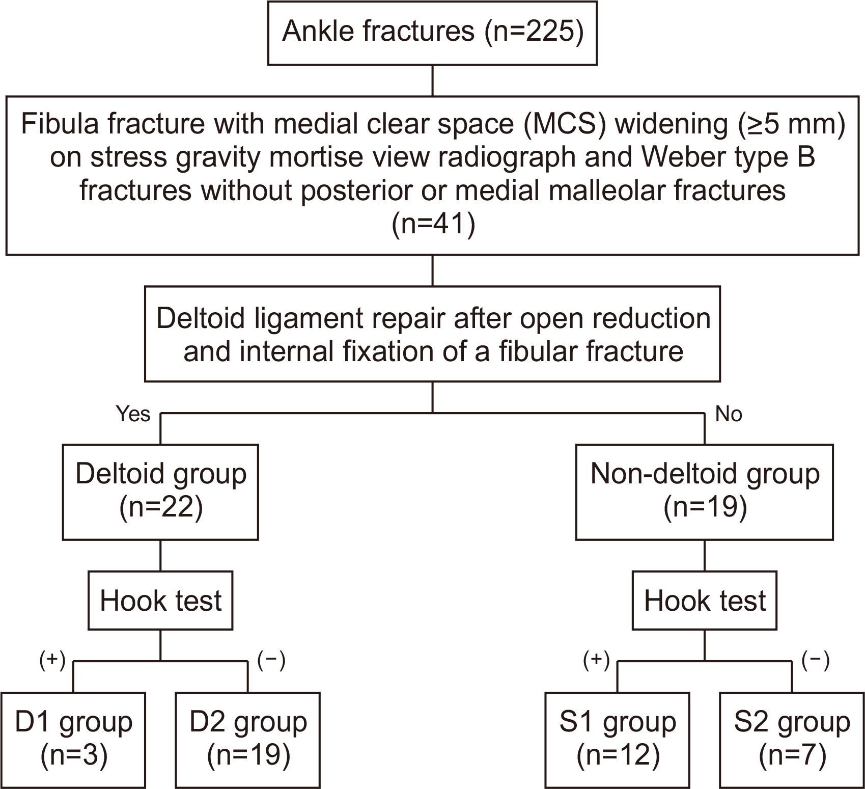
A total of 225 operatively treated ankle fractures were identified during this period in our institute; 41 (41 ankles) of them were enrolled in this study based on the inclusion and exclusion criteria (Fig. 1). The inclusion criteria were: patients 1) with fibular fracture with medial clear space (MCS) widening (≥5 mm) on gravity stress mortise view radiographs; 2) with Weber type B fractures; 3) with fractures that occurred within 2 weeks of presentation; 4) ≥18 years, and 5) who had at least undergone 12 months of follow-up. The exclusion criteria were: patients with 1) posterior or medial malleolar fractures; 2) previous ankle fractures or abnormalities; 3) open fractures; 4) neurovascular injury; 5) pathologic fractures; and 6) connective tissue diseases, such as Marfan syndrome or Ehlers-Danlos syndrome.
All patients first underwent open reduction and internal fixation of the fibular fracture. They were divided into two groups: those who underwent deltoid ligament repair (deltoid group) and those who did not (non-deltoid group). The former group consisted of 22 patients (15 men and seven women) with a mean age of 39.7 years (range 22∼63 years), who were further divided into two subgroups: those who underwent syndesmotic screw fixation (D1 group) and those who did not (D2 group). The D1/D2 groups consisted of 3/19 patients (three men and 0 women/12 men and seven women) with a mean age of 32/40.9 years (range 22∼46/24∼63 years). The non-deltoid group consisted of 19 patients (13 men and six women) with a mean age of 40.2 years (range 21∼63 years), and was also further divided into two subgroups: those who underwent syndesmotic screw fixation (S1 group) and those who did not (S2 group). The S1/S2 group consisted of 12/7 patients (eight men and four women/five men and two women) with a mean age of 39.8/40.9 years (range 21∼60/25∼63 years) (Fig. 2).
Preoperative anteroposterior (AP), lateral, and gravity stress mortise view radiographs were obtained routinely.8,9) In this study, ruptured deltoid and syndesmotic ligaments were considered if the MCS was greater than 5 mm on gravity stress mortise view radiographs. When the talus was not centralized after a fibular open reduction and internal fixation, the hook test was performed as an intraoperative stress test. The hook test was considered positive if the lateral shift of the fibula was greater than 2 mm.10) Syndesmotic fixation was indicated if 1) the talus was not centered on AP and mortise views and 2) if the hook test was positive.
MCS on the mortise view, the anterior fibular line (AFL) ratio (Fig. 3A), and posterior fibular line (PFL) distance (Fig. 3B) were measured at the final follow-up examination. Lateral clear space were measured by tibiofibular clear space (TFCS) on the AP view, pre- and postoperatively (Fig. 3C). The AFL ratio and PFL distance were measured to assess syndesmotic reduction.11) AFL, PFL, and inferior tibial line (ITL) are lines drawn on the flat anterior border of the fibula, on the flat posterior border of the fibula, and from the anterior to the posterior lip of the distal tibia on the true lateral view of the ankle, respectively. The AFL ratio was calculated from the intersection between the AFL and ITL to the entire ITL length. PFL distance was measured from the most posterior ITL point to the intersection between PFL and ITL. A positive PFL distance value is defined as anterior fibular displacement, whereas a negative value is defined as posterior fibular displacement. All radiographs were retrospectively reviewed and documented for data analysis, being blinded to the clinical information. To assess the reproducibility of the findings, an author (DWK) who had not been involved in the initial treatment of patients examined all radiographs twice with a between-measurement interval of 2 weeks. The values were averaged and statistically analyzed. All radiographs were digitally obtained through the Picture Archiving Communication System (Marosis Enterprise PACS; Infinitt, Seoul, Korea). The 10-point visual analog scale (VAS), American Orthopaedic Foot and Ankle Society (AOFAS) ankle-hindfoot score,12) and the Foot Function Index (FFI)13) were used to evaluate the postoperative clinical outcomes at the final follow-up examination (DWK and HJC). The 100-point AOFAS scoring system evaluated subjective and objective parameters consisting of pain (40 points), function (45 points), and alignment (15 points). The 230-point FFI consists of pain (90 points), disability (90 points), and activity limitation (50 points).
All statistical analyses were performed using SPSS Statistics version 25.0 (IBM Corp, Armonk, NY, USA). Statistical analyses of differences among the groups were performed using the Mann–Whitney test. The Kruskal–Wallis and Mann–Whitney test were applied to compare variables among the subgroups. A p-value of ≤0.05 was considered statistically significant.
All operative procedures were performed by the corresponding author (HJC). A thigh pneumatic tourniquet was inflated to 300 mmHg under spinal or general anesthesia. Patients were placed in the supine position, and fibular fractures were first treated with open reduction and internal fixation with a 2.8-mm lag screw; thereafter, a neutralization plate (LCP; DePuy Synthes, Oberdorf, Switzerland) was placed in all patients.
Twenty-two ankles treated with deltoid ligament repair (deltoid group). In this group, if the talus remained uncentered after the anatomical reduction and appropriate fixation of the fibula, an anterior curved incision was made on the medial aspect of the ankle. After confirming and extracting the ruptured deltoid ligament and anteromedial joint capsule in the medial gutter of the ankle, a bleeding cancellous surface was prepared at the medial malleolus using a rongeur. Two 1.4-mm soft anchors (JuggerKnot, Zimmer Biomet, Warsaw, IN, USA) were placed at the medial malleolus, and the full thickness of the deep deltoid ligament was advanced to the prepared bleeding surface and repaired with the horizontal mattress method. The superficial deltoid complex was directly sutured. After stabilizing both columns, syndesmotic stability was assessed using the lateral hook test. Based on these indications for syndesmotic fixation, a 3.5-mm cortical screw was inserted between 2.5 and 3 cm above the tibial plafond with the screw purchasing three cortices. Instead of clamp, gentle manual compression was used for syndesmotic reduction during syndesmotic fixation.
In 19 ankles, if the talus remained uncentered after the anatomical reduction and fixation of the fibula, the lateral hook test was performed to assess syndesmotic stability. A 3.5-mm cortical screw was inserted in the same manner as previously indicated for syndesmotic fixation. When soft tissue impingement of the medial gutter was suspected, the impinged tissue was extracted through a small incision.
All patients followed the same postoperative rehabilitation schedule. They were kept non-weight bearing with a posterior splint for 4 weeks postoperatively. Thereafter, the splint was replaced with a walking boot brace to allow tolerable weight bearing at 4 weeks postoperatively. During the first 2 weeks postoperatively, home exercises with a gentle active range of motion were allowed, and physical therapy was started. Full weight bearing was permitted at 6 weeks postoperatively. In all cases, the syndesmotic screw was removed between 12 and 14 weeks postoperatively.
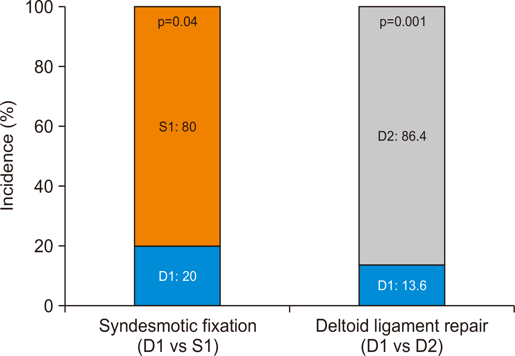
No significant differences in the demographics were found between the groups (Table 1). The incidence of syndesmotic fixation was significantly different between the D1 and S1 groups (p=0.04) and between the D1 and D2 groups (p=0.001) (Fig. 4). The postoperative MCS, TFCS, AFL ratio, and PFL distance were not significantly different between the groups (p=0.59, p=0.89, p=0.22, and p=0.08, respectively). The mean postoperative VAS, AOFAS score, and FFI were also not significantly different between the groups (p=0.21, p=0.83, and p=0.16, respectively) (Table 2).
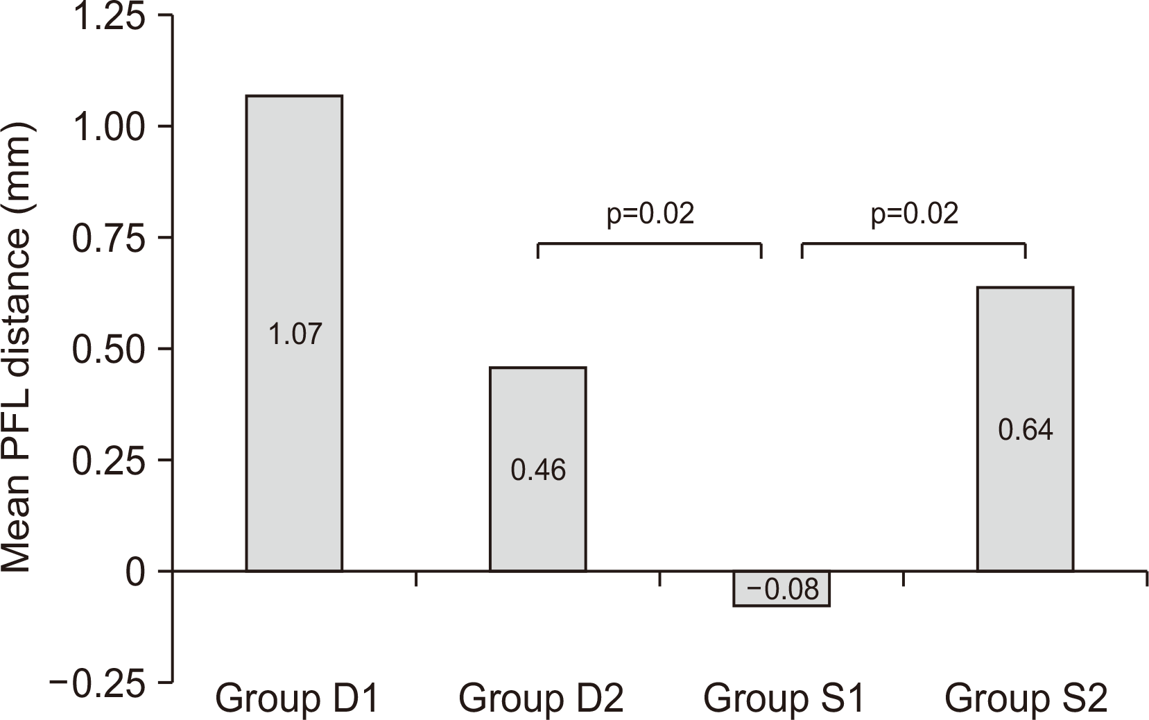
The postoperative MCS, TFCS, and AFL ratios were not significantly different between the subgroups (p=0.82, p=0.45, p=0.25, respectively). However, the postoperative PFL distance was 0.46 mm (range 0∼1.5 mm) in the D2 group, ―0.08 mm (range ―1.0∼1.0 mm) in the S1 group, and 0.64 mm (range 0∼1.5 mm) in the S2 group. The postoperative PFL distance was different among 4 subgroups (p=0.02, respectively). Each paired subgroup was analyzed by Mann–Whitney test (Table 3). The postoperative PFL distance was significantly different between the D2 and S1 groups and between the S1 and S2 groups (p=0.02, p=0.02, respectively) (Fig. 5). TFCS were significantly different between before and after the surgery in D2 and S1 groups (p=0.01, p=0.03, respectively) (Table 4). The mean postoperative VAS, AOFAS scores, and FFI were also not significantly different between the subgroups (p=0.44, p=0.40, and p=0.46, respectively) (Table 5). The number of patients who underwent syndesmotic fixation was significantly smaller in the D1 group (3/15)—those who underwent deltoid repair—compared to that in the S1 group (12/15)—those who did not undergo deltoid repair (p=0.04). Furthermore, when the deltoid ligament was repaired, the number of patients who required syndesmotic fixation (D1: 3/22) was also significantly smaller than those who did not (D2: 19/22) (p=0.001).
In this study, we aimed to evaluate the role of deltoid ligament repair on syndesmotic stabilization in patients with acute ankle fractures with ruptured deltoid and syndesmotic ligaments. We found that our results corroborated the hypothesis that deltoid ligament repair will restore ankle stability without having to manage the syndesmosis in Weber type B ankle fractures with rupture of the deltoid and syndesmotic ligaments.
Syndesmotic instability causes a lateral shift of the talus by inducing diastasis between the tibia and fibula. Due to the talar shift, the contact area decreases, and the peak pressure increases at the tibiotalar joint, which causes posttraumatic arthritis in the future.14) Therefore, syndesmotic fixation is performed during operative treatment of acute ankle fracture to restore the normal tibiotalar relationship by grasping the lateral shift of the talus. The deltoid ligament plays an important role in syndesmotic stability as a primary structure to prevent a lateral shift of the talus, even though the deltoid ligament is not one of the syndesmotic ligaments.15,16) Proper reconstruction of the medial and lateral columns of the ankle joint is important to prevent lateral talar subluxation during the operative treatment of ankle fractures, and this has been recognized for several decades.17) Recently, Feller et al.18) again emphasized the importance of the deltoid ligament in syndesmosis by confirming tibiofibular diastasis using arthroscopy after an isolated deltoid ligament transection using cadavers. Burns et al.19) concluded in their biomechanical study that the tibiotalar contact area decreased and the tibiotalar peak pressure increased only when a ruptured deltoid ligament or unrepaired medial malleolar fracture was combined with a ruptured syndesmotic ligament; however, if both columns were appropriately stabilized, syndesmotic fixation would not be required because of the stable tibiotalar contact area. Recently, Massri-Pugin et al.20) and Massri-Pugin et al.15) reported that syndesmosis remained stable when the anterior inferior tibiofibular ligament and interosseous ligament (IOL) were transected without including the deltoid ligament when assessing the contribution of the deltoid ligament to syndesmotic stability.
In our study, the postoperative PFL distances between the D2 and S1 groups and between the S1 and S2 groups differed significantly. The mean PFL distance in the S1 group was ―0.08 mm, whereas the mean PFL distances in the D2 and S2 groups were 0.46 mm and 0.64 mm, respectively, which indicates that posterior fibular displacement occurred more frequently in the S1 group than in the D2 and S2 groups. Syndesmotic malreduction is a complication of syndesmotic fixation.11,21) Sagi et al.22) reported that approximately 28% of rotational malposition occurred after syndesmotic fixation. This complication may occur if the syndesmotic joint is over-compressed during syndesmotic reduction. However, because postoperative CT scans were not performed and syndesmotic fixation was not assessed using arthroscopy, the results of this study do not suggest that the S1 group has an apparent malreduction state. Although clamps were not used to reduce the syndesmosis to avoid over-compression, a trend of posterior displacement was observed in the S1 group compared to that in other subgroups. However, no significant differences were detected among the subgroups on clinical evaluation or through other radiographic parameters.
In this study, although no significant differences were noted between the clinical and radiologic results of the D1 and D2 groups, the authors considered the patients in the D1 group to be those that underwent unnecessary syndesmotic fixation. Nonetheless, the reason why three of the 22 patients in the deltoid group underwent syndesmotic fixation might be a subjective error due to the excessive concern about syndesmotic issues. The intraoperative stress test may have been influenced by the surgeons’ preferences on the amount of force applied to the fibula without using a force gauge or the actual measurement of displacement with an MCS of ≥5 mm without using a ruler in the general operating room.23) We believe that excessive emphasis on the syndesmosis might cause subjective errors when assessing syndesmotic stability; however, this cannot be concluded decisively because of the limited clinical data in this study.
The numbers of patients in the S1 and S2 groups were not significantly different (p=0.36); however, seven of the 19 patients in the non-deltoid group did not require syndesmotic fixation, although deltoid ligament repair was not performed in advance (S2 group). This means that good fibular reduction and fixation will result in talar centralization in Weber type B fractures. However, the degree of rupture in syndesmotic ligaments and the state of the IOL were not assessed in the S1 and S2 groups because only plain radiographs were used for preoperative radiographic evaluation in this study. Even if magnetic resonance imaging (MRI) was obtained preoperatively, the detailed status of the number of fibers with ruptured syndesmotic ligaments could hardly be determined. Additionally, because ruptured syndesmotic ligaments detected on MRI do not indicate syndesmotic instability,24) plain radiographs were used as a standard approach in this study. Therefore, it remains unclear whether syndesmotic fixation in the S1 group is unnecessary, as in the D1 group, or necessary, in contrast to the D1 group. However, considering the results of Massri-Pugin et al.’s study,15) the S1 and S2 groups might differ owing to the degree of syndesmotic ligament rupture from different injury mechanisms. This is a retrospective clinical study; therefore, biomechanical studies should be conducted to identify the different degrees of syndesmotic ligament rupture based on the injury mechanism.
Lateral side widening of syndesmosis structure increases TFCS. In this study, TFCS were significantly decreased in the D2 and S1 group (p=0.01, p=0.03). This implies that not only syndesmotic fixation, also deltoid ligament repair may restore syndesmosis widening. Although tibiofibular overlap (TFO) also suggests syndesmosis widening, TFO were hard to evaluate due to interference of fibula lag screw position in some cases.
This study has several limitations. First, it is a retrospective study with a small number of cases. A power analysis using a post-hoc calculation to prove the conclusion that there were significant differences between the groups revealed that 67 patients in both groups needed to have 80.0% of power with 95.0% of a two-sided confidence interval. This study excluded patients with medial and posterior malleolar fractures to highlight the importance of the deltoid ligament. Although this study did not have sufficient statistical power because of the small sample size, we recommend that the timing of syndesmotic stability evaluation should be considered so that deltoid ligament repair can be performed first during the operative treatment of patients with acute ankle fractures with MCS widening on plain radiographs. However, future research regarding operative treatment strategies based on various injury mechanisms should be conducted with a biomechanical or a prospective study with a larger number of cases to yield more meaningful results. Second, only plain radiographs were used for evaluation. Recently, preoperative MRI has been performed to establish a treatment plan, and efforts have been made to evaluate the syndesmotic stability using arthroscopy during operative treatment.21) However, performing MRI for patients with acute fractures remains controversial because it cannot be used to assess dynamic syndesmotic instability.24) Arthroscopy is technically demanding, and not all physicians can perform an arthroscopic operation for ankle fractures; moreover, it is sometimes limited by the patient’s condition, such as significant swelling associated with acute fractures. On the contrary, because plain radiographs have been the traditional diagnostic method for fractures, they were used as a basic tool for fracture evaluation and decision-making. Therefore, we believe that plain radiographs remain a universally valid method as long as there are no clinical problems during the preparation of the treatment plan and there are no postoperative clinical problems during follow-up. MCS measurement to evaluate the deltoid and syndesmotic ligaments has sometimes been reported to have a high false-positive rate;25) however, MCS widening has been reported to be usually correlated with ruptured syndesmotic and deep deltoid ligaments and can be easily measured clinically, compared to TFCS and TFO.26) Although the reference value of MCS widening was ≥5 mm in this study, we believe that the physicians’ immediate and unified recognition of MCS widening is more reliable when evaluating the plain radiograph. Plain radiographs were used as the basic tool during the postoperative evaluation in this study because CT scans were only performed when the patient reported discomfort or when abnormalities were found on plain radiographs during the postoperative follow-up.
Deltoid ligament repair may restore ankle stability without having to address the syndesmosis in Weber type B ankle fractures with rupture of both the deltoid and syndesmotic ligaments. Therefore, deltoid ligament repair could be an effective option for syndesmosis stabilization in Weber type B ankle fracture with ruptured deltoid ligaments.
REFERENCES
1. Jeong MS, Choi YS, Kim YJ, Kim JS, Young KW, Jung YY. 2014; Deltoid ligament in acute ankle injury: MR imaging analysis. Skeletal Radiol. 43:655–63. doi: 10.1007/s00256-014-1842-5. DOI: 10.1007/s00256-014-1842-5. PMID: 24599341.

2. Tornetta P 3rd, Axelrad TW, Sibai TA, Creevy WR. 2012; Treatment of the stress positive ligamentous SE4 ankle fracture: incidence of syndesmotic injury and clinical decision making. J Orthop Trauma. 26:659–61. doi: 10.1097/BOT.0b013e31825cf39c. DOI: 10.1097/BOT.0b013e31825cf39c. PMID: 23100079.
3. Brosky T, Nyland J, Nitz A, Caborn DN. 1995; The ankle ligaments: consideration of syndesmotic injury and implications for rehabilitation. J Orthop Sports Phys Ther. 21:197–205. doi: 10.2519/jospt.1995.21.4.197. DOI: 10.2519/jospt.1995.21.4.197. PMID: 7773271.

4. Chissell HR, Jones J. 1995; The influence of a diastasis screw on the outcome of Weber type-C ankle fractures. J Bone Joint Surg Br. 77:435–8. doi: 10.1302/0301-620X.77B3.7744931. DOI: 10.1302/0301-620X.77B3.7744931.

5. Kennedy JG, Soffe KE, Dalla Vedova P, Stephens MM, O'Brien T, Walsh MG, et al. 2000; Evaluation of the syndesmotic screw in low Weber C ankle fractures. J Orthop Trauma. 14:359–66. doi: 10.1097/00005131-200006000-00010. DOI: 10.1097/00005131-200006000-00010. PMID: 10926245.

6. Jones CR, Nunley JA 2nd. 2015; Deltoid ligament repair versus syndesmotic fixation in bimalleolar equivalent ankle fractures. J Orthop Trauma. 29:245–9. doi: 10.1097/BOT.0000000000000220. DOI: 10.1097/BOT.0000000000000220. PMID: 25186845.

7. Woo SH, Bae SY, Chung HJ. 2018; Short-term results of a ruptured deltoid ligament repair during an acute ankle fracture fixation. Foot Ankle Int. 39:35–45. doi: 10.1177/1071100717732383. DOI: 10.1177/1071100717732383. PMID: 29078057.

8. Michelson JD, Varner KE, Checcone M. Diagnosing deltoid injury in ankle fractures: the gravity stress view. Clin Orthop Relat Res. 2001; (387):178–82. doi: 10.1097/00003086-200106000-00024. DOI: 10.1097/00003086-200106000-00024. PMID: 11400880.
9. Schock HJ, Pinzur M, Manion L, Stover M. 2007; The use of gravity or manual-stress radiographs in the assessment of supination-external rotation fractures of the ankle. J Bone Joint Surg Br. 89:1055–9. doi: 10.1302/0301-620X.89B8.19134. DOI: 10.1302/0301-620X.89B8.19134. PMID: 17785745.

10. Cotton FJ. 1924; Dislocations and joint-fractures. Ann Surg. 80:286. DOI: 10.1097/00000658-192408000-00013.

11. Loizou CL, Sudlow A, Collins R, Loveday D, Smith G. 2017; Radiological assessment of ankle syndesmotic reduction. Foot (Edinb). 32:39–43. doi: 10.1016/j.foot.2017.05.002. DOI: 10.1016/j.foot.2017.05.002. PMID: 28675813.

12. Kitaoka HB, Alexander IJ, Adelaar RS, Nunley JA, Myerson MS, Sanders M, et al. 1997; Clinical rating systems for the ankle-hindfoot, midfoot, hallux, and lesser toes. Foot Ankle Int. 18:187–8. doi: 10.1177/107110079701800315. DOI: 10.1177/107110079701800315. PMID: 28799420.

13. Budiman-Mak E, Conrad KJ, Roach KE. 1991; The Foot Function Index: a measure of foot pain and disability. J Clin Epidemiol. 44:561–70. doi: 10.1016/0895-4356(91)90220-4. DOI: 10.1016/0895-4356(91)90220-4. PMID: 2037861.

14. Earll M, Wayne J, Brodrick C, Vokshoor A, Adelaar R. 1996; Contribution of the deltoid ligament to ankle joint contact characteristics: a cadaver study. Foot Ankle Int. 17:317–24. doi: 10.1177/107110079601700604. DOI: 10.1177/107110079601700604. PMID: 8791077.

15. Massri-Pugin J, Lubberts B, Vopat BG, Wolf JC, DiGiovanni CW, Guss D. 2018; Role of the deltoid ligament in syndesmotic instability. Foot Ankle Int. 39:598–603. doi: 10.1177/1071100717750577. DOI: 10.1177/1071100717750577. PMID: 29320936.

16. Watanabe K, Kitaoka HB, Berglund LJ, Zhao KD, Kaufman KR, An KN. 2012; The role of ankle ligaments and articular geometry in stabilizing the ankle. Clin Biomech (Bristol, Avon). 27:189–95. doi: 10.1016/j.clinbiomech.2011.08.015. DOI: 10.1016/j.clinbiomech.2011.08.015. PMID: 22000065.

17. Klossner O. Late results of operative and non-operative treatment of severe ankle fractures. A clinical study. Acta Chir Scand Suppl. 1962; Suppl 293:1–93.
18. Feller R, Borenstein T, Fantry AJ, Kellum RB, Machan JT, Nickisch F, et al. 2017; Arthroscopic quantification of syndesmotic instability in a cadaveric model. Arthroscopy. 33:436–44. doi: 10.1016/j.arthro.2016.11.008. DOI: 10.1016/j.arthro.2016.11.008. PMID: 28160934.

19. Burns WC 2nd, Prakash K, Adelaar R, Beaudoin A, Krause W. 1993; Tibiotalar joint dynamics: indications for the syndesmotic screw--a cadaver study. Foot Ankle. 14:153–8. doi: 10.1177/107110079301400308. DOI: 10.1177/107110079301400308. PMID: 8491430.

20. Massri-Pugin J, Lubberts B, Vopat BG, Guss D, Hosseini A, DiGiovanni CW. 2017; Effect of sequential sectioning of ligaments on syndesmotic instability in the coronal plane evaluated arthroscopically. Foot Ankle Int. 38:1387–93. doi: 10.1177/1071100717729492. DOI: 10.1177/1071100717729492. PMID: 28884593.

21. Chan KB, Lui TH. 2016; Role of ankle arthroscopy in management of acute ankle fracture. Arthroscopy. 32:2373–80. doi: 10.1016/j.arthro.2016.08.016. DOI: 10.1016/j.arthro.2016.08.016. PMID: 27816101.

22. Sagi HC, Shah AR, Sanders RW. 2012; The functional consequence of syndesmotic joint malreduction at a minimum 2-year follow-up. J Orthop Trauma. 26:439–43. doi: 10.1097/BOT.0b013e31822a526a. DOI: 10.1097/BOT.0b013e31822a526a. PMID: 22357084.

23. Candal-Couto JJ, Burrow D, Bromage S, Briggs PJ. 2004; Instability of the tibio-fibular syndesmosis: have we been pulling in the wrong direction? Injury. 35:814–8. doi: 10.1016/j.injury.2003.10.013. DOI: 10.1016/j.injury.2003.10.013. PMID: 15246807.

24. Hermans JJ, Wentink N, Beumer A, Hop WC, Heijboer MP, Moonen AF, et al. 2012; Correlation between radiological assessment of acute ankle fractures and syndesmotic injury on MRI. Skeletal Radiol. 41:787–801. doi: 10.1007/s00256-011-1284-2. DOI: 10.1007/s00256-011-1284-2. PMID: 22012479. PMCID: PMC3368108.

25. Schuberth JM, Collman DR, Rush SM, Ford LA. 2004; Deltoid ligament integrity in lateral malleolar fractures: a comparative analysis of arthroscopic and radiographic assessments. J Foot Ankle Surg. 43:20–9. doi: 10.1053/j.jfas.2003.11.005. DOI: 10.1053/j.jfas.2003.11.005. PMID: 14752760.

26. Cheung Y, Perrich KD, Gui J, Koval KJ, Goodwin DW. 2009; MRI of isolated distal fibular fractures with widened medial clear space on stressed radiographs: which ligaments are interrupted? AJR Am J Roentgenol. 192:W7–12. doi: 10.2214/AJR.08.1092. DOI: 10.2214/AJR.08.1092. PMID: 19098171. PMCID: PMC5585779.

27. Gardner MJ, Demetrakopoulos D, Briggs SM, Helfet DL, Lorich DG. 2006; Malreduction of the tibiofibular syndesmosis in ankle fractures. Foot Ankle Int. 27:788–92. doi: 10.1177/107110070602701005. DOI: 10.1177/107110070602701005. PMID: 17054878.

28. Song DJ, Lanzi JT, Groth AT, Drake M, Orchowski JR, Shaha SH, et al. 2014; The effect of syndesmosis screw removal on the reduction of the distal tibiofibular joint: a prospective radiographic study. Foot Ankle Int. 35:543–8. doi: 10.1177/1071100714524552. DOI: 10.1177/1071100714524552. PMID: 24532699.
29. Manjoo A, Sanders DW, Tieszer C, MacLeod MD. 2010; Functional and radiographic results of patients with syndesmotic screw fixation: implications for screw removal. J Orthop Trauma. 24:2–6. doi: 10.1097/BOT.0b013e3181a9f7a5. DOI: 10.1097/BOT.0b013e3181a9f7a5. PMID: 20035170.

Figure 1
Preoperative radiograph of a Weber type B ankle fracture in a patient with suspected ruptured deltoid and syndesmotic ligaments.
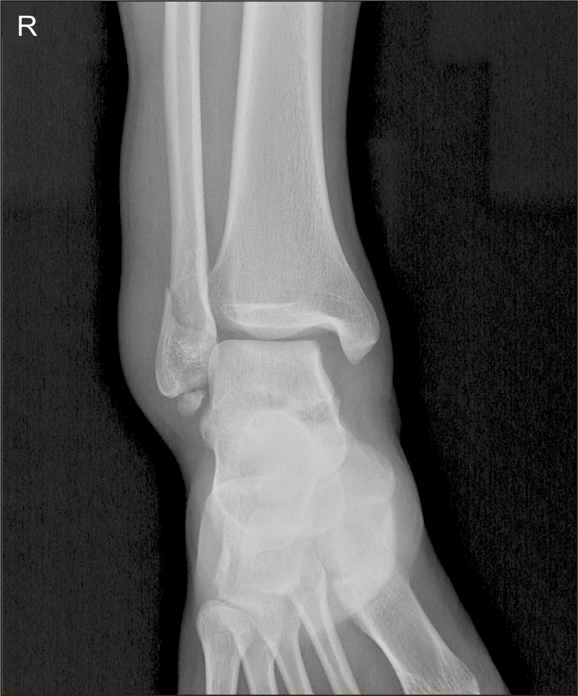
Figure 3
(A) The anterior fibular line (AFL) ratio was calculated from the intersection between AFL and inferior tibial line (ITL) to the entire ITL length. (B) The posterior fibular line (PFL) distance was measured from the most posterior ITL point to the intersection point between PFL and ITL. (C) The tibiofibular clear space (TFCS) was measured from the distance between the medial border of the fibula and the lateral border of the posterior tibia as it extends into the incisura fibularis at 10 mm above the tibial plafond.
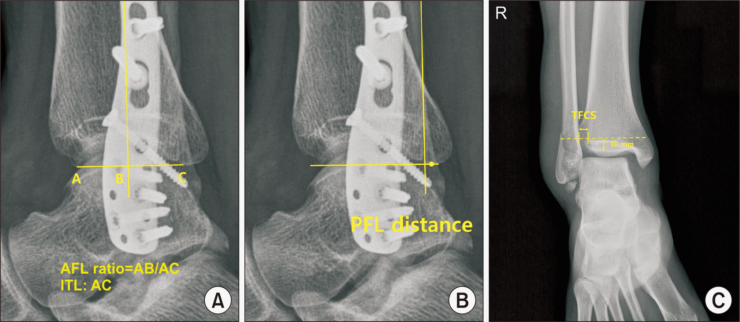
Table 1
Patient Demographics (41 Ankles in 41 Patients)
Table 2
Results of Radiographic and Clinical Evaluations between the Groups (41 Ankles in 41 Patients)
Table 3
Post Hoc Analysis of PFL Distance between Subgroups
| Postoperative PFL distance | D1 vs. D2 | D1 vs. S1 | D1 vs. S2 | D2 vs. S1 | D2 vs. S2 | S1 vs. S2 |
|---|---|---|---|---|---|---|
| p-value | 0.15 | 0.07 | 0.36 | 0.02 | 0.21 | 0.02 |
Table 4
Pre- and Postoperative TFCS in the Subgroups (41 Ankles in 41 Patients)
Table 5
Results of Radiographic and Clinical Evaluations between the Subgroups (41 Ankles in 41 Patients)


 PDF
PDF Citation
 Print
Print


 XML Download