

 PDF
PDF Citation
 Print
Print


Introduction
Methods
Patient selection
Imaging protocol
Endovascular reperfusion procedure
Image analyses
Statistical analysis
Results

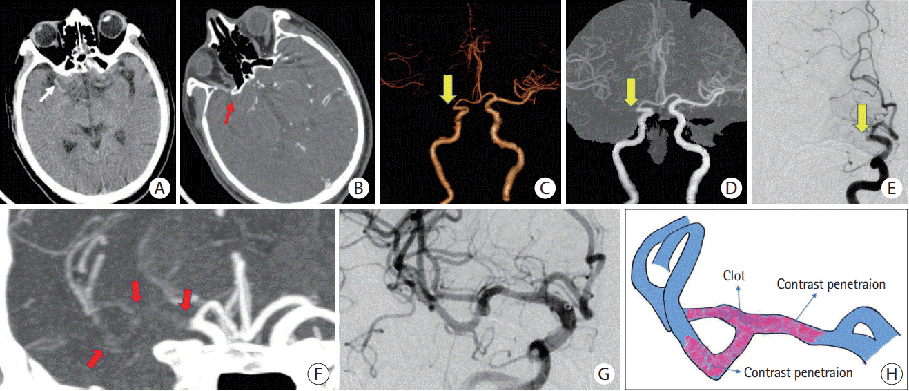 | Figure 2.Embolism-related LVO with a positive TES. (A) NCCT reveals HAS in the M1 segment of the right MCA (white arrow). (B) Original axial slice of CTA indicates a thrombus filling defect at the M1 segment of the right MCA (red arrow). (C and D) Volume rendering and full-slab MIP image reconstruction of CTA showing the occluded site (yellow arrow). (E) Anteroposterior angiogram shows the occlusion site at the M1 segment of the right MCA (yellow arrow). (F) Thin slab MIP image reconstruction of CTA shows the TES (red arrows) at right MCA. (G) Complete recanalization of the right MCA without residual stenosis after a single attempt of stent retrieval. (H) Schematic diagram of the pathogenesis of embolism-related LVO. LVO, large vessel occlusion; TES, thrombus enhancement sign; NCCT, non-contrast computed tomography; HAS, hyperdense artery sign; MCA, middle cerebral artery; CTA, computed tomography angiography; MIP, maximum intensity projection.
|
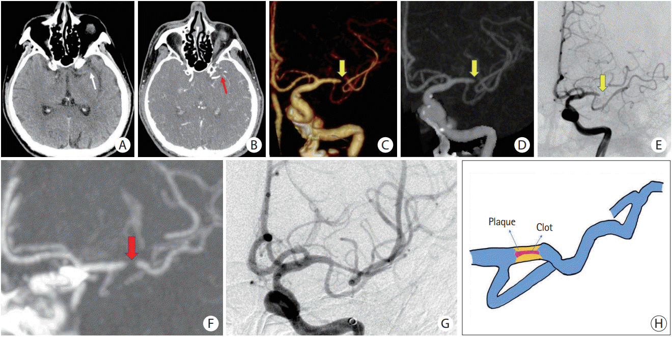 | Figure 3.ICAS-related LVO with a negative TES. (A) NCCT does not reveal HAS in the M1 segment of the left MCA (white arrow). (B) Original axial slice of CTA indicates thrombus filling defect at the M1 segment of the left MCA (red arrow). (C and D) Volume rendering and full-slab MIP image reconstruction of CTA showing the occluded site (yellow arrow). (E) Anteroposterior angiogram shows occlusion site at the M1 segment of the left MCA (yellow arrow). (F) Thin slab MIP image reconstruction of CTA shows a negative TES (red arrow) at the left MCA. (G) After attempting stent retrieval, rescued angioplasty and stenting was applied. Residual stenosis remains, but distal flow significantly improves. (H) Schematic diagram of the pathogenesis of ICAS-related LVO. ICAS, intracranial atherosclerotic stenosis; LVO, large vessel occlusion; TES, thrombus enhancement sign; NCCT, non-contrast computed tomography; HAS, hyperdense artery sign; MCA, middle cerebral artery; CTA, computed tomography angiography; MIP, maximum intensity projection.
|
Table 1.
| Characteristic | Positive TES (n=205) | Negative TES (n=83) | P |
|---|---|---|---|
| Age (yr) | 71.9±11.7 | 71.0±11.4 | 0.520 |
| Male sex | 121 (59.0) | 43 (51.8) | 0.294 |
| Baseline NIHSS | 16.5±5.3 | 15.3±5.0 | 0.074 |
| Baseline ASPECTS | 9.6±0.8 | 9.7±0.6 | 0.348 |
| Risk factors | |||
| Diabetes mellitus | 54 (26.3) | 18 (21.7) | 0.455 |
| Hypertension | 119 (58.0) | 52 (62.7) | 0.509 |
| Atrial fibrillation | 138 (67.3) | 28 (33.7) | <0.001* |
| Coronary artery disease | 65 (31.7) | 18 (21.7) | 0.114 |
| Dyslipidemia | 39 (19.0) | 19 (22.9) | 0.517 |
| Current smoking | 37 (18.0) | 12 (14.5) | 0.495 |
| Occlusion sites | 0.123 | ||
| MCA | 136 (66.3) | 63 (75.9) | |
| ICA | 69 (33.7) | 20 (24.1) | |
| CBS | 6.6±1.1 | 6.2±1.2 | 0.007 |
| IVT | 86 (42.0) | 31 (37.3) | 0.509 |
| Onset to puncture (min) | 276.0±155.5 | 253.6±106.8 | 0.230 |
| Puncture to recanalization (min) | 55.6±26.3 | 52.0±23.8 | 0.278 |
| Procedure time (min) | 70.1±27.4 | 93.6±31.3 | <0.001* |
| Stroke subtypes | <0.001* | ||
| Embo-LVO | 197 (96.1) | 38 (45.8) | |
| ICAS-LVO | 8 (3.9) | 45 (54.2) | |
| 2b-3 Final mTICI achieved | 160 (78.0) | 67 (80.7) | 0.750 |
| mRS ≤2 at 90 days | 70 (34.1) | 35 (42.2) | 0.225 |
| sICH | 20 (9.8) | 4 (4.8) | 0.239 |
Values are presented as mean±standard deviation or number (%).
TES, thrombus enhancement sign; NIHSS, National Institutes of Health Stroke Scale; ASPECTS, Alberta Stroke Program Early CT Score; MCA, middle cerebral artery; ICA, internal carotid artery; CBS, clot burden score; IVT, intravenous thrombolysis; embo-LVO, embolism-related large vessel occlusion; ICAS-LVO, intracranial atherosclerotic stenosis-related large vessel occlusion; mTICI, modified Thrombolysis in Cerebral Infarction; mRS, modified Rankin Scale; sICH, symptomatic intracerebral hemorrhage.
![]()
Table 2.
| Embo-LVO (n=235) | ICAS-LVO (n=53) | P | |
|---|---|---|---|
| Age (yr) | 71.3±11.7 | 73.1±11.2 | 0.331 |
| Male sex | 138 (58.7) | 26 (49.1) | 0.221 |
| Baseline NIHSS | 16.4±5.3 | 14.8±4.6 | 0.042* |
| Baseline ASPECTS | 9.6±0.8 | 9.7±0.4 | 0.373 |
| Risk factors | |||
| Diabetes mellitus | 61 (26.0) | 11 (20.8) | 0.486 |
| Hypertension | 135 (57.4) | 36 (67.9) | 0.168 |
| Atrial fibrillation | 157 (66.8) | 9 (17.0) | <0.001* |
| Coronary artery disease | 72 (30.6) | 11 (20.8) | 0.180 |
| Dyslipidemia | 43 (18.3) | 15 (28.3) | 0.128 |
| Current smoking | 41 (17.4) | 8 (15.1) | 0.840 |
| Occlusion sites | 0.073 | ||
| MCA | 161 (68.5) | 38 (71.7) | |
| ICA | 74 (31.5) | 15 (28.3) | |
| TES | 197 (83.8) | 8 (15.1) | <0.001* |
| CBS | 6.5±1.2 | 6.4±1.2 | 0.268 |
| IVT | 94 (40.0) | 23 (43.4) | 0.646 |
| Onset to puncture (min) | 271.8±150.1 | 259.7±109.2 | 0.582 |
| Puncture to recanalization (min) | 55.3±25.8 | 51.3±24.6 | 0.300 |
| Procedure time (min) | 68.1±25.6 | 115.4±17.3 | <0.001* |
| 2b-3 Final mTICI achieved | 189 (80.4) | 38 (71.7) | 0.192 |
| mRS ≤2 at 90 days | 88 (37.4) | 17 (32.1) | 0.529 |
| sICH | 22 (9.4) | 2 (3.8) | 0.271 |
Values are presented as mean±standard deviation or number (%).
embo-LVO, embolism-related large vessel occlusion; ICAS-LVO, intracranial atherosclerosis-related large vessel occlusion; NIHSS, National Institutes of Health Stroke Scale; ASPECTS, Alberta Stroke Program Early CT Score; MCA, middle cerebral artery; ICA, internal carotid artery; TES, thrombus enhancement sign; CBS, clot burden score; IVT, intravenous thrombolysis; mTICI, modified Thrombolysis in Cerebral Infarction; mRS, modified Rankin Scale; sICH, symptomatic intracerebral hemorrhage.
![]()
Table 3.
![]()
Table 4.
![]()
 XML Download