Journal List > Int J Stem Cells > v.16(2) > 1516082834

Park, Kim, Jang, and Cha: Transition Substitution of Desired Bases in Human Pluripotent Stem Cells with Base Editors: A Step-by-Step Guide

Abstract

The recent advances in human pluripotent stem cells (hPSCs) enable to precisely edit the desired bases in hPSCs to be used for the establishment of isogenic disease models and autologous ex vivo cell therapy. The knock-in approach based on the homologous directed repair with Cas9 endonuclease, causing DNA double-strand breaks (DSBs), produces not only insertion and deletion (indel) mutations but also deleterious large deletions. On the contrary, due to the lack of Cas9 endonuclease activity, base editors (BEs) such as adenine base editor (ABE) and cytosine base editor (CBE) allow precise base substitution by conjugated deaminase activity, free from DSB formation. Despite the limitation of BEs in transition substitution, precise base editing by BEs with no massive off-targets is suggested to be a prospective alternative in hPSCs for clinical applications. Considering the unique cellular characteristics of hPSCs, a few points should be considered. Herein, we describe an updated and optimized protocol for base editing in hPSCs. We also describe an improved methodology for CBE-based C to T substitutions, which are generally lower than A to G substitutions in hPSCs.

Introduction

Brief overview

Precise genome editing in human pluripotent stem cells (hPSCs) holds great potential for both isogenic disease modeling and autologous ex vivo stem cell therapy (1-3). Generally, induced pluripotent stem cells (iPSCs) with different genetic backgrounds have been used as controls to characterize iPSCs from patients harboring pathogenic mutations. Correcting pathogenic mutations allows using iPSCs from the same patients to precisely compare cellular characteristics to understand disease mechanisms (1) and autologous ex vivo stem cell therapy, thus avoiding immune rejection (4).
With the advancements in genome editing tool kits (5, 6), hPSCs have been subjected to newly developed editing tools such as base editors (BEs) (7) and prime editors (PEs) (7-9) as well as conventional knock-in with Cas9 (8, 10). Despite extensive efforts to improve the efficiency of knock-in with Cas9 in hPSCs (8, 10-12), in which endonuclease activity of Cas9 results in massive p53-dependent cell death due to DSBs induction (13), the frequent incidence of deleterious large deletions by endonuclease activity of Cas9 in hPSCs (14) has become a serious technical issue. Instead, BEs with nickase Cas9 (nCas9) or catalytically dead Cas9 (dCas9) are capable to induce base transitions by conjugated deaminase activity without creating DSBs (15, 16). Thus, BEs have been applied to establish isogenic hPSC models for disease modeling (7) and gene correction in patient-derived iPSCs (17), and to edit pathogenic transition mutations accounting for 30% of disease-associated human genetic variants (18). In this regard, optimization of BEs, newly developed base editing tools to extend the editing scope, becomes highly important for application in hPSCs due to higher editing of pathogenic point mutations (58%) (19), including transversion mutations than that of BEs.

Mechanisms of base editing

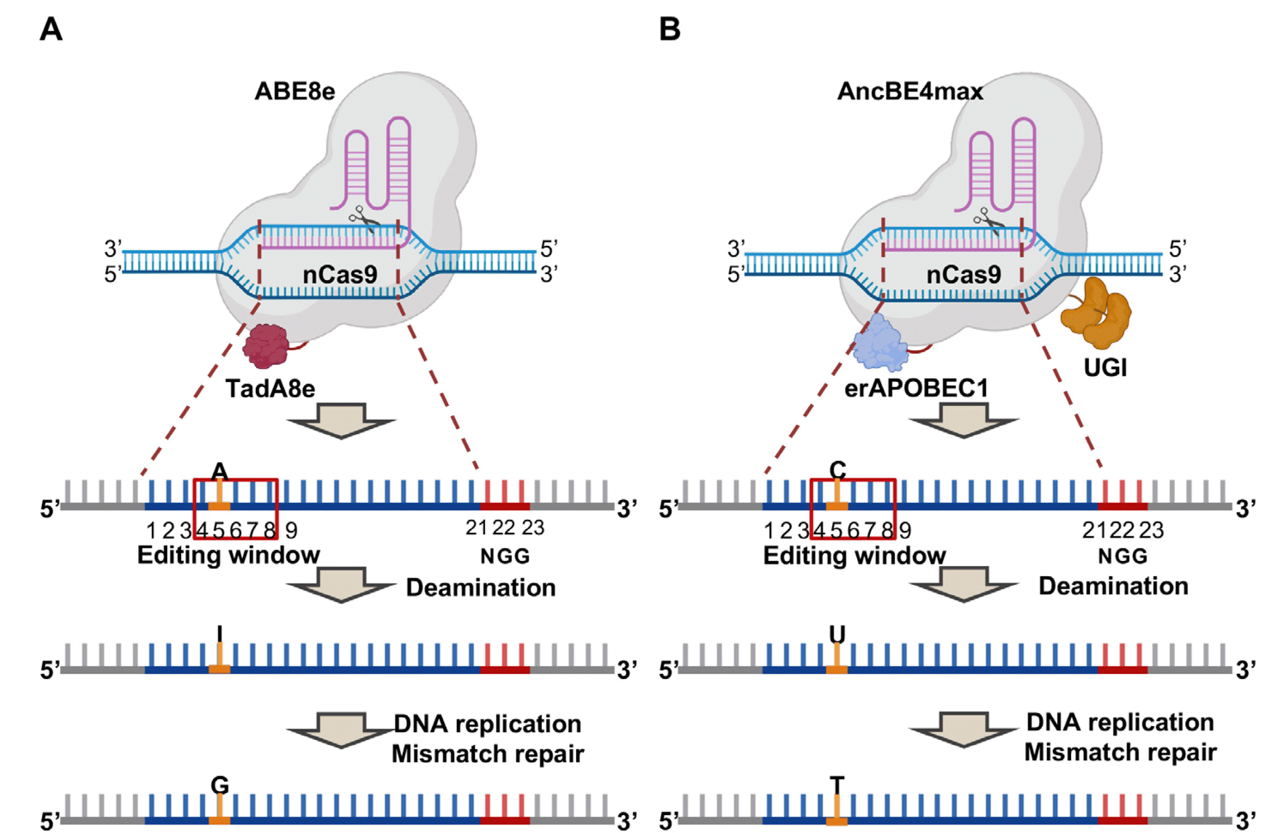
Diverse versions of BEs exist: this protocol is optimized with ABE8e for ABE (addgene# 138489) (20) and AncBE4max for CBE (addgene #112094) (21). ABE8e is composed by engineered TadA (TadA8e) adenine deaminase linked to nickase Cas9 (nCas9) (Fig. 1A). AncBE-4max consists of modified rat APOBEC1 (erAPOBEC1), via ancestral reconstruction, linked to nCas9 and uracil glycosylase inhibitors (UGI) (Fig. 1B). Most base editors including ABE8e and AncBE4max adopt nCas9 because its nickase activity makes single strand break (SSB) on the non-editing strand. This helps ensuring the repair of the non-editing strand, not that of the editing strand where the deaminated base is located. This repair is mediated by the endogenous mismatch repair (MMR) system, that recognizes a nick on the non-editing strand and activates to produce the desirable base editing. Instead, the editing strand is repaired by base excision repair machinery like Uracil DNA glycosylase (UNG), that recognize and recover the deaminated base.
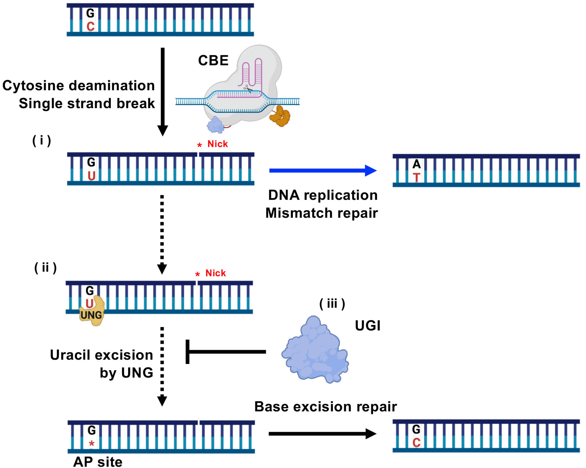
The deaminase in BEs catalyzes deamination within a small range of bases called editing window (15). This editing window is where the majority of deamination occurs and is different for each BE (19). For instance, the editing window of AncBE4max is positioned 4 to 8 nucleotides (nt) in the protospacer sequences (i.e., complementary cleaved target sequences, red box in Fig. 1) counting the protospacer adjacent motif (PAM) as positions 21∼23, that of ABE7.10 is 4 to 7 nt, (Fig. 1) (20). ABE8e induce A:T to G:C conversion by catalyze deamination on adenine (A), turning it into inosine (I) which is recognized as guanine (G) during DNA replication and mismatch repair (MMR) (Fig. 1A) (22). Similarly, AncBE4max turns cytosine (C) to uracil (U) by deaminase activity of APOBEC1 (Fig. 1B) (15). U in DNA is recognized as thymine (T) in DNA replication and MMR, therefore the deamination of C induces C:G to T:A conversion (Fig. 1B) (15). Since deamination of C occurs frequently in nature, DNA glycosylases such as uracil DNA glycosylase (UNG) efficiently recognize and remove the deaminated C (i.e., U) in DNA to maintain genome integrity (23). BE4 adopted additional UGIs compared to BE3 to inhibit UNG activity (Fig. 2), because cellular UNG activity, triggered by deamination of cytosine, impairs C:G to T:A mutation by CBE (24). Due to high expression of UNG in hPSCs, additional depletion of UNG with small interfering RNA improves the efficiency and product purity of AncBE4max in hPSCs (25).

Prerequisites for base editing

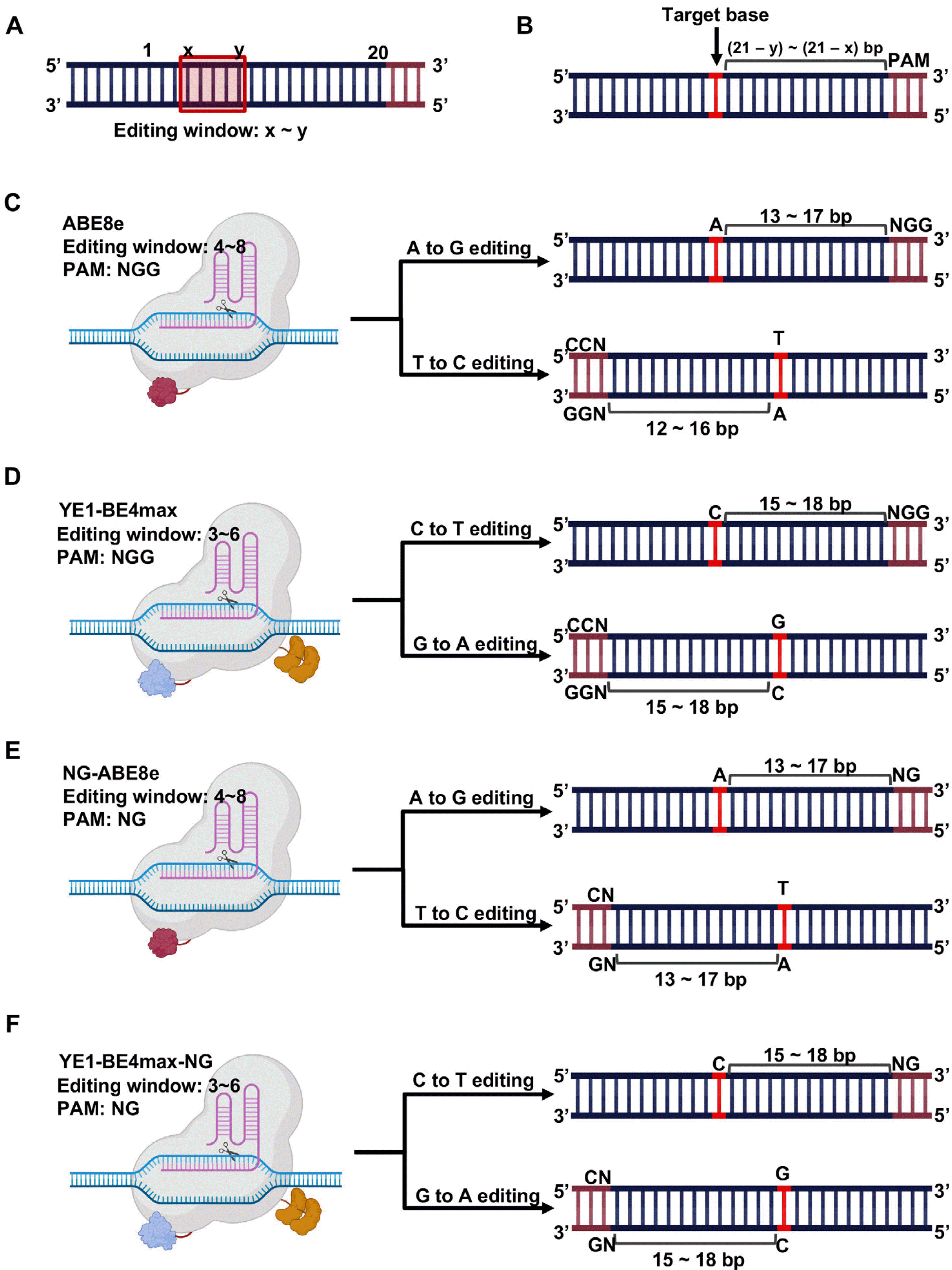
To apply BEs for precise genome editing, three main factors need to be considered: i) base editor for mutation type (i.e., ABE or CBE), ii) existence of protospacer adjacent motif (PAM), and iii) possible occurrence of bystander base editing. Among the twelve possible types of transition and transversion point mutations, ABE and CBE are able to edit only transition mutations. Thus, A to G (or T to C) base substitution is conducted by ABE and C to T (or G to A) conversion is achieved by CBE. The second prerequisite for application of BEs is the presence of the PAM sequence [i.e., 5’-NGG-3’ for BEs based on Cas9 from S. pyogenes (SpCas9)] at an appropriate distance from the target base. If the editing window ranges from x to y nt in the spacer sequence, the PAM sequence should be placed (21 – y) to (21 – x) nt away from the target base (Figs. 3A and 3B). For instance, as ABE8e, whose editing window is positioned 4∼8 nucleotides of protospacer sequences, targets A, the PAM sequence of ABE8e should be located 13 to 17 bp away from the target base (Fig. 3C) (20). Similarly, as the editing window of YE1-BE4max, a narrow editing window version of BE4max, is 3 to 6 nt, YE1-BE4max requires NGG at 15 to 18 bp distance from the target base (Fig. 3D) (26).
The requirement that the PAM sequence be located at a specific region for desired base substitutions largely limits the application of BEs. Due to this restriction, only around 15% (i.e., 9 out of 63 patients) and 13% (i.e., 16 out 121 patients) of pathogenic mutations associated with GNE myopathy (OMIM #605820) (7) and Tay-Sachs disease (OMIM#272800) (unpublished data) respectively are covered by ABE or CBE. The limited genome-targeting scope of conventional BE due to the PAM sequence is relieved by improved BEs based on engineered SpCas9 (i.e., SpCas9-NG), such as NG-ABE (27) and NG-CBE (28). By including a PAM-released version of BEs, the editing accessibility of point mutations is extended to 38% for GNE myopathy (7) and 24% for Tay-Sachs disease (unpublished data) respectively.
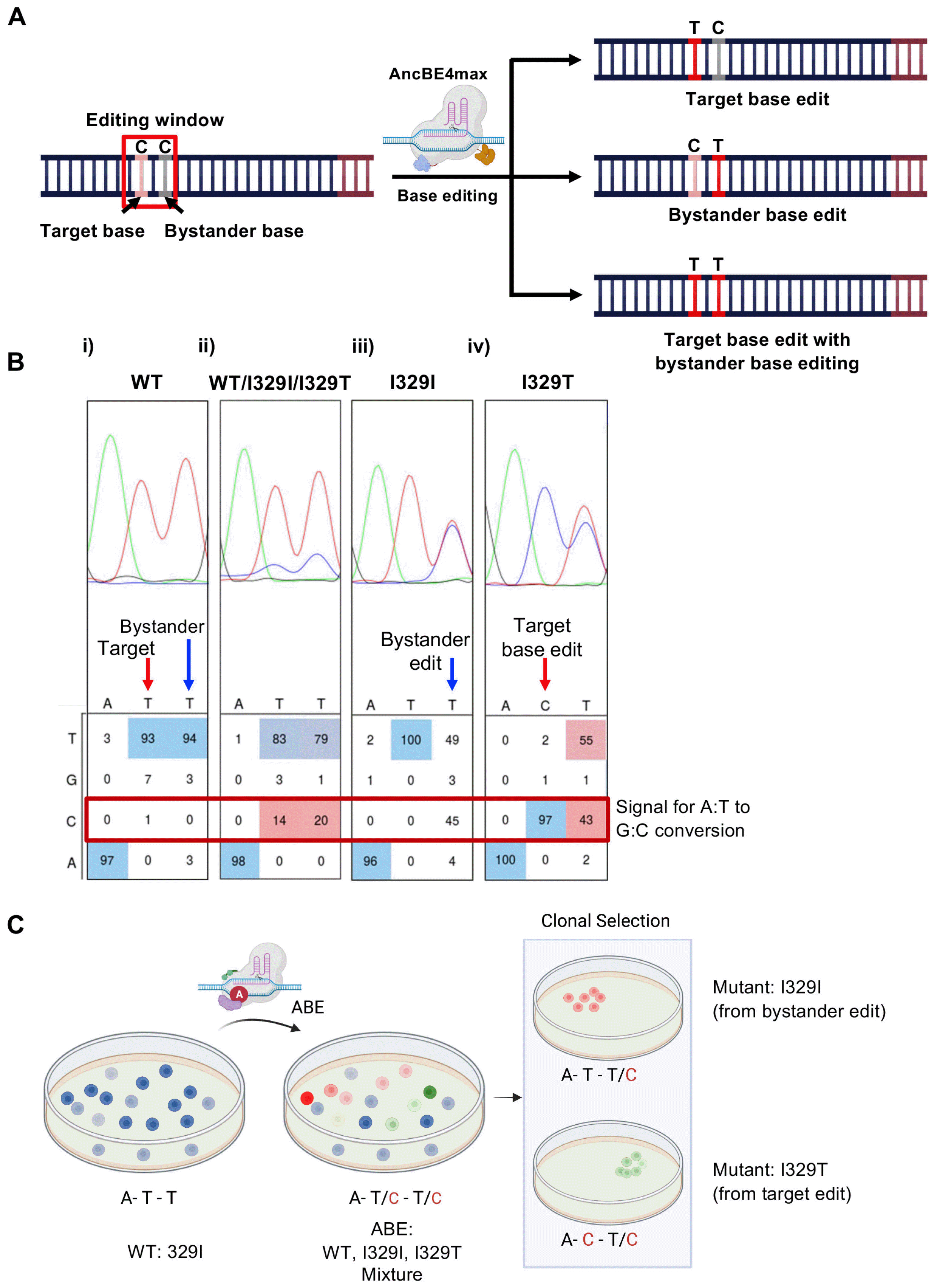
Bystander editing from unintended bases within the editing window is inevitably produced by BEs due to the capacity of deaminases to access any base in the target range. The possibility of bystander editing requires a laborious clonal selection to produce the desirable clone (Fig. 4A). For instance, while hESCs mutants harboring I329T mutation in GNE was produced by ABE (7), mixture of mutants including target (T in the middle to C) as well as bystander (T in the right to C) edit was observed (Fig. 4B). After clonal selection, I329I (from bystander edit) and I329T (from target edit) mutant clones were established (Fig. 4C). Targets without bystander bases are suitable for accurate base editing. Otherwise, narrow-editing window CBEs such as YE1-BE3 (29) and YE1-BE4max (25) can selectively edit a target C over a neighboring C, which are alternatives for BE3, or BE4 max to reduce bystander editing. Also, using a ABE7.10 rather than using ABE8e can reduce bystander editing with similar editing window, since ABE7.10 has lower processivity than ABE8e (20). Computational predictions such as those provided by DeepBaseEditor (30) are useful methods to choose accurate target by predicting editing outcome of BEs.

Materials and Methods

Single guide RNA (sgRNA) vector synthesis

Equipment

  • Alcohol Lamp.

  • Spreader.

  • 42℃ Heat block.

  • 36℃ shaking incubator.

  • 1L erlenmeyer flask.

  • Polymerase chain reaction (PCR) equipment (Agillent Tcehnologies, SureCycler 8800).

  • Micropipette.

    • - P10 (1∼10 μl).

    • - P20 (2∼20 μl).

    • - P200 (20∼200 μl).

    • - P1000 (200∼1000 μl).

  • 500 ml beaker.

  • Gel electrophoresis equipment (Takara, AD110).

  • Agarose gel tray (Takara, AD210).

  • 100 ml Erlenmeyer flask.

  • Microwave machine.

  • Autoclave.

  • 1.5 ml E-tube (Axygen, MCT-150-C).

  • 50 ml centrifuge tube (SPL, 50050).

  • 100 mm petri dish (SPL, 10090).

  • Micropipette tip P10 (Neptune, BT10XLS3).

  • Micropipette tip P20 (Neptune, BT20).

  • Micropipette tip P200 (Neptune, BT200).

  • Micropipette tip P1000 (Neptune, BT1250).

  • 10 ml serological pipette (Corning, 4101).

  • Latex glove (Ultratex, UF).

Reagents

  • 50x TAE buffer (Biosesang TR2002-100-00).

  • BsaI (Enzynomics, R0725).

  • T4 DNA ligase (Solgent, SDL01-R40k).

  • T4 PNK (Enzynomics, M0055).

  • 10x T4 DNA ligase buffer (Solgent, SDL01-R40k).

  • Taq DNA polymerase (Solgent, STD95-E500).

  • 10x Taq buffer (Solgent, STD22-B12h).

  • 10 mM dNTP (Solgent, SDN12-B10h).

  • Ultra pure water (Biosesang, WR4006-100-00).

  • CIP (BioLabs #M20905).

  • LB broth stick (LPS, LB-250).

  • LB agar broth powder (BioSesang LR3004-250-02).

  • Fragment DNA purification kit (iNtRON, 17290).

  • Agarose (Young Sciences, Y50004).

  • Red safe (iNtRON, 21141).

  • 100 bp DNA ladder (iNtRON, 24073).

  • 1,000 bp DNA ladder (iNtRON, 24074).

  • Ampicillin (BioSesang, AC1043-005-00).

  • 6x DNA buffer (iNtRON, 21162).

  • Xtra Midi EF Kit (NucleoBond, 740420.50).

  • 8-well Strip Plates (Thermo Fisher Scientific, 15031).

  • pRG2 vector (addgene, 104174).

  • DH5α E.coli (Real Biotech, RH617).

  • pRG2_Rvs primer (5’ gagtcagtgagcgaggaagc 3’).

  • 99% ethyl alcohol (DUKSAN, UN1170).

hPSCs culture, transfection and single cell line establishment

Equipment

  • Clean bench.

  • CO2 incubator (37℃, 5% CO2) (Thermo Scientific, 311).

  • Microscope.

  • Centrifuge machine.

  • Hemocytometer.

  • Micropipette.

    • - P10 (1∼10 μl).

    • - P20 (2∼20 μl).

    • - P200 (20∼200 μl).

    • - P1000 (200∼1000 μl).

  • NEPA21 electroporator (NEPAGENE, NPG-NEPA).

  • Cell freezing container (NALGENE, 5100-0001).

  • 1.5 ml E-tube (Axygen, MCT-150-C).

  • 60 mm cell culture dish (Corning, 353802).

  • 6 well cell culture dish (FALCON, 353046).

  • 24 well cell culture dish (FALCON, 353047).

  • Micropipette tip P10 (Neptune, BT10XLS3).

  • Micropipette tip P20 (Neptune, BT20).

  • Micropipette tip P200 (Neptune, BT200).

  • Micropipette tip P1000 (Neptune, BT1250).

  • 10 ml serological pipette (Corning, 4101).

  • 5 ml serological pipette (Corning, 4051).

  • Latex glove (Ultratex, UF).

  • Electroporation tube (NEPAGENE, EC-002S).

Reagents

  • StemFit04 complete media (AJBASIC04CT).

  • Y-27632 (Sigma, Y0503-5MG).

  • iMatrix (891-012, ATRIXOME).

  • Accutase (562527, BD).

  • Opti-MEM (11058021, gibco).

  • AccuPrep genomic DNA extraction kit (BIONEER, K-3032).

  • Dulbecco’s phosphate buffered saline (DPBS) (Gibco, 14190-250).

  • Taq DNA polymerase (STD95-E500).

  • 10x Taq buffer (STD22-B127).

  • 10 mM dNTP (SDN12-B10h).

  • ABE8e vector (addgene, 138489).

  • NG-ABE8e vector (addgene, 124163).

  • AncBE4max vector (addgene, 112094).

  • YE1-BE4max-NG vector (addgene, 138159).

  • siRNA targeting UNG (BIONEER, 7374-2).

Procedure

Designing and cloning sgRNA

Material Preparation

  • Preparation of LB broth with ampicillin (100 μg/ml).

    • - Dissolve ampicillin powder to 100 mg/ml with UPW.

    • - Add two LB broth powder stick and 500 ml of distilled water (DW) to 1 L Erlenmeyer flask.

    • - Wrap top of 1 L Erlenmeyer flask with aluminum foil.

    • - Autoclave LB broth and cool down to room temperature.

    • - Add 500 μl of ampicillin (100 mg/ml) in front of flamed alcohol lamp (Note. Flame micropipette tip before use).

  • Preparation of LB agar broth plate with ampicillin (100 μg/ml).

    • - Add LB agar broth powder 20 g and 500 ml of DW in 1 L Erlenmeyer flask.

    • - Wrap top of 1 L Erlenmeyer flask with aluminum foil.

    • - Autoclave LB agar broth and cool down to 40∼50℃ then add 500 μl of ampicillin (100 mg/ml) in front of flamed alcohol lamp (Note. Flame micropipette tip before use).

    • - Add 15 ml of LB agar broth to 100 mm petri dish in front of flamed alcohol lamp (Note. Flame serological pipette before use) and set in room temperature for overnight.

sgRNA cloning

  • Editing tool for C:G to T:A or A:T to G:C: Base editor is selected as described in Figure 2 among AncBE4max (editing window: 4∼8 nt), NG-AncBE4max (editing window: 4∼8 nt), ABE8e (editing window: 4∼8 nt), and NG-ABE8e (editing window: 4∼7 nt).

  • Order DNA oligo for sgRNA cloning: Add CACC sequence to 5’ of forward oligo and AAAC sequence to 5’ of reverse oligo as Table 1 in order to match the sticky end of pRG2 vector cleaved by BsaI.

  • Preparation of Insert.

    • - Dilute each oligo to 10 pmol/μl.

    • - Add 1 μl of each oligo, 1 μl of 10x T4 ligation buffer, 1 μl of T4 PNK, 6 μl of UPW to 8-well strip plate.

    • - React as Table 2 follow with PCR machine (melt and re-annealing).

    • - Dilute reacted solution to UPW with 1/50 ratio in e-tube.

  • Digestion of pRG2 vector with enzymes.

    • - Add 40 μl of UPW, 5 μl of 10×IV buffer, 1 μl of BsaI, 1 μl of CIP, and 3 μl of pRG2 vector (500 ng/μl) to 8-well strip plate at 37℃ for 4 hour (hr).

  • Preparation of Enzyme-digested pRG2 vector.

    • - Dilute 50x TAE buffer to 1x concentration with UPW.

    • - Mix 1x TAE buffer and agarose for 1% (weight/volume) agarose gel in 100 ml Erlenmeyer flask.

    • - Melt agarose with microwave, then add red safe and mix gently by shaking flask.

    • - Set agarose gel in gel tray.

    • - Mix solution from 1.2.d with 6X DNA loading buffer. Load the solution into a well of 1% agarose gel, and 1,000 bp DNA ladder into other well of the gel.

    • - Set the gel into electrophoresis equipment and run the equipment.

    • - Cut the gel at 2,500 bp position, checking a clear band being loaded within the cut gel fragments. Also, check the unclear band at 700 bp position within uncut part of the gel.

    • - Extract DNA from the cut gel fragment with DNA purification kit.

  • Add 0.7 μl of solution from 1.2.c, 30 ng of purified DNA from 1.2.e, 1 μl of 10×ligation buffer, 1 μl of T4 DNA ligase, and UPW up to 10 μl to 8-well strip plate, react at 16℃ for overnight.

  • Add 5 μl of solution from 1.2.f to 50 μl of DH5α E.coli, then, heat-shock at 42℃ for 1 minute (min). (Note. Keep DH5α in ice).

  • Place DH5α from 1.2.g in ice for 10 min.

  • Spread DH5α from 1.2.h to LB agar plate with ampicillin by spreader. (Note. Keep spreader in 99% EtOH, flame before spread DH5α).

  • Incubate the LB agar plate from 1.2.i at 37℃ for overnight.

  • Colony PCR.

    • - Inoculate colony formed on LB agar plate from 1.2.m to 1 ml LB broth with ampicillin for 4 hr in 37℃.

    • - Add 1 μl of DH5α, 2.5 μl of 10×Taq buffer, 0.25 μl of Taq DNA polymerase, 0.5 μl of dNTP, 2 μl of forward oligomer from 1.2.b, 2 μl of pRG2_Rvs primer, and 16.75 μl of UPW to 8-well strip plate. PCR as Table 3.

  • Mix PCR product with 6×DNA buffer and run with 100 bp DNA ladder in 2 % agarose gel by gel electrophoresis equipment. If DNA band with 250 bp size is observed, sgRNA cloning can be considered as completed.

  • Add DH5α from 1.2.k to 500 ml of LB broth with ampicillin, culture in 37℃ with shaking incubator for overnight.

  • Prep sgRNA vector from cultured DH5α with Xtra Midi EF Kit. Sequence prepped plasmid DNA with pRG2_Rvs primer for confirmation of cloning.

hPSCs culture, transfection and single cell line establishment

Material preparation

  • Cell culture media preparation.

    • - Aliquot StemFit 04 media with 50 ml volume in 50 ml centrifuge tube, store at 4℃.

    • Y27632 10 μM culture media preparation.

    • - Dilute Y27632 powder to 10 mM with UPW

    • - Add 50 μl 10 mM Y27632 to 50 ml of StemFit 04 media from 2.1.a.

  • Cell transfer culture media preparation.

    • - Just before transfer, add 0.5% volume of iMatrix to Y27632 10 μM culture media from 2.1.b.

    • - Add 3 ml of cell transfer culture media to 60 mm cell culture dish, 1.5 ml to 6 well cell culture dish, and 350 μl to 24 well cell culture dish.

hPSCs culture and transfer

  • Among various hPSCs culture media and coating materials, we adopted StemFit 04 complete media and iMatrix for clinical grade culture condition.

  • When cells grew up to 70∼80% of confluency, suction culture media and add 1 ml of Accutase for 60 mm cell culture dish, 500 μl for 6 well cell culture dish, and 150 μl for 24 well cell culture dish. Incubate at 37℃ for 10 min in CO2 incubator.

  • Transfer detached cells to e-tube, centrifuge with 1,000 rpm for 1 min at room temperature.

  • Suction supernatant, wash with DPBS for two times.

  • Resuspend cells with 200 μl of Y27632 10 μM culture media.

  • Seed 5∼20 μl of cells to well plate with cell transfer culture media.

  • Wash out cell transfer culture media with DPBS and add cell culture media after 1 day from transfer.

  • Change media every other day.

hPSCs transfection

  • Transfection requires 1×10⁶ cells.

  • Detach and wash cells followed by process 2.2.b∼2.2.d.

  • Resuspend cells with opti-MEM and count with hemocytometer.

  • Dilute cells to 1×10⁶ cell/100 μl.

  • Add 3.5 μg of base editor vector, 1.5 μg of sgRNA vector to 100 μl of cell (1×10⁶ cell/100 μl). (Note. For CBE application, add 2 μg of siRNA targeting UNG to increase editing efficiency and product purity).

  • After pipetting, transfer solution from 2.3.e to electroporation tube.

  • Electroporate cell with NEPA 21 with condition described in Table 4. (Instead of NEP21, other electroporation system, such as Amaxa 4D Nucleofector system (Lonza) was also used as described (31)).

  • Transfer electroporated cells to e-tube with 200 μl of Y27632 10 μM culture media, using pipette contained within electroporation tube set.

  • Seed cells to well plate with cell transfer culture media.

Single cell picking

  • After 4 days from transfection, detach and wash cells followed by the previous step from 2.2b to 2.2e.

  • Count cells with hemocytometer and dilute 1 cell/1 μl.

  • Seed 10 μl of diluted cells from 2.4.b to 60 mm culture dish with cell transfer culture media. Change media every other day.

  • After colony formation (usually takes 1∼2 week), mechanically detach each colony with 1 ml micropipette tip, and transfer to e-tube with 50 μl of cell transfer culture media.

  • After breaking colony with pipetting, transfer cells to 24 well plate filled with cell transfer culture media. Culture up to 10∼30% confluency.

Colony genotyping

  • Transfer cells from 2.4.e followed by process 2.2.b∼2.2.f.

  • Extract genomic DNA from left cells from 2.5.a with AccuPrep genomic DNA extraction kit.

  • Design primer for target gene amplification with primer3web (https://primer3.ut.ee/).

  • Add 1 μl of gDNA, 2.5 μl of 10×Taq buffer, 0.25 μl of Taq DNA polymerase, 0.5 μl of dNTP, 2 μl of forward primer, 2 μl of reverse primer, and 16.75 μl of UPW to 8-well strip plate. PCR as follow.

    • - 94℃ 2 min → [94℃ 20 sec → 58℃ 20 sec → 72℃ 60 sec]×34 cycle → 72℃ 5 min → store at 4℃.

  • Analyze amplified target gene with Sanger sequencing.

Notes

Potential Conflict of Interest

The authors have no conflicting financial interest.

References

1. Merkle FT, Eggan K. 2013; Modeling human disease with pluripotent stem cells: from genome association to function. Cell Stem Cell. 12:656–668. DOI: 10.1016/j.stem.2013.05.016. PMID: 23746975.
crossref
2. Musunuru K. 2013; Genome editing of human pluripotent stem cells to generate human cellular disease models. Dis Model Mech. 6:896–904. DOI: 10.1242/dmm.012054. PMID: 23751357. PMCID: PMC3701209. PMID: 0597c183c52c4f5399dcbb9af1a35746.
crossref
3. Yamanaka S. 2020; Pluripotent stem cell-based cell therapy-promise and challenges. Cell Stem Cell. 27:523–531. DOI: 10.1016/j.stem.2020.09.014. PMID: 33007237.
crossref
4. Rowe RG, Daley GQ. 2019; Induced pluripotent stem cells in disease modelling and drug discovery. Nat Rev Genet. 20:377–388. DOI: 10.1038/s41576-019-0100-z. PMID: 30737492. PMCID: PMC6584039.
crossref
5. Anzalone AV, Koblan LW, Liu DR. 2020; Genome editing with CRISPR-Cas nucleases, base editors, transposases and prime editors. Nat Biotechnol. 38:824–844. DOI: 10.1038/s41587-020-0561-9. PMID: 32572269.
crossref
6. Jeong YK, Song B, Bae S. 2020; Current status and challenges of DNA base editing tools. Mol Ther. 28:1938–1952. DOI: 10.1016/j.ymthe.2020.07.021. PMID: 32763143. PMCID: PMC7474268.
crossref
7. Park JC, Kim J, Jang HK, Lee SY, Kim KT, Kwon EJ, Park S, Lee HS, Choi H, Park SY, Choi HJ, Park SJ, Moon SH, Bae S, Cha HJ. 2022; Multiple isogenic GNE-myopathy modeling with mutation specific phenotypes from human pluripotent stem cells by base editors. Biomaterials. 282:121419. DOI: 10.1016/j.biomaterials.2022.121419. PMID: 35202935.
crossref
8. Kim KT, Park JC, Jang HK, Lee H, Park S, Kim J, Kwon OS, Go YH, Jin Y, Kim W, Lee J, Bae S, Cha HJ. 2020; Safe scarless cassette-free selection of genome-edited human pluripotent stem cells using temporary drug resistance. Biomaterials. 262:120295. DOI: 10.1016/j.biomaterials.2020.120295. PMID: 32916603.
crossref
9. Chemello F, Chai AC, Li H, Rodriguez-Caycedo C, Sanchez-Ortiz E, Atmanli A, Mireault AA, Liu N, Bassel-Duby R, Olson EN. 2021; Precise correction of Duchenne muscular dystrophy exon deletion mutations by base and prime editing. Sci Adv. 7:eabg4910. DOI: 10.1126/sciadv.abg4910. PMID: 33931459. PMCID: PMC8087404.
crossref
10. Martin RM, Ikeda K, Cromer MK, Uchida N, Nishimura T, Romano R, Tong AJ, Lemgart VT, Camarena J, Pavel-Dinu M, Sindhu C, Wiebking V, Vaidyanathan S, Dever DP, Bak RO, Laustsen A, Lesch BJ, Jakobsen MR, Sebastiano V, Nakauchi H, Porteus MH. 2019; Highly efficient and marker-free genome editing of human pluripotent stem cells by CRISPR-Cas9 RNP and AAV6 donor-mediated homologous recombination. Cell Stem Cell. 24:821–828.e5. DOI: 10.1016/j.stem.2019.04.001. PMID: 31051134.
crossref
11. Yu C, Liu Y, Ma T, Liu K, Xu S, Zhang Y, Liu H, La Russa M, Xie M, Ding S, Qi LS. 2015; Small molecules enhance CRISPR genome editing in pluripotent stem cells. Cell Stem Cell. 16:142–147. DOI: 10.1016/j.stem.2015.01.003. PMID: 25658371. PMCID: PMC4461869.
crossref
12. Li S, Akrap N, Cerboni S, Porritt MJ, Wimberger S, Lundin A, Möller C, Firth M, Gordon E, Lazovic B, Sieńska A, Pane LS, Coelho MA, Ciotta G, Pellegrini G, Sini M, Xu X, Mitra S, Bohlooly-Y M, Taylor BJM, Sienski G, Maresca M. 2021; Universal toxin-based selection for precise genome engineering in human cells. Nat Commun. 12:497. Erratum in: Nat Commun 2021;12:2832. DOI: 10.1038/s41467-020-20810-z. PMID: 33479216. PMCID: PMC7820243. PMID: 5902061ff2c8407598c33e5d4d157a46.
crossref
13. Ihry RJ, Worringer KA, Salick MR, Frias E, Ho D, Theriault K, Kommineni S, Chen J, Sondey M, Ye C, Randhawa R, Kulkarni T, Yang Z, McAllister G, Russ C, Reece-Hoyes J, Forrester W, Hoffman GR, Dolmetsch R, Kaykas A. 2018; p53 inhibits CRISPR-Cas9 engineering in human pluripotent stem cells. Nat Med. 24:939–946. DOI: 10.1038/s41591-018-0050-6. PMID: 29892062.
crossref
14. Simkin D, Papakis V, Bustos BI, Ambrosi CM, Ryan SJ, Baru V, Williams LA, Dempsey GT, McManus OB, Landers JE, Lubbe SJ, George AL Jr, Kiskinis E. 2022; Homozygous might be hemizygous: CRISPR/Cas9 editing in iPSCs results in detrimental on-target defects that escape standard quality controls. Stem Cell Reports. 17:993–1008. DOI: 10.1016/j.stemcr.2022.02.008. PMID: 35276091. PMCID: PMC9023783.
crossref
15. Komor AC, Kim YB, Packer MS, Zuris JA, Liu DR. 2016; Programmable editing of a target base in genomic DNA without double-stranded DNA cleavage. Nature. 533:420–424. DOI: 10.1038/nature17946. PMID: 27096365. PMCID: PMC4873371.
crossref
16. Eid A, Alshareef S, Mahfouz MM. 2018; CRISPR base editors: genome editing without double-stranded breaks. Biochem J. 475:1955–1964. DOI: 10.1042/BCJ20170793. PMID: 29891532. PMCID: PMC5995079.
crossref
17. Osborn MJ, Newby GA, McElroy AN, Knipping F, Nielsen SC, Riddle MJ, Xia L, Chen W, Eide CR, Webber BR, Wandall HH, Dabelsteen S, Blazar BR, Liu DR, Tolar J. 2020; Base editor correction of COL7A1 in recessive dystrophic epidermolysis bullosa patient-derived fibroblasts and iPSCs. J Invest Dermatol. 140:338–347.e5. DOI: 10.1016/j.jid.2019.07.701. PMID: 31437443. PMCID: PMC6983342.
18. Anzalone AV, Randolph PB, Davis JR, Sousa AA, Koblan LW, Levy JM, Chen PJ, Wilson C, Newby GA, Raguram A, Liu DR. 2019; Search-and-replace genome editing without double-strand breaks or donor DNA. Nature. 576:149–157. DOI: 10.1038/s41586-019-1711-4. PMID: 31634902. PMCID: PMC6907074.
crossref
19. Rees HA, Liu DR. 2018; Base editing: precision chemistry on the genome and transcriptome of living cells. Nat Rev Genet. 19:770–788. Erratum in: Nat Rev Genet 2018;19:801. DOI: 10.1038/s41576-018-0059-1. PMID: 30323312. PMCID: PMC6535181.
crossref
20. Richter MF, Zhao KT, Eton E, Lapinaite A, Newby GA, Thuronyi BW, Wilson C, Koblan LW, Zeng J, Bauer DE, Doudna JA, Liu DR. 2020; Phage-assisted evolution of an adenine base editor with improved Cas domain compatibility and activity. Nat Biotechnol. 38:883–891. Erratum in: Nat Biotechnol 2020;38:901. DOI: 10.1038/s41587-020-0453-z. PMID: 32433547. PMCID: PMC7357821.
crossref
21. Koblan LW, Doman JL, Wilson C, Levy JM, Tay T, Newby GA, Maianti JP, Raguram A, Liu DR. 2018; Improving cytidine and adenine base editors by expression optimization and ancestral reconstruction. Nat Biotechnol. 36:843–846. DOI: 10.1038/nbt.4172. PMID: 29813047. PMCID: PMC6126947.
crossref
22. Gaudelli NM, Komor AC, Rees HA, Packer MS, Badran AH, Bryson DI, Liu DR. 2017; Programmable base editing of A•T to G•C in genomic DNA without DNA cleavage. Nature. 551:464–471. Erratum in: Nature 2018;559:E8. DOI: 10.1038/nature24644. PMID: 29160308. PMCID: PMC5726555.
crossref
23. Duncan BK, Miller JH. 1980; Mutagenic deamination of cytosine residues in DNA. Nature. 287:560–561. DOI: 10.1038/287560a0. PMID: 6999365.
crossref
24. Komor AC, Zhao KT, Packer MS, Gaudelli NM, Waterbury AL, Koblan LW, Kim YB, Badran AH, Liu DR. 2017; Improved base excision repair inhibition and bacteriophage Mu Gam protein yields C:G-to-T:A base editors with higher efficiency and product purity. Sci Adv. 3:eaao4774. DOI: 10.1126/sciadv.aao4774. PMID: 28875174. PMCID: PMC5576876.
crossref
25. Park JC, Jang HK, Kim J, Han JH, Jung Y, Kim K, Bae S, Cha HJ. 2021; High expression of uracil DNA glycosylase determines C to T substitution in human pluripotent stem cells. Mol Ther Nucleic Acids. 27:175–183. DOI: 10.1016/j.omtn.2021.11.023. PMID: 34976436. PMCID: PMC8688811.
crossref
26. Doman JL, Raguram A, Newby GA, Liu DR. 2020; Evaluation and minimization of Cas9-independent off-target DNA editing by cytosine base editors. Nat Biotechnol. 38:620–628. DOI: 10.1038/s41587-020-0414-6. PMID: 32042165. PMCID: PMC7335424.
crossref
27. Huang TP, Zhao KT, Miller SM, Gaudelli NM, Oakes BL, Fellmann C, Savage DF, Liu DR. 2019; Circularly permuted and PAM-modified Cas9 variants broaden the targeting scope of base editors. Nat Biotechnol. 37:626–631. Erratum in: Nat Biotechnol 2019;37:820. DOI: 10.1038/s41587-019-0134-y. PMID: 31110355. PMCID: PMC6551276.
crossref
28. Thuronyi BW, Koblan LW, Levy JM, Yeh WH, Zheng C, Newby GA, Wilson C, Bhaumik M, Shubina-Oleinik O, Holt JR, Liu DR. 2019; Continuous evolution of base editors with expanded target compatibility and improved activity. Nat Biotechnol. 37:1070–1079. Erratum in: Nat Biotechnol 2019;37:1091. DOI: 10.1038/s41587-019-0193-0. PMID: 31332326. PMCID: PMC6728210.
crossref
29. Kim YB, Komor AC, Levy JM, Packer MS, Zhao KT, Liu DR. 2017; Increasing the genome-targeting scope and precision of base editing with engineered Cas9-cytidine deaminase fusions. Nat Biotechnol. 35:371–376. DOI: 10.1038/nbt.3803. PMID: 28191901. PMCID: PMC5388574.
crossref
30. Song M, Kim HK, Lee S, Kim Y, Seo SY, Park J, Choi JW, Jang H, Shin JH, Min S, Quan Z, Kim JH, Kang HC, Yoon S, Kim HH. 2020; Sequence-specific prediction of the efficiencies of adenine and cytosine base editors. Nat Biotechnol. 38:1037–1043. DOI: 10.1038/s41587-020-0573-5. PMID: 32632303.
crossref
31. Habib O, Habib G, Hwang GH, Bae S. 2022; Comprehensive analysis of prime editing outcomes in human embryonic stem cells. Nucleic Acids Res. 50:1187–1197. DOI: 10.1093/nar/gkab1295. PMID: 35018468. PMCID: PMC8789035.
crossref

Fig. 1
Composition and mechanisms of ABE8e and AncBE4max. Graphical scheme for composition and mechanisms of (A) ABE8e and (B) AncBE4max, Red box indicates editing window, bases, colored in blue indicates spacer sequence, PAM sequence is colored in red, and the target base for each base editor is colored in yellow. Number indicates the position in the spacer sequence. A for Adenine, I for Inosine, G for Guanine, C for Cytosine, U for Uracil land T for Thymine. Created with BioRender.com.
ijsc-16-2-234-f1.tif
Fig. 2
Role of DNA replication, mismatch repair, base excision repair and UNG in C to T conversion. (i) Nickase activity of nCas9 in CBE induces nick on the editing strand and deaminase activity in CBE produces G:U mismatch. G:U mismatch is converted to A:T via DNA replication followed by mismatch repair. (ii) Alternatively, G:U mismatch, recognized by base excision repair (BER), is removed by UNG to produce AP site, forming G:C. (iii) UNG activity to impair the G-to-A substitution is the co-expression of UNG inhibitor (UGI). Created with BioRender.com.
ijsc-16-2-234-f2.tif
Fig. 3
PAM requirement for base editing. (A) Graphical scheme for editing window of BEs, Red box indicates editing window. ‘x’ and ‘y’ indicates start and end position of editing window respectively. (B) Target base is colored in red and PAM sequence is colored in brown. Graphical scheme of PAM requirement for, (C) ABE8e, (D) YE1-BE4max, (E) NG-ABE8e, and (F) YE1-BE4max-NG. Target base is colored in red and PAM sequence is colored in brown. Created with BioRender.com.
ijsc-16-2-234-f3.tif
Fig. 4
Bystander base editing of AncBE4max. (A) Graphical scheme for the bystander base in the editing window (colored in red) and possible outcomes. (B) Sequences of GNE encoding 329 isoleucine (329I) in WT hESCs (i), GNE mutants hESCs after ABE application (ii), isolated hESCs with I329I silence mutation due to bystander editing (blue arrow) and GNE mutant hESCs with I329T mutation from target base edit (red arrow) (C) Graphical scheme for GNE mutant hPSCs, WT (blue), I329I mutant (red), and I329T mutant hESCs (green). Created with BioRender.com.
ijsc-16-2-234-f4.tif
Table 1
Example of DNA oligo for sgRNA cloning
F/R 5’ add 20nt spacer sequence without PAM (5’ to 3’)
Gene of interest F CACC GN1N2…Ni (i=19 or 20)
R AAAC NiNi-1…N1C

The N represents nucleotide A, T, G, or C. The red Ni represents reverse complementary sequence of the Ni. Note that a spacer sequence must start with G, because transcription driven by U6 promoter starts with G. In this case, the produced sgRNA is called GX19, which has 20 nucleotides of spacer sequences with 5’ G matched with target site. If a spacer sequence does not start with G, additional G nucleotide must be added upstream of spacer sequence for appropriate transcription. In this case, the produced sgRNA is called GX20 or gX20, which has 20 nucleotides of spacer sequences with additional 5’ G matched or unmatched with target site.

Table 2
PCR setting for oligo phosphorylationand annealing
Temperature Time
37.0℃ 60 min
95.0℃ 5 min
−2.0℃/ 1 sec 5 sec
−0.1℃/ 1 sec 600 sec
20.0℃ Until ready to process
Table 3
PCR setting for colony PCR
Step Temperature Time
Initial denaturation 94.0℃ 2 min
Denaturation 94.0℃ 20 sec
Annealing 58.0℃ 20 sec
Extension 72.0℃ 20 sec, repeat from denaturation step
Final extension 72.0℃ 5 min
Hold 4.0℃ Until ready to process
Table 4
Condition for electroporation used in NEPA21
V Length (ms) Interval (ms) No. D.Rate (%) Polarity
Poring Pulse 175 2.5 50 2 10 +
Transfer Pulse 20 50 50 5 40 +/−
TOOLS
Similar articles