Journal List > Korean J Gastroenterol > v.81(5) > 1516082820

Arora, Singh, Prasad, Rahul, and Singh: Case Reports on Black Fungus of the Gastrointestinal Tract: A New Complication in COVID-19 Patients

Abstract

Gastrointestinal mucormycosis is a rare disease with a significant mortality rate, even when promptly diagnosed and treated. An unusual complication was observed in India during the second wave of coronavirus disease 2019 (COVID-19). Two incidences of gastric mucormycosis were found. A 53-year-old male patient with a history of COVID-19 one month earlier came into the intensive care unit. After admission, the patient developed hematemesis, which was initially treated with blood transfusions and digital subtraction angiography embolization. Esophagogastroduodenoscopy (EGD) revealed a large ulcer with a clot in the stomach. During an exploratory laparotomy, the proximal stomach was necrotic. Histopathological examination confirmed mucormycosis. The patient was started on antifungals, but despite rigorous therapy, the patient died on the tenth postoperative day. Another patient, an 82-year-old male with a history of COVID-19, arrived with hematemesis two weeks earlier and was treated conservatively. EGD revealed a large white-based ulcer with abundant slough along the larger curvature of the body. Mucormycosis was verified by biopsy. He was treated with amphotericin B and isavuconazole. He was discharged after two weeks in a stable condition. Despite quick detection and aggressive treatment, the prognosis is poor. In the second case, prompt diagnosis and treatment saved the patient’s life.

INTRODUCTION

Mucormycosis is extremely uncommon and almost always fatal. The disease is caused by a fungus called Zygomycetes.1 Mucormycosis manifests differently depending on where it occurs.2 Most patients with invasive mucormycosis are immunocompromised because of underlying diseases, such as transplant recipients, hematological illnesses, diabetes mellitus, and immunosuppressive medication. Although the majority of mucormycosis infections are sinus, pulmonary, and cutaneous, and rarely impact the gastrointestinal tract. The mortality rates were high for those suffering from gastrointestinal infections.3
The first case of a severe acute respiratory syndrome coronavirus 2 (SARS-CoV-2) infection was reported in Wuhan in December 2019,4 but there is very little information on infections caused by SARS-CoV-2. This paper presents two cases of mucormycosis following SARS-CoV-2 infection in this study.

CASE REPORT

1. Case 1

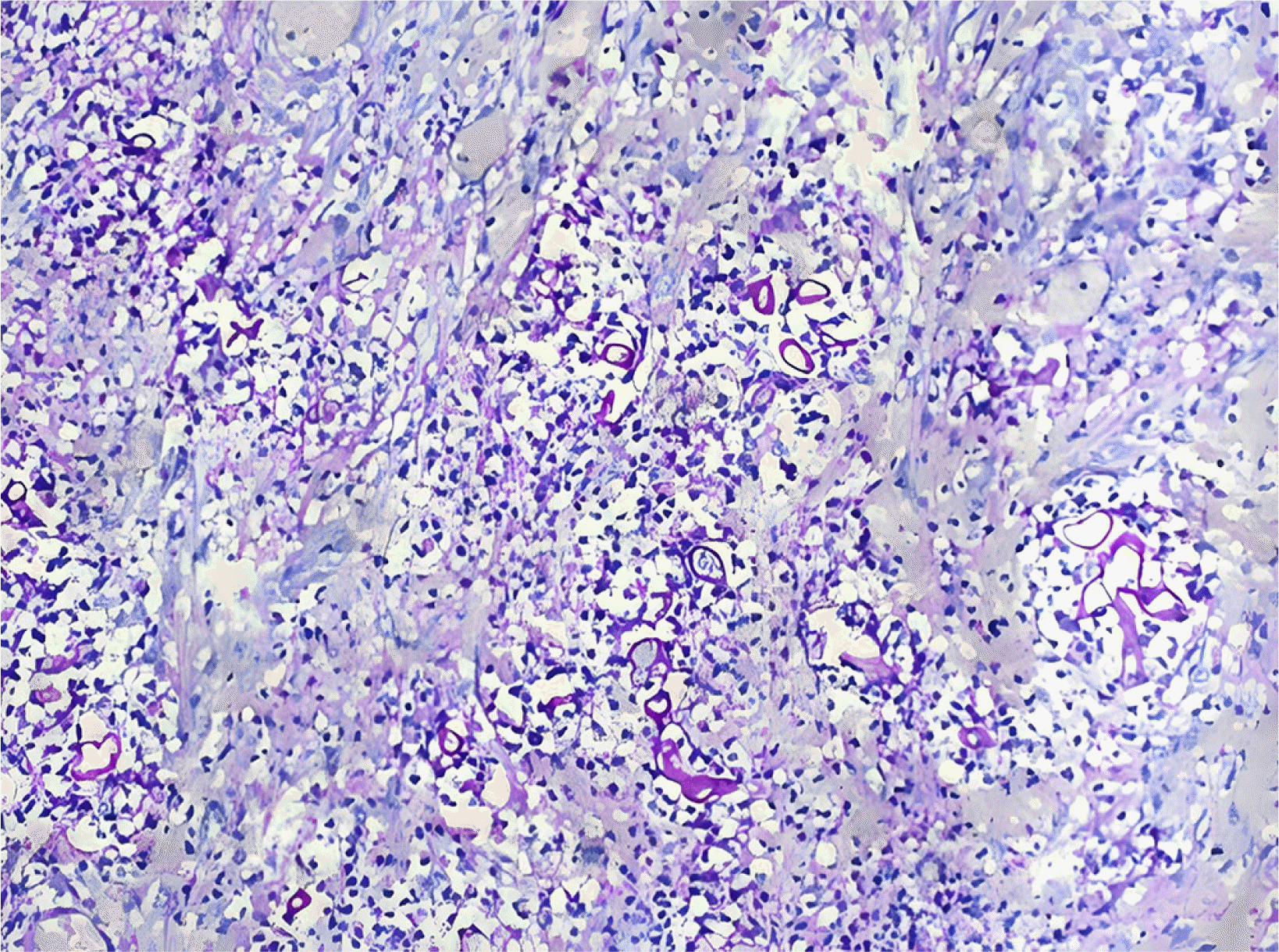
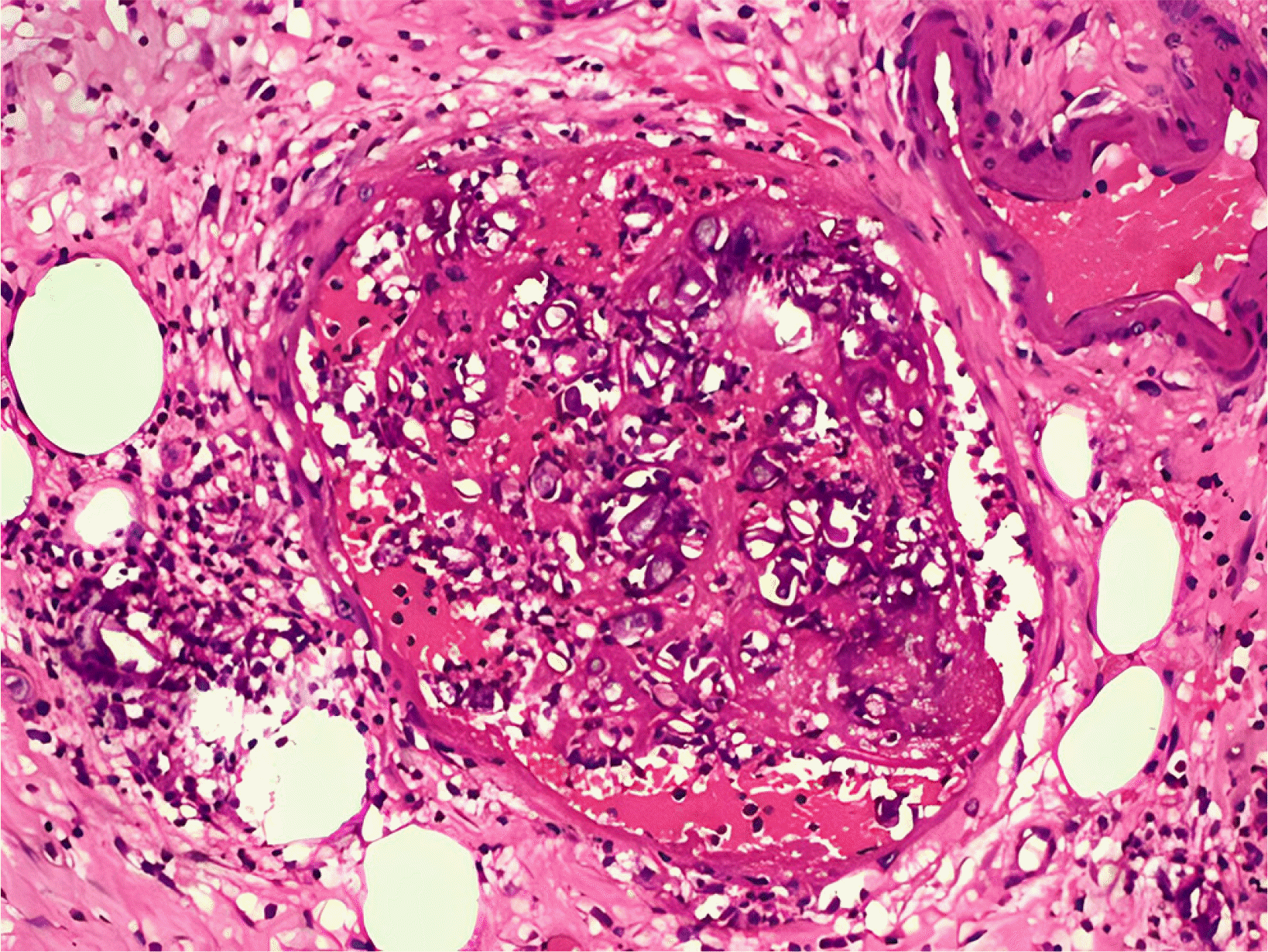
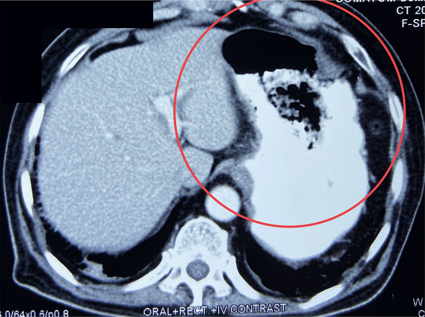
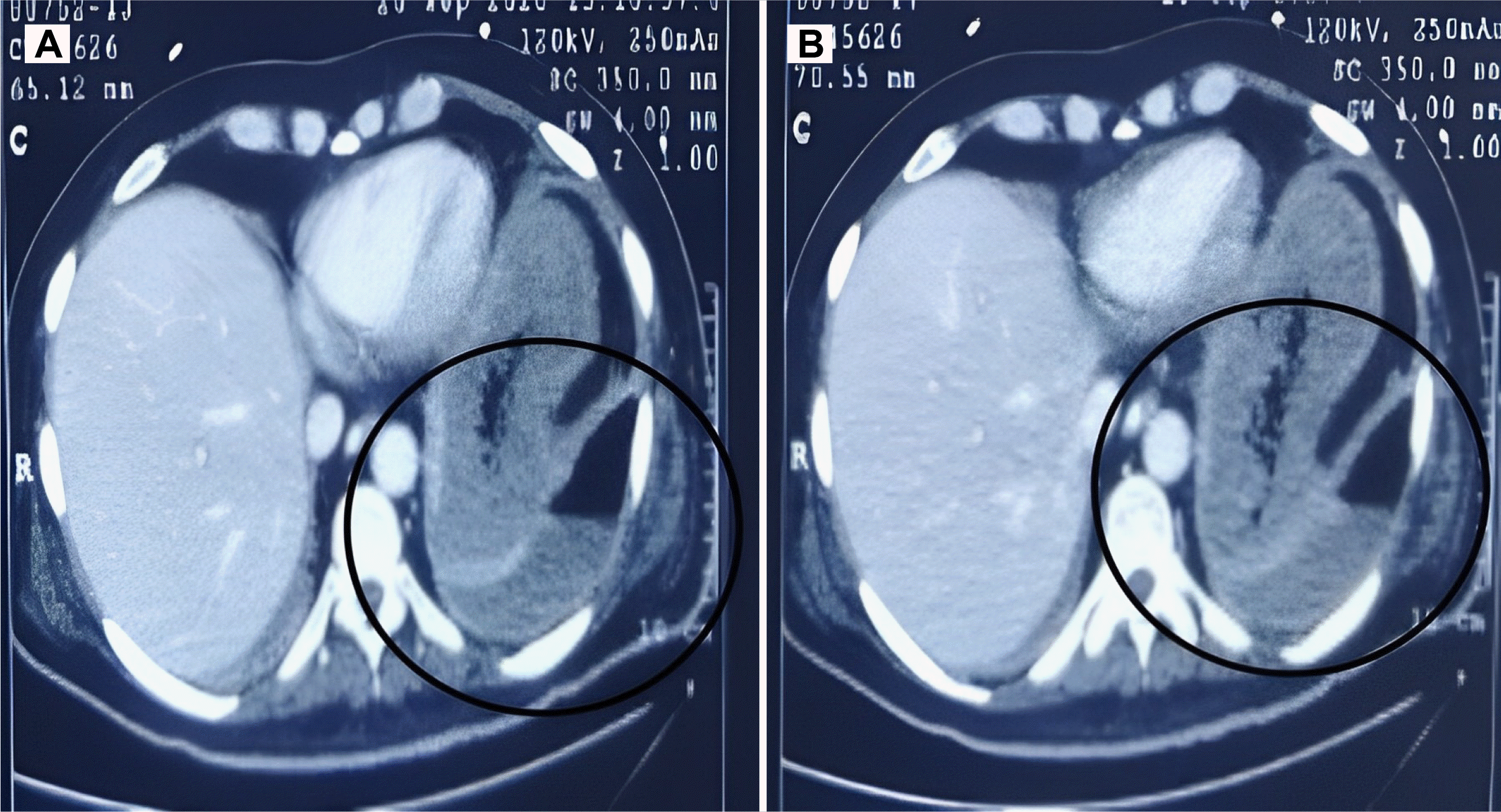
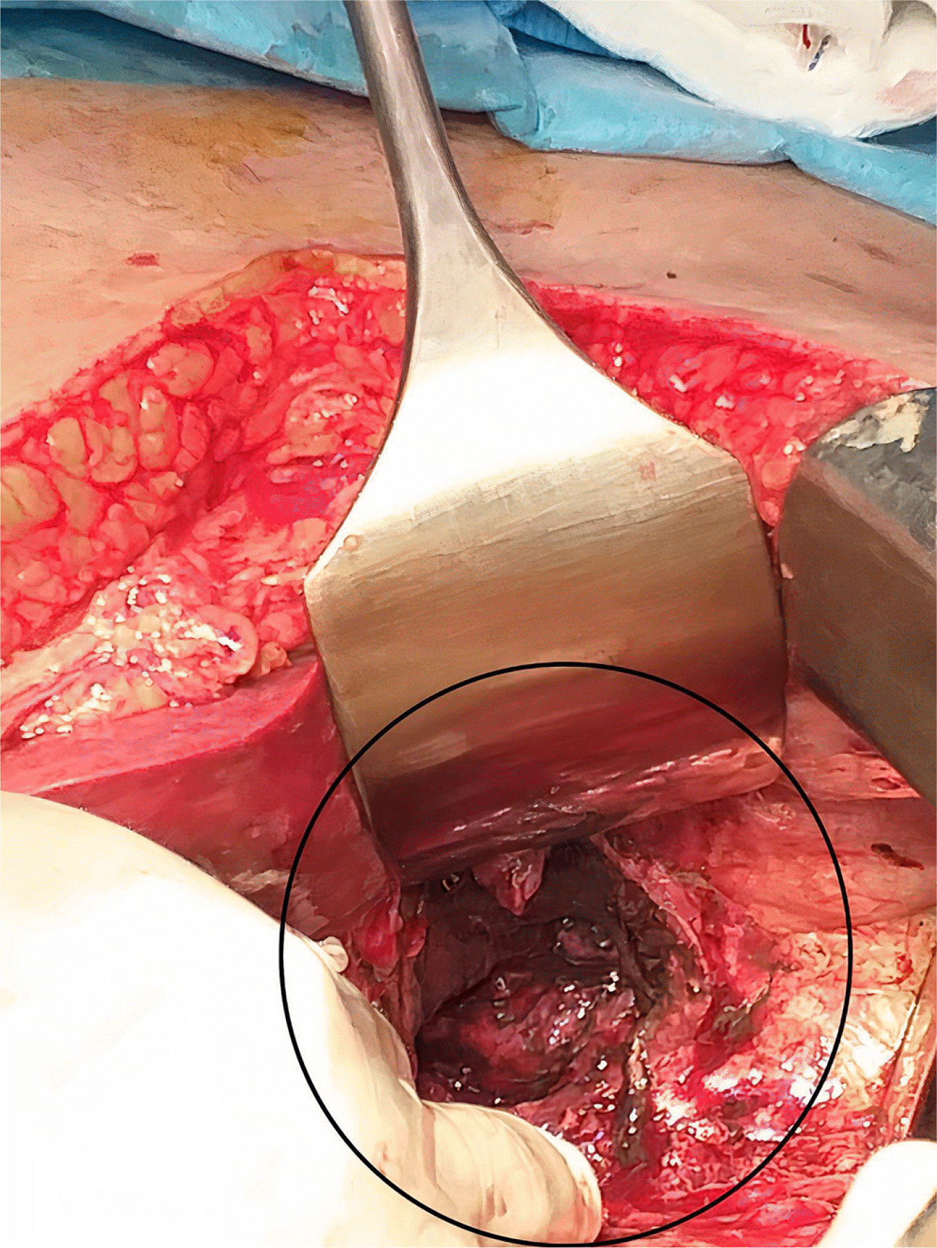
A 53-year-old male patient presented with fever, cough, and a saturation of 92% on room air; a throat swab demonstrated coronavirus disease 2019 (COVID-19). The patient was admitted to the intensive care unit. He required non-invasive ventilatory assistance for approximately ten days and was later switched to a face mask with an oxygen requirement of two liters per minute. During his one-month hospital stay, he was given ceftriaxone, remdesivir, hydrocortisone, and azithromycin. A CT chest scan revealed several patchy areas of interstitial thickening with ground glass opacities in the subpleural and peribronchial regions of the bilateral lung fields, as well as air space densities and fibrosis, particularly in the posterior aspect and basal segments of bilateral lung fields. With 94% saturation, the patient stayed home for 15 days with nasal prongs assistance. At home, the room air saturation was 86– 88%. After 15 days, the patient complained of significant left-side chest discomfort and was hospitalized. A chest CT revealed a left-sided pneumothorax. Intercostal drainage was performed immediately, and the patient was intubated on the same day. After 15 days in that hospital, he was referred to the authors’ hospital because of an inability to wean from ventilatory support and renal failure necessitating dialysis. He had abnormal blood sugar levels following the covid infection. After three days, the patient presented with significant upper gastrointestinal (GI) bleeding and hypotension, necessitating vasopressors. Three units of packed red blood cells (PRBCs) and omeprazole were administered. Emergency angiography revealed active extravasation from the distal splenic artery, which was embolized with metallic micro coils. After stabilization, the patient underwent esophagogastroduodenoscopy (EGD), which revealed a large ulcer with a clot in the fundus. A triple-phase computed tomography of the abdomen showed no obvious contrast extravasation or pseudoaneurysm, with a non-enhancing stomach wall at the fundus, adherent to the left hemidiaphragm, and gastro pleural connection with left-sided hydropneumothorax (Fig. 1). Bile and necrotic debris were emerging from the intercostal tube. The fluid sample was sent for a bacterial and fungal culture. The following observations were made on emergency exploration. Blood clots (500 g) were observed in the left subdiaphragmatic region. The proximal stomach was necrotic, which included the cardia, fundus, and proximal section of the greater curvature (Fig. 2). A diaphragmatic defect with a diameter of approximately 5 cm is evident. The pancreas was plastered to the posterior wall of the stomach. A proximal gastrectomy, tube esophagostomy, vented gastrostomy, and feeding jejunostomy were necessary. Mucormycosis was demonstrated by pathology (Figs. 3, 4). Liposomal amphotericin B was administered to the patient. The patient later experienced severe respiratory failure because of pneumonia. Bloodstream cultures revealed Klebsiella pneumoniae. Unfortunately, the patient passed away on the tenth postoperative day.

2. Case 2

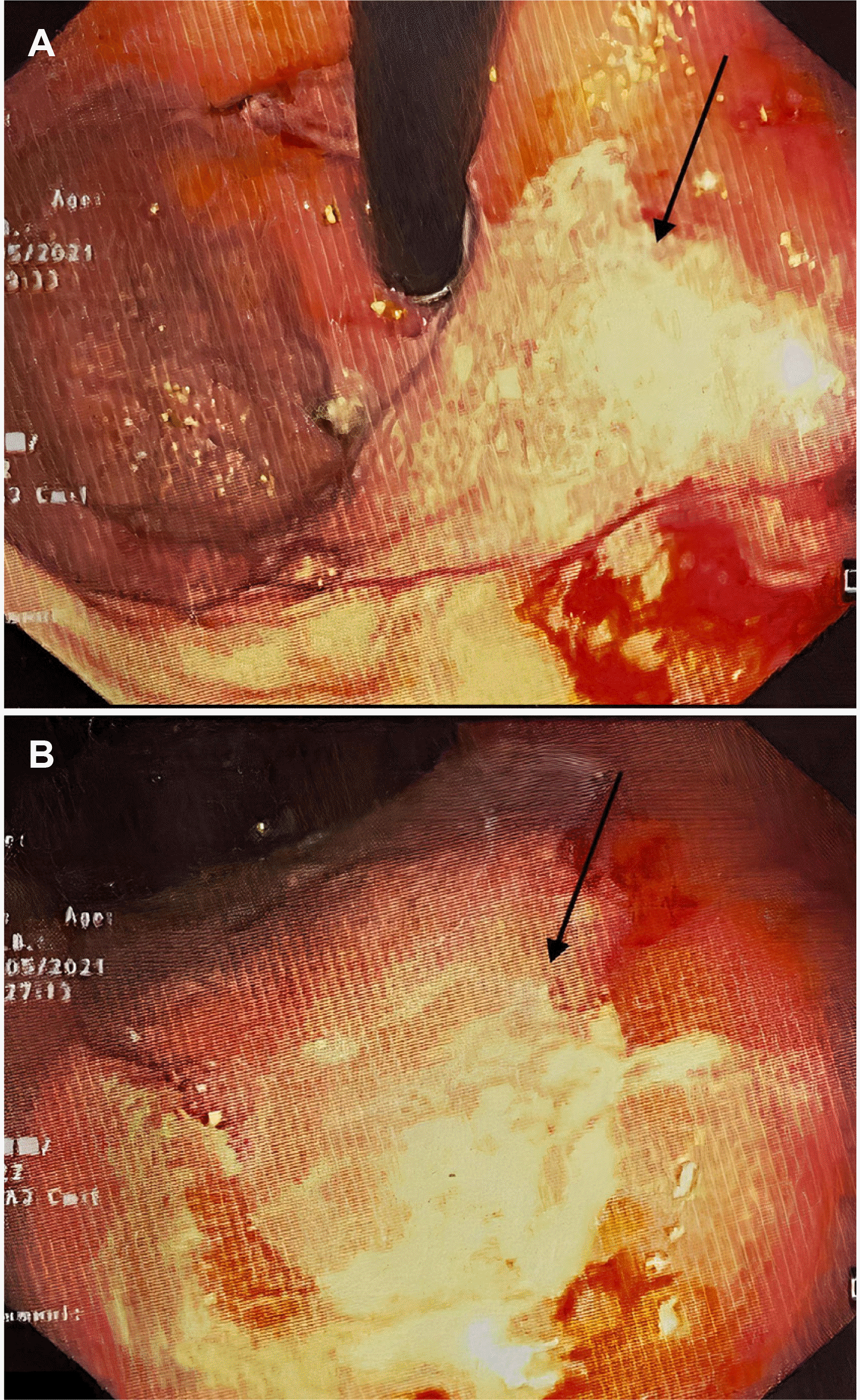
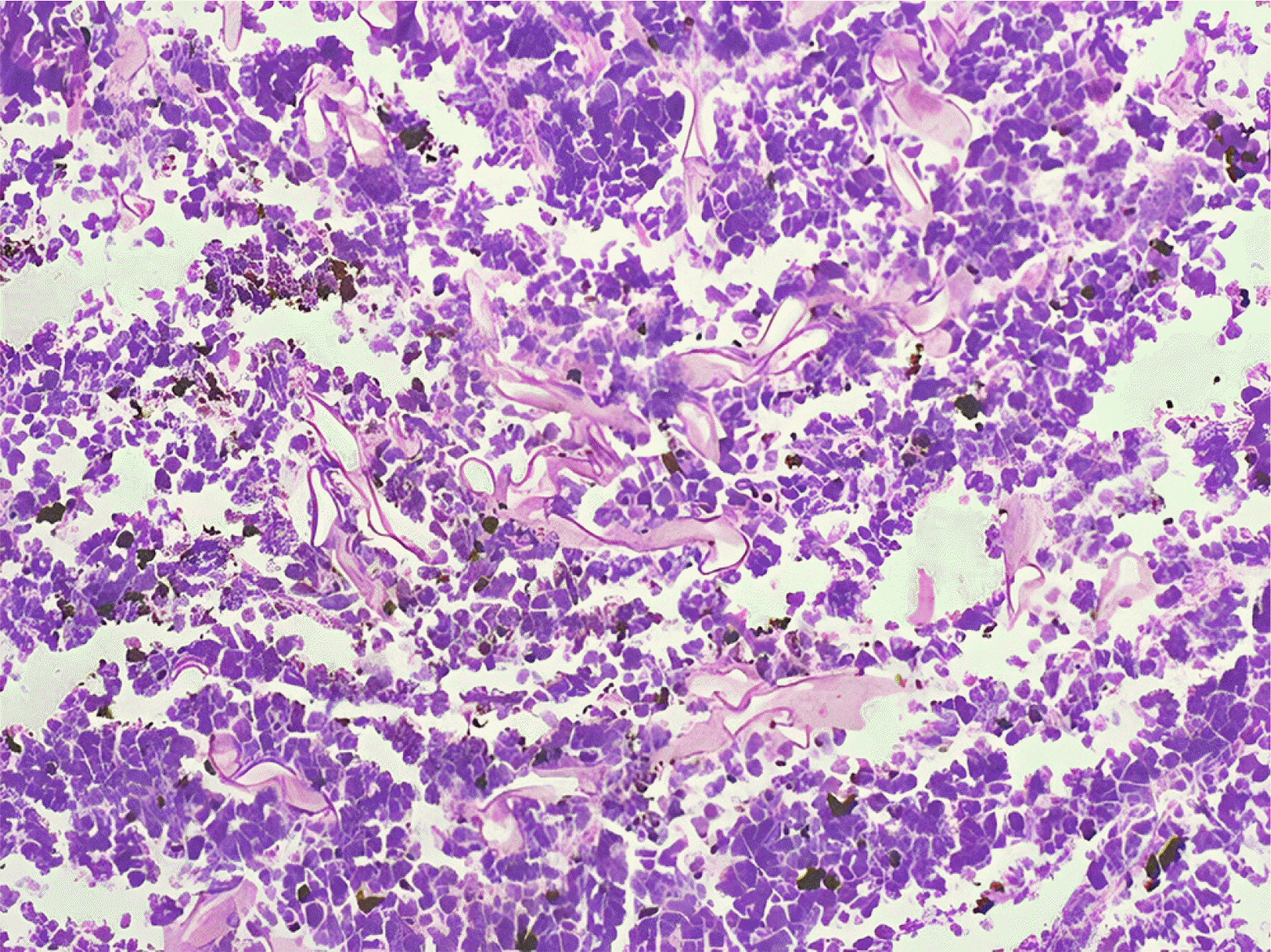
An 82-year-old male patient with a history of a COVID-19 infection (two weeks earlier) arrived with upper stomach pain and two episodes of severe hematemesis linked to dizziness and syncope. The patient was admitted to the intensive care unit, and there was no hemodynamic instability at the time of admission. He was started on a pantoprazole infusion and received two PRBC transfusions. The patient underwent an emergency Esophagogastroduodenoscopy that revealed a large white-based ulcer with many sloughs along the larger curvature of the body (Fig. 5), and biopsies were taken. Histopathology revealed mucormycosis (Fig. 6). The contrast- enhanced chest and abdomen CT revealed ground-glass attenuation in the peripheral parts of the lower lobes of both lungs, mild wall thickening along the greater curvature of the stomach, and a hypodense non-enhancing mottled air-containing slough-like lesion adhered to the gastric wall along the greater curvature (Fig. 7). The patient was given a cumulative dose of 2.5 gm of intravenous liposomal Amphotericin B before being transferred to oral isavuconazole 200 mg once per day for two weeks. The patient was eating normally, and there were no further episodes of hematemesis. After four weeks, the patient was advised to come to the out patient department (OPD) for a repeat EGD.

DISCUSSION

COVID-19 was discovered in Wuhan, China in December 2019 and was declared a pandemic by World Health Organization (WHO) in March 2020. The second wave of this epidemic began in India in March 2021. The sickness was severe during this second wave, and deaths occurred even among the younger population. There was a trend of delayed complications throughout this wave, including mucormycosis and thrombo-embolic events. Mucormycosis typically affects the paranasal sinus, which extends to the orbit, mouth, face, brain, and lungs.5 Mucormycosis of the gastrointestinal system is uncommon. In India, a few cases of gastrointestinal mucormycosis have been reported.6,7 Mucormycosis is characterized by tissue necrosis because of the inherent capacity of angioinvasion.8 Most patients are immunocompromised or suffer from poor glycemic control. COVID-19 patients who are immunocompromised due to a viral infection and are receiving steroids concurrently have poor glycemic control.9 Both patients received high doses of steroids and broad-spectrum antibiotics, increasing their susceptibility to fungal infections. The stomach and colon are the most commonly affected organs in GI mucormycosis, and mortality can reach 85%. Non-specific symptoms, GI bleeding, or perforation can also occur. A high level of suspicion is warranted, particularly if there is a history of COVID-19 infection, use of drugs, or diabetes. The identification of fungal hyphae in biopsy specimens is required for further confirmation.10 Various fungi-specific stains have been used to diagnose mucormycosis, e.g., Hematoxylin and eosin or Periodic acid-Schiff. Antifungals and surgical debridement of the necrosed/involved tissues are necessary for the treatment. Liposomal amphotericin B is the drug of choice for mucormycosis. The recommended dose is 5 mg/kg body weight administered over one hour. The renal function and potassium and magnesium levels in all patients should be monitored for at least once a week because the most prevalent adverse effects are nephrotoxicity, hypokalemia, and hypomagnesemia. Oral posaconazole or isavuconazole can be considered. Mucormycosis is difficult to diagnose and requires prompt treatment. The delays in detection and treatment can increase mortality considerably.11 Monte Junior et al.12 reported a case of stomach mucormycosis in a COVID-19 patient. A one-week delay in diagnosis resulted in the patient's death.12 Even with timely diagnosis and aggressive management, the prognosis is not good. In the first case, the patient could not be saved despite vigorous treatment.
In conclusion, mucormycosis is difficult to diagnose and must be treated immediately. Delays in detection and treatment can result in a twofold increase in mortality. Nevertheless, the prognosis is poor despite quick detection and aggressive treatment. In the second case, prompt diagnosis and treatment saved his life.
Notes

Financial support

None.

Conflict of interest

None.

REFERENCES

1. Adhikari S, Gautam AR, Paudyal B, Sigdel KR, Basnyat B. 2019; Case Report: Gastric Mucormycosis- a rare but important differential diagnosis of upper gastrointestinal bleeding in an area of Helicobacter pylori endemicity. Wellcome Open Res. 4:5. DOI: 10.12688/wellcomeopenres.15026.1. PMID: 31131332. PMCID: PMC6518441.
crossref
2. Kauffman CA, Malani AN. 2007; Zygomycosis: an emerging fungal infection with new options for management. Curr Infect Dis Rep. 9:435–440. DOI: 10.1007/s11908-007-0066-4. PMID: 17999877.
crossref
3. Roden MM, Zaoutis TE, Buchanan WL, et al. 2005; Epidemiology and outcome of zygomycosis: a review of 929 reported cases. Clin Infect Dis. 41:634–653. DOI: 10.1086/432579. PMID: 16080086.
crossref
4. Rothan HA, Byrareddy SN. 2020; The epidemiology and pathogenesis of coronavirus disease (COVID-19) outbreak. J Autoimmun. 109:102433. DOI: 10.1016/j.jaut.2020.102433. PMID: 32113704. PMCID: PMC7127067.
crossref
5. Riley TT, Muzny CA, Swiatlo E, Legendre DP. 2016; Breaking the mold: A review of mucormycosis and current pharmacological treatment options. Ann Pharmacother. 50:747–757. DOI: 10.1177/1060028016655425. PMID: 27307416.
6. Singh RP, Gupta N, Kaur T, Gupta A. 2021; Rare case of gastrointestinal mucormycosis with colonic perforation in an immunocompetent patient with COVID-19. BMJ Case Rep. 14:e244096. DOI: 10.1136/bcr-2021-244096. PMID: 34215642. PMCID: PMC8256735.
crossref
7. Jain M, Tyagi R, Tyagi R, Jain G. 2022; Post-COVID-19 gastrointestinal invasive mucormycosis. Indian J Surg. 84:545–547. DOI: 10.1007/s12262-021-03007-6. PMID: 34177157. PMCID: PMC8219178.
crossref
8. Petrikkos G, Skiada A, Lortholary O, Roilides E, Walsh TJ, Kontoyiannis DP. 2012; Epidemiology and clinical manifestations of mucormycosis. Clin Infect Dis. 54 Suppl 1:S23–34. DOI: 10.1093/cid/cir866. PMID: 22247442.
crossref
9. Song G, Liang G, Liu W. 2020; Fungal Co-infections associated with Global COVID-19 pandemic: A clinical and diagnostic perspective from China. Mycopathologia. 185:599–606. DOI: 10.1007/s11046-020-00462-9. PMID: 32737747. PMCID: PMC7394275.
crossref
10. Spellberg B, Walsh TJ, Kontoyiannis DP, Edwards J Jr, Ibrahim AS. 2009; Recent advances in the management of mucormycosis: from bench to bedside. Clin Infect Dis. 48:1743–1751. DOI: 10.1086/599105. PMID: 19435437. PMCID: PMC2809216.
crossref
11. Werthman-Ehrenreich A. 2021; Mucormycosis with orbital compartment syndrome in a patient with COVID-19. Am J Emerg Med. 42:264.e5–264.e8. DOI: 10.1016/j.ajem.2020.09.032. PMID: 32972795. PMCID: PMC7493738.
crossref
12. Monte Junior ESD, Santos MELD, Ribeiro IB, et al. 2020; Rare and fatal gastrointestinal mucormycosis (zygomycosis) in a COVID-19 patient: a case report. Clin Endosc. 53:746–749. DOI: 10.5946/ce.2020.180. PMID: 33207116. PMCID: PMC7719411.
crossref

Fig. 1
CT images showing a gastropleural fistula with left-sided hydropneumothorax (A and B). A triple-phase computed tomography of the abdomen demonstrated no obvious contrast extravasation or pseudoaneurysm, with a non-enhancing stomach wall at the fundus, adherent to the left hemidiaphragm, and gastro pleural connection with a left-sided hydropneumothorax. Circle in the picture showing gastro pleural connection with left-sided hydropneumothorax.
kjg-81-5-221-f1.tif
Fig. 2
Intra-operative image showing a necrosed proximal stomach and large diaphragmatic defect. Blood clots (500 g) were observed in the left subdiaphragmatic region. The proximal stomach was necrotic, which included the cardia, fundus, and proximal section of the greater curvature. A diaphragmatic defect with a diameter of around 5 cm is evident. Circle in the picture showing the diaphragmatic defect.
kjg-81-5-221-f2.tif
Fig. 3
Section from a gastrectomy specimen showing numerous broad, aseptate fungal profiles, suggesting mucormycosis (PAS, ×20).
kjg-81-5-221-f3.tif
Fig. 4
Photomicrograph showing angioinvasion by the funga elements. (H&E, ×40).
kjg-81-5-221-f4.tif
Fig. 5
Large white-based ulcer with plenty of sloughs along the greater curvature in the body region (A and B). Arrow in the picture showing white based ulcer with slough.
kjg-81-5-221-f5.tif
Fig. 6
Section from gastric biopsy showing numerous, broad, aseptate, hyaline fungal profiles identified in an area of necrosis. (H&E, ×40).
kjg-81-5-221-f6.tif
Fig. 7
CT image showing hypodense non-enhancing mottled air containing a slough-like lesion adherent to the gastric wall along the greater curvature. Circle in the picture showing hypodense non-enhancing mottled air containing a slough-like lesion.
kjg-81-5-221-f7.tif
TOOLS
Similar articles