INTRODUCTION
Class III malocclusions are typically challenging because of the complexity of treatment, and unpredictable skeletal growth and treatment outcomes.
1,2 According to Guyer et al.,
3 57% of patients with either a normal or prognathic mandible have a deficiency in the maxilla. The application of a maxillary protraction (MP) facemask (FM) with or without rapid palatal expansion (RPE) was popular for the early intervention of skeletal Class III children with midface deficiency.
4-6 More recently, a new protocol entitled “alternate rapid maxillary expansion and constriction” (Alt-RAMEC) was introduced by Liou and colleagues
7,8 to achieve maximum disarticulation of the circummaxillary sutures without over-expansion. Some authors have reported that Alt-RAMEC increased skeletal effects during MP,
9-11 while other studies found no significant or clinically relevant difference between conventional RPE/FM and Alt-RAMEC/FM protocols in terms of MP effectiveness.
12-15
The upper airway, including the nasal cavity (NC), pharyngeal airway (PA), and paranasal sinuses (e.g., maxillary sinuses, MS), not only supports breathing but is also involved in speech and swallowing. In growing Class III patients, the underdeveloped or retrognathic maxilla is expected to have an impact on airway development.
3,16-18 Many studies have examined the effects of either Alt-RAMEC only or Alt-RAMEC/FM on the PA in Class III patients and have reported divergent results,
15,19-22 which may be due to the different methodologies used (two-dimensional [2D] vs. three-dimensional [3D]) or the absence of a control group. Unlike the PA, the NC and MS are surrounded by bony structures and are relatively dimensionally stable. With the buccal tipping of the molars and lateral movements of the alveolar process during palatal expansion, distortion of the lower border of the sinuses might result in an increase in the MS volume after RPE,
23,24 RPE/FM,
17 or Alt-RAMEC.
21 To the best of our knowledge, no study has documented the continuous upper airway volumetric changes of the Alt-RAMEC protocol at different stages, after expansion and protraction, and compared the treatment outcomes with those of the RPE/FM.
Thus, the aim of this retrospective study was to evaluate and compare differences in maxillary advancement and volumetric changes in the NC, PA, and MS at different treatment stages between matched Alt-RAMEC/FM and RPE/FM groups, using cone-beam computed tomography (CBCT).
Go to :

MATERIALS AND METHODS
This single-center, single-blind, retrospective study was approved by the Institutional Review Board (IRB) of the University of British Columbia (H19-01744) and inform consent from the patient was waived. The sample size calculation was based on a previous study,
24 which indicated that a minimum of 20 subjects in each group would be needed to detect a 3,660 mm
3 difference in the change in NC volume, with 80% power (α = 0.05). CBCT records of 20 Chinese patients (age range: 7–12 years; mean age, 10.28 ± 1.45 years) who had undergone Alt-RAMEC before FM treatment were collected from the archives of the Department of Orthodontics at Peking University, School and Hospital of Stomatology (Beijing, China), from 2010 to 2015. The inclusion criteria were as follows: (1) cervical vertebral maturation stage 1–3 at the initial stage; (2) skeletal Class III relationship (ANB < 0°, Wits appraisal < –2 mm) as a result of maxillary retrusion (A point to N perpendicular < 0 mm), with no functional shift detected; and (3) availability of CBCT scans obtained pretreatment, directly after expansion, and after FM treatment, with complete imaging of the cranial base, maxilla, mandible, and upper airway. The exclusion criteria were as follows: (1) previous orthodontic/orthopedic treatment; (2) mandible guided to an edge-to-edge bite; (3) known systematic diseases, craniofacial anomalies, or temporomandibular joint disorders; (4) history of adenotonsillectomy; (5) movement artifacts; (6) major variation in the head or craniocervical orientation > 5° between serial CBCT scans; and (7) compliance issues recorded on the chart. Once the subjects in the Alt-RAMEC/FM group had been identified, the same inclusion and exclusion criteria were applied again to obtain an RPE/FM group, and 20 subjects (age range 7–12 years; mean age, 9.66 ± 1.23 years), who had been successfully matched to the Alt-RMEC/FM group for skeletal age and sex, were selected.
Patients in both groups received a Hyrax-type expander with four bands and an expansion screw (Dentaurum, Pforzheim, Germany). In the Alt-RAMEC group, parents or guardians were instructed to activate/open by two turns per day (0.5 mm/day) for the first 2 weeks and to deactivate/close in the next 2 weeks. Such alternate opening and closing were repeated for five consecutive cycles or 10 weeks. At the end of the 10th week, expansion was discontinued, and the screw was fixed with a 0.012” ligature wire. In the RPE group, the expander was activated twice a day (0.5 mm/day) for 2 weeks. In both groups, a Delaire-type face mask was delivered for MP and the patients were instructed to use it at least 14 h/day immediately after Alt-RAMEC or RPE. A protraction force ranging from 400 to 500 g/side was directed 15–30° downward from the occlusal plane. The treatment was completed when a positive overjet with a Class II or Class I molar relationship was achieved. Cephalometric analyses of skeletal features at baseline, including SNA, SNB, ANB, and FMA, were performed on cephalometric radiographs generated from CBCT.
All images at pretreatment (T1), postexpansion (T2), and postprotraction (T3) were acquired using a Vatech CBCT machine (DCTPRO-050Z; Vatech Co., Ltd., Hwaseong, Korea). The following parameters were used: 90 kV, 7 mA, 15 cm × 15 cm field-of-view, 0.4-mm voxel, and a 12-second scan time. Data were saved in the Digital Imaging and Communication in Medicine format and uploaded to the Dolphin Imaging software (version 11.9; Dolphin Imaging and Management Solutions, Chatsworth, CA, USA). Prior to landmark identification and airway/sinus volume measurements, all CBCT images were oriented based on the skeletal midline, a line passing through the left and right bottom rims of the orbit and the Frankfort horizontal plane.
In the 2D coronal tomographic window, landmarks were marked on the right and left spinous foramens, and the first reference point (ML) was placed in the middle of the line connecting the two points. In the midsagittal view, at the ML point, a horizontal line was drawn passing through the subspinale point (Point A). The anteroposterior (AP) and vertical positions of Point A relative to the ML were measured as the distances between A and MLʹ and ML and MLʹ, respectively (
Figure 1).
25
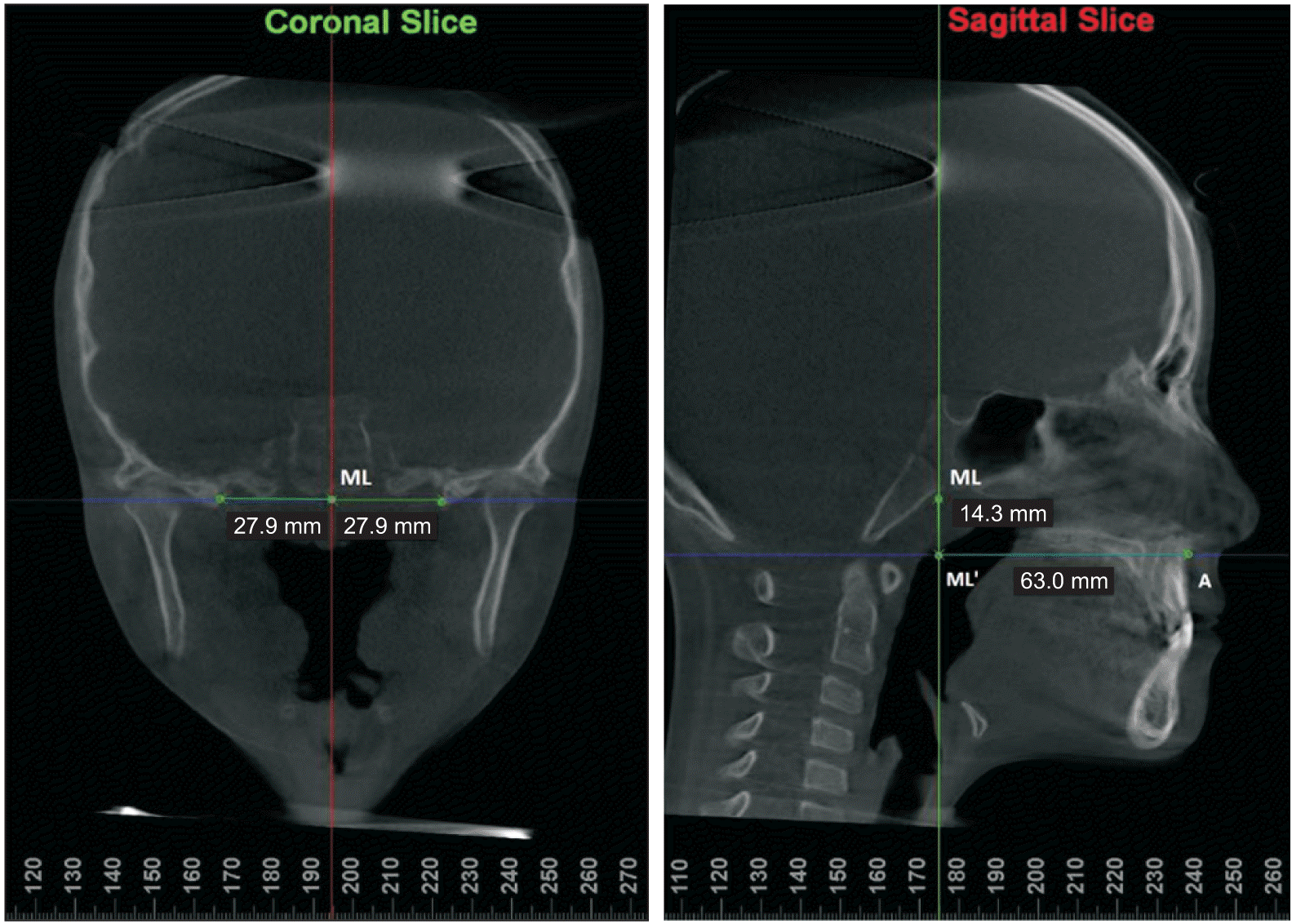 | Figure 1The coronal and mid-sagittal sections on cone-beam computed tomography used to measure the antero-posterior and vertical position of Point A relative to the midline reference point (ML). 
|
The volumetric measurements of the NC, nasopharyngeal, oropharyngeal, and MS were semi-automatically calculated by segmenting the area-of-interest and locating “seed points” at sagittal, coronal, and axial views by visual inspection using threshold values adjusted for each scan.
26,27 The boundaries and parameters used to measure the volumes of NC, PA, and MS in this study are shown in
Figure 2.
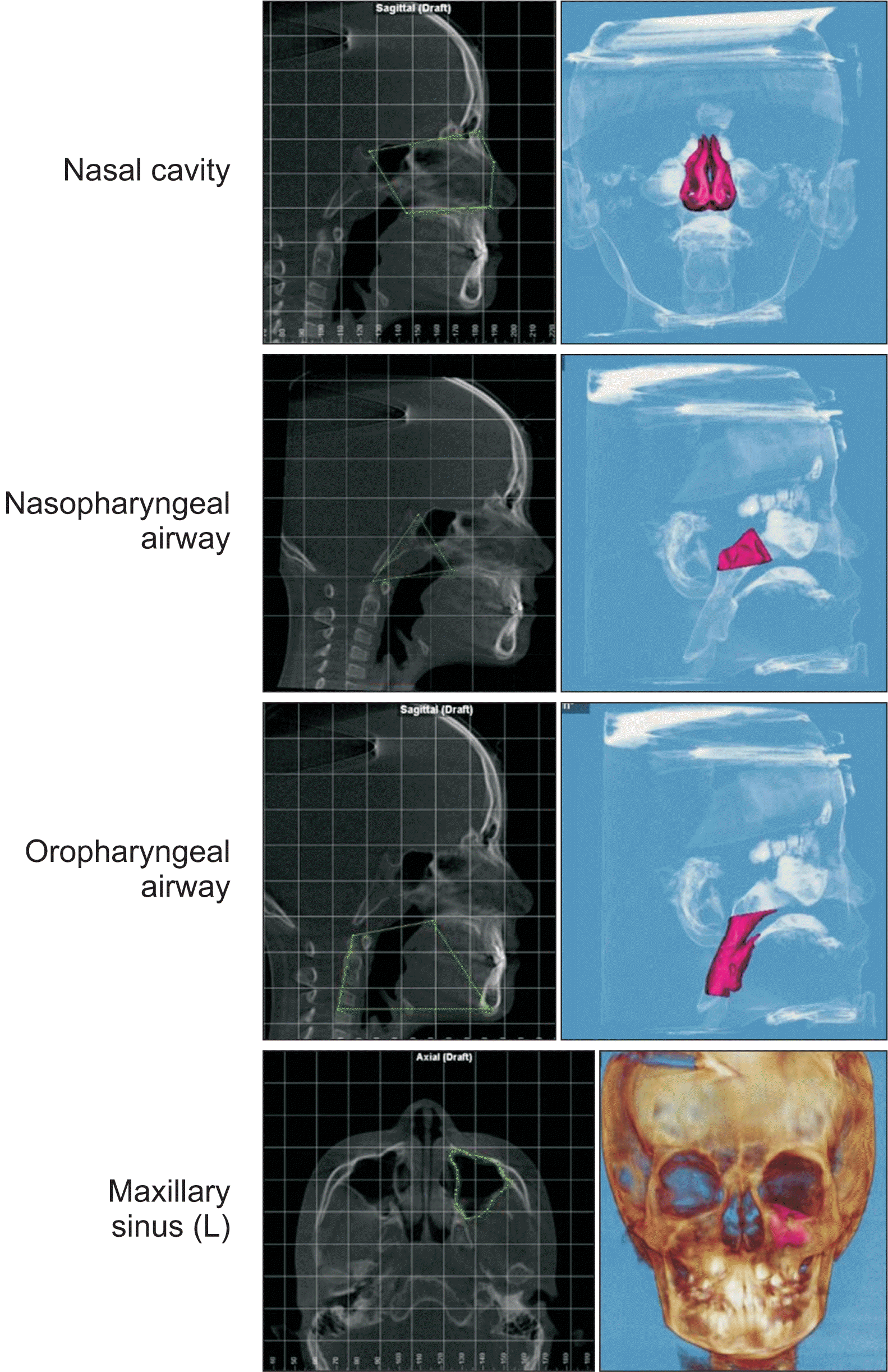 | Figure 2Boundaries and volume measurements used in this study.
|
All the measurements were recorded by a single investigator. Twenty random CBCT scans were re-evaluated by the same investigator at an interval of 4 weeks for intraobserver reliability testing, and by another investigator for interobserver reliability testing.
Statistical analysis
IBM SPSS Statistics for Windows, version 27.0 (IBM Corp., Armonk, NY, USA) was used for data analyses. Intraclass correlation coefficients (ICCs) were used to assess intra- and inter-observer agreements. The Shapiro–Wilk test was used to test the normality of distribution of the cephalometric (SNA, SNB, ANB, and FMA), and linear (A-MLʹ and ML-MLʹ) and volumetric (NC, nasopharynx, oropharynx, and MS) CBCT parameters. An independent t-test or Mann–Whitney U test (nonparametric test) was used for intergroup comparisons of the parameters at different time points. Intragroup differences in each variable at T2 vs. T1, T3 vs. T2, and T3 vs. T1 were analyzed using either the paired t-test or Wilcoxon signed-rank test. All tests were considered statistically significant at p < 0.05.
Go to :
RESULTS
ICCs showed a range of 0.85–0.98 for intraobserver and 0.78–0.96 for interobserver agreement, which indicated a substantial to almost perfect level of reliability for all parameters.
All parameters were normally distributed, except for the oropharyngeal volume at T3 in the RPE/FM group and the ML –ML’, NC volume, and nasopharyngeal volume at T2 in the Alt/RAMEC group after the Shapiro–Wilk test.
Table 1 shows no statistically significant differences in age, sex, skeletal age, treatment time, and cephalometric, and linear and volumetric CBCT parameters between the two groups before treatment, indicating a perfect match.
Table 1
Comparison of the parameters for RPE/FM and Alt-RAMEC/FM groups at pretreatment (T1)
|
Variable |
RPE/FM (n = 20) |
Alt-RAMEC/FM (n = 20) |
p-value |
|
Age (yr) |
9.66 ± 1.23 |
10.28 ± 1.45 |
0.143 |
|
Sex (F/M) |
14/6 |
14/6 |
1.000†
|
|
CVMS |
|
|
|
|
1 |
3 |
2 |
0.844†
|
|
2 |
5 |
6 |
|
|
3 |
12 |
12 |
|
|
Treatment time (mo) |
10.59 ± 4.36 |
10.84 ± 2.65 |
0.828 |
|
SNA (°) |
80.66 ± 2.88 |
80.33 ± 4.58 |
0.782 |
|
SNB (°) |
81.97 ± 3.18 |
81.52 ± 4.70 |
0.714 |
|
ANB (°) |
−1.31 ± 2.08 |
−1.18 ± 1.23 |
0.807 |
|
FH-MP (°) |
29.99 ± 4.41 |
28.94 ± 5.15 |
0.488 |
|
Maxillary width (mm) |
63.0 ± 1.8 |
63.3 ± 2.0 |
0.146 |
|
A-ML’ (mm) |
63.0 ± 3.3 |
63.4 ± 3.4 |
0.663 |
|
ML-ML’ (mm) |
19.0 ± 2.7 |
18.6 ± 3.5 |
0.705 |
|
NC volume (mm3) |
19,456 ± 3,115 |
20,808 ± 4,007 |
0.241 |
|
Nasopharynx volume (mm3) |
3,913 ± 1,539 |
3,456 ± 1,951 |
0.416 |
|
Oropharynx volume (mm3) |
11,248 ± 4,119 |
12,229 ± 4,646 |
0.484 |
|
Maxillary sinus R volume (mm3) |
11,488 ± 3,400 |
11,702 ± 3,171 |
0.838 |
|
Maxillary sinus L volume (mm3) |
11,284 ± 2,780 |
11,978 ± 2,874 |
0.443 |

The AP and vertical positions of Point A relative to the cranial base and airway volume, measured at three different time points, i.e., T1, T2, and T3, are listed in
Tables 2 and
3 for the RPE/FM and Alt-RAMEC/FM groups respectively. There was a significant increase in the advancement of Point A in all groups after expansion and protraction. Point A moved forward by 1.3 mm after expansion in both groups, while it moved even further forward after protraction in Alt-RAMEC/FM (by 2.4 mm) than in RPE/FM, (1.1 mm); this difference was statistically significant (
p < 0.05,
Table 4). Point A was advanced by 3.7 mm in Alt-RAMEC/FM, and 2.4 mm in RPE/FM across the entire treatment period, which also showed statistical significance (
p < 0.05,
Table 4). In terms of the vertical position of Point A, it had a significant increase posttreatment in RPE/FM (by 1.1 mm), and postexpansion in the Alt-RAMEC group (by 1.3 mm), but there was no difference between the two groups at any time points observed (
Table 4).
Table 2
Parameters for the RPE/FM group (n = 20) at pretreatment (T1), postexpansion (T2), and postprotraction (T3)
|
Parameter |
T1 |
T2 |
T3 |
T2-T1 |
p-value |
T3-T2 |
p-value |
T3-T1 |
p-value |
|
A-ML’ (mm) |
63.0 ± 3.3 |
64.2 ± 3.5 |
65.3 ± 3.6 |
1.3 ± 1.3 |
0.000***
|
1.1 ± 1.5 |
0.005**
|
2.4 ± 1.8 |
0.000***
|
|
ML-ML’ (mm) |
19.0 ± 2.7 |
19.8 ± 2.4 |
20.1 ± 2.8 |
0.8 ± 1.8 |
0.068 |
0.4 ± 1.4 |
0.271 |
1.1 ± 2.0 |
0.020*
|
|
NC volume (mm3) |
19,456 ± 3,115 |
22,950 ± 4,586 |
23,021 ± 4,908 |
3,495 ± 3,840 |
0.001**
|
701.1 ± 3,698 |
0.933 |
3,565 ± 4,674 |
0.003**
|
|
Nasopharynx volume (mm3) |
3,913 ± 1,539 |
4,505 ± 1,620 |
5,403 ± 2,232 |
591 ± 710 |
0.001**
|
899 ± 1,373 |
0.009**
|
1,490 ± 1,323 |
0.000***
|
|
Oropharynx volume (mm3) |
11,248 ± 4,119 |
12,060 ± 3,920 |
14,721 ± 6,861 |
811 ± 4,507 |
0.431 |
2,661 ± 4,977 |
0.027a*
|
3,473 ± 5,810 |
0.015a*
|
|
Maxillary sinus volume (mm3) |
22,773 ± 5,986 |
22,361 ± 8,058 |
24,767 ± 7,016 |
−411 ± 14,475 |
0.686 |
2,405 ± 3,071 |
0.002**
|
1,993 ± 4,044 |
0.040*
|
Table 3
Parameters for the Alt-RAMEC/FM group (n = 20) at pretreatment (T1), postexpansion (T2), and postprotraction (T3)
|
Parameter |
T1 |
T2 |
T3 |
T2-T1 |
p-value |
T3-T2 |
p-value |
T3-T1 |
p-value |
|
A-ML’ (mm) |
63.4 ± 3.4 |
64.7 ± 3.2 |
67.0 ± 2.9 |
1.3 ± 1.6 |
0.001**
|
2.4 ± 2.0 |
0.000***
|
3.7 ± 1.8 |
0.000***
|
|
ML-ML’ (mm) |
18.6 ± 3.5 |
20.0 ± 2.4 |
19.7 ± 2.9 |
1.3 ± 3.0 |
0.049a*
|
−0.3 ± 1.8 |
0.469a
|
0.8 ± 2.8 |
0.124 |
|
NC volume (mm3) |
20,808 ± 4,007 |
23,414 ± 4,097 |
25,481 ± 4,597 |
2,904 ± 2,690 |
0.001a**
|
2,067 ± 3,438 |
0.015a*
|
4,710 ± 3,367 |
0.000***
|
|
Nasopharynx volume (mm3) |
3,456 ± 1,951 |
4,212 ± 2,331 |
4,830 ± 2,446 |
688 ± 1,002 |
0.004a**
|
618 ± 939 |
0.008a**
|
1,317 ± 1,286 |
0.000***
|
|
Oropharynx volume (mm3) |
12,229 ± 4,646 |
12,817 ± 4,271 |
14,932 ± 5,831 |
480 ± 2,804 |
0.373 |
2,114 ± 5,433 |
0.098 |
1,861 ± 4,370 |
0.045*
|
|
Maxillary sinus volume (mm3) |
23,681 ± 5,861 |
24,371 ± 7,587 |
27,169 ± 6,551 |
690 ± 2,895 |
0.300 |
2,797 ± 5,037 |
0.022*
|
3,488 ± 4,001 |
0.001**
|
Table 4
Comparison of the parameter difference (RPE/FM - Alt-RAMEC/FM) at different time points: postexpansion (T2-T1), postprotraction (T3-T2), and posttreatment (T3-T1)
|
Parameter |
Postexpansion T2 vs. T1 |
|
Postprotraction T3 vs. T2 |
|
Posttreatment T3 vs.T1 |
|
Mean (95% CI) |
p-value |
Mean (95% CI) |
p-value |
Mean (95% CI) |
p-value |
|
A-ML’ (mm) |
–0.03 (–0.95, 0.89) |
0.948 |
|
–1.27 (–2.40, –0.14) |
0.028*
|
|
–1.30 (–2.43, –0.17) |
0.026*
|
|
ML-ML’ (mm) |
–0.52 (–2.08,1.04) |
0.504a
|
|
0.66 (–0.38, 1.69) |
0.209a
|
|
0.28 (–1.25, 1.81) |
0.713 |
|
NC volume (mm3) |
590 (–1,532, 2,712) |
0.505a
|
|
–1,997 (–4,282, 289) |
0.085a
|
|
–1,145 (–3,753, 1,462) |
0.380 |
|
Nasopharynx volume (mm3) |
–97 (–653, 461) |
0.726a
|
|
280 (–472, 1,034) |
0.455a
|
|
172.4 (–662, 1,007) |
0.678 |
|
Oropharynx volume (mm3) |
330 (–2,087, 2,749) |
0.782 |
|
547 (–2,787, 3,883) |
0.741a
|
|
1,611 (–1,679, 4,902) |
0.328a
|
|
Maxillary sinus volume (mm3) |
–1,101 (–3,514, 1,310) |
0.361 |
|
–393 (–3,063, 2,278) |
0.768 |
|
–1,494 (–4,069, 1,080) |
0.247 |
Although the NC and nasopharyngeal airway volumes increased significantly in both groups after expansion, postprotraction, and post-treatment, with the exception of the NC in RPE/FM postprotraction (
Tables 2 and
3), no intergroup differences were observed at any time point (
Table 4). The oropharyngeal volume appeared to increase after treatment in both groups; however, the difference was not statistically significant (
Table 4). For MS volume, both groups showed a significant increase post-traction and post-treatment (
Tables 2 and
3); nonetheless, no intergroup difference was observed (
Table 4).
Go to :
DISCUSSION
To ascertain whether there are any beneficial effects during the protraction period after different expansion protocols, it is essential to evaluate airway changes after expansion/before protraction. Previous studies have investigated the sole effects of Alt-RAMEC
20 or its combination with FM
15,21,22 on the upper airway. Those studies were limited by a lack of controls
20 or use of a 2D cephalometric method
15,22 to study a 3D airway structure. To date, no information is available on the serial effects of different expansion protocols on airways after expansion and protraction. In this retrospective study, we used CBCT to assess maxillary advancement and volume changes in the NC, nasopharyngeal and oropharyngeal airways, and MS associated with Alt-RAMEC/FM and RPE/FM at different periods of treatment, such as postexpansion, postprotraction, and throughout the treatment, which has not been addressed previously. Therefore, the findings of this study are significant.
We used relatively stable reference points on the cranial base to measure the maxillary advancement at Point A and the distance between the bilateral spinous foramina was fixed at all tomographic time periods within each patient.
25 This method is more accurate than the traditional 2D measurement.
11,13 Immediately after expansion, we found a significant intragroup change of 1.3 mm for Point A advancement in both groups. Liou and Tsai
7 reported a significant horizontal movement of Point A in the Alt-RAMEC/FM group (3.0 mm) after expansion, as compared to that in the RPE/FM group (1.6 mm) on cephalograms, which differed from the findings of other studies. The participants in their study were patients with unilateral cleft lip and palate (UCLP), whose anatomy differ from that of other patients without any bony defect in the maxilla. In another UCLP study,
28 no significant difference (0.71 mm,
p > 0.05) was found between Alt-RAMEC/FM and RPE/FM patients during the expansion period in a study that used a 2-week Alt-RAMEC protocol with a Haas-type expander. However, Isci et al.
9 detected a significant difference (1.17 mm,
p < 0.05; 3.2 mm vs. 2.03 mm) between the activation-deactivation/reverse headgear (RH) and RPE/RH groups in a 2D study, which could be explained by their T2 observation time point, which included both expansion and the first 6 months of MP. Other studies focusing on the Alt-RAMEC phase reported different results. Çelebi and Çelikdelen’s
29 2D study reported that Point A moved forward by 0.9 mm in the RPE group, and by a smaller amount (0.44 mm) in the Alt-RAMEC group, which might partly be due to their modified Alt-RAMEC protocol (a 4-weekly sequence). Yilmaz et al.
20 observed a significant forward movement of Point A (0.89 mm) after Alt-RAMEC, excluding the control and comparison groups. Celikoglu and Buyukcavus
22 compared two different Alt-RAMEC protocols (5 weeks vs. 9 weeks) with 2D analysis and found similar amounts of forward movement (0.93 mm in 5 weeks, 0.85 in 9 weeks). Their concerns regarding the 9-week protocol as compared to the 5-week period included potential periodontal damage to the anchor teeth with prolonged expansion. Lemos Rinaldi et al.
30 evaluated the buccal bone plate after different maxillary expansion appliances and protocols using CBCT. They found a significant periodontal attachment loss after Hyrax/Alt-RAMEC (5.09 mm, 4 turns/day for 7 weeks) compared with Haas-type 2/4 (1.28 mm, 2 turns/day for 18 days), Haas-type 4/4 (0.23 mm, 4 turns/day or 9 days), and Hyrax-type 2/4 (1.80 mm, 2 turns/day for 18 days) groups. It remains unclear whether this attachment loss is permanent or reversible, warranting future prospective clinical studies with long-term observation.
During the protraction stage (T3 vs. T2), Point A was further advanced by 2.4 mm in the Alt-RAMEC/FM group, which was more than double that of the RPE/FM group (1.1 mm), and the difference was statistically significant (1.27 mm,
p < 0.05). Liou and Tsai’s UCLP study
7 reported a significant anterior displacement in both groups, with an amount three times greater in the Alt-RAMEC/FM than in the RPE/FM group (0.9 mm in RPE, 2.9 mm in Alt-RAMEC, respectively). They used a compliance-free intraoral MP spring, which was made of 0.036” β-nickel–titanium and which delivered the MP force bilaterally. However, in Da Luz Vieira et al.’s UCLP study,
28 the authors failed to find a significant difference (1.62 mm,
p > 0.05) between the two groups during the protraction period. Isci et al.
9 investigated the second 6 months of RH after applying different expansion protocols and found almost no extra advancement of Point A in RPE/RH, but did find a significant change in Alt-RAMEC/RH (0.93 mm,
p < 0.05), which might imply that the maxillary advancement happened mainly in the early stage (first 6 months) after expansion. Baccetti et al.
31 reported a 1.3-mm advancement of Point A during MP without any expansion compared to a 1.2-mm backward movement in an untreated Class III control group. Taking the above as baseline, it is difficult to conclude that the Alt-RAMEC protocol has a more positive effect on maxillary advancement during MP, with limited heterogeneity, particularly when weighing complexity, cost, and risks.
During the overall treatment (T3 vs. T1), Point A advanced significantly in both groups: 3.7 mm in the Alt-RAMEC/FM vs. 2.4 mm in the RPE/FM group, with a significant intergroup difference of 1.3 mm (
p < 0.05). This result was in line with those of previous 2D studies,
9,10,12 which showed that MP with Alt-RAMEC could positively affect the forward movement of the maxilla, as compared to traditional RPE/FM, in the early treatment of patients with maxillary retrusion without cleft lip and palate. In other 2D
32 and 3D studies,
13,21 researchers found that Alt-RAMEC/FM did not affect the forward movement of the maxilla. This discrepancy is mainly due to the diversity of the protocols, age of the study subjects, and methods used. Even with statistically significant differences between the two groups in maxillary advancement during protraction and throughout the entire treatment (both 1.3 mm and
p < 0.05), we could not conclude that Alt-RAMEC has a beneficial effect on MP, since the difference was too small to be clinically relevant. Canturk and Celikoglu
32 suggested an immediate load of FM with Alt-RAMEC, without the need to wait until the Alt-RAMEC procedure was completed. Özbilen et al.
14 also supported the early loading of FM due to a decrease in bone height and thickness on the anchor teeth because of a lack of orthopedic response. They claimed that it was essential to start MP as early as possible, regardless of the protocol. Recently, additional modifications, such as Alt-RAMEC throughout the entire MP course,
11 addition of Class III elastics,
33,34 temporary anchorage devices supported by FM
33,35,36 or expanders,
34 have been made to the traditional Alt-RAMEC/FM protocol. A long-term study
31 stated that Alt-RAMEC, if performed at the right time with a Liou-type expander, followed by full-time intraoral Class III springs or elastic wearing, would allow for stable long-term results. However, due to the scarcity of randomized clinical trials with long-term observations, it is impossible to conclude which MP protocol is superior.
In this study, two groups of samples were retrospectively matched according to skeletal age, sex, and severity of skeletal malocclusions. The comparison (
Table 1) showed that there was no significant difference in any of the airway parameters before treatment, indicating the homogeneity of the study subjects between the two groups. After the expansion/FM combined treatment (T3 vs. T1), both groups showed significant increases in the volumes of the NC, nasopharyngeal airway, oropharyngeal airway, and MS, whereas all volumetric changes for both groups were comparable (
p > 0.05). Özbilen et al.’s study
21 was the only 3D study to compare the PA and MS volume changes after Alt-RAMEC/FM and RPE/FM. They reported that different expansion devices and protocols did not seem to affect PA volumes, although a significant increase in both lower and total PA was exclusively detected in the Alt-RAMEC group, without a significant intergroup difference. However, they did find an increase in MS volume in the Alt-RAMEC/FM group. Kale and Buyukcavus’s 2D study
15 showed that Alt-RAMEC/FM yielded significantly larger nasopharyngeal, oropharyngeal, and total pharyngeal area changes than did RPE/FM, except in the hypopharyngeal area. Moreover, the above-mentioned changes in the Alt-RAMEC group were comparable to those in the skeletal anchorage (miniplate) group, which they thought to be the most effective method in terms of PA dimensions, particularly in the nasopharynx. When comparing the two different Alt-RAMEC protocols, that is, 5-week vs. 9-week cycles, Celikoglu and Buyukcavus’s
22 2D findings were in agreement with those of Kale and Buyukcavus,
15 who also showed significant increases in the nasopharyngeal and upper airway dimensions and insignificant changes in the lower pharyngeal dimension in both groups. The contrasting findings regarding the upper airway and MS dimensional changes could be attributed to different ethnicities of the study population, different Alt-RAMEC protocols applied, resultant amount of anterior movement of the maxilla, 2D vs. 3D methods, and 3D software used for airway/sinus segmentation and measurements. In the present study, we did not measure the hypopharyngeal airway because a previous study
27 proved the low reliability of upper airway analysis for the hypopharynx and the excellent intra- and inter-examiner reliability for oropharyngeal volume.
The NC and nasopharynx volumes significantly increased in both groups after expansion, but the observed increase in oropharyngeal volume was insignificant. No significant differences were found between the two groups. Yilmaz and Kucukkeles
20 also found a significant increase in the anterior nasal compartment, nasal compartment, and total airway volume after the Alt-RAMEC procedure, but they were not able to compare their findings with those of any control group. A systematic review and meta-analysis
37 on 3D analyses of short- and long-term effects of RPE on the NC and upper airway also concluded that RPE had only a short-term positive effect on increasing the volume of the NC and upper part of the airway. As there have been few studies showing airway volume changes from T2 to T3 after Alt-RAMEC treatment to date, this study provides information missing from the current literature. The NC increased significantly in both groups after expansion. However, during protraction, the Alt-RAMEC/FM group showed a greater volume increase (2,067 mm
3) than did the RPE/FM group (701 mm
3), although the difference was not statistically significant. During FM, both groups showed a significant increase in MS volume, but without an intergroup difference. Conversely, no significant increase in MS volume was found in either group immediately after expansion, which is in agreement with the results of a previous study.
24 The MS in both groups increased significantly during the protraction stage and throughout the entire treatment rather than during the expansion period, possibly due to the shorter observation time from T1 to T2 (2 weeks to 10 weeks) than from T2 to T3 (8.3 months to 10 months on average), or from T1 to T3, when normal growth was observed.
The present study had three main limitations. First, the sample size was relatively small and the sex distribution was not even, with more female than male subjects included. Second, there was a lack of an untreated Class III control group, for ethical reasons. Third, the retrospective nature of the study resulted in some unavoidable biases in terms of treatment and sample selection. We attempted to match the two groups according to skeletal age, sex, and severity of skeletal malocclusions with full CBCT records from a relatively large Class III patient pool and investigated the difference between the two protocols in maxilla advancement and airway volume changes at different treatment stages. Given the substantial reduction in CBCT radiation dose and the introduction of new methodologies, randomized clinical trials with larger sample sizes and long-term observations should be conducted in future.
Go to :


 PDF
PDF Citation
 Print
Print


 XML Download