Journal List > Korean J Orthod > v.53(3) > 1516082809

Xiong, Liu, Wu, Ye, Zhang, Zhu, Yang, and Wang: Association between the severity of hypodontia and the characteristics of craniofacial morphology in a Chinese population: A cross-sectional study

Abstract

Objective

To investigate craniofacial differences in individuals with hypodontia and explore the relationship between craniofacial features and the number of congenitally missing teeth.

Methods

A cross-sectional study was conducted among 261 Chinese patients (males, 124; females, 137; age, 7–24 years), divided into four groups (without hypodontia no teeth missing, mild one or two missing teeth, moderate three to five missing teeth, severe six or more missing teeth) according to the number of congenitally missing teeth. Differences in cephalometric measurements among the groups were analyzed. Further, multivariate linear regression and smooth curve fitting were performed to evaluate the relationship between the number of congenitally missing teeth and the cephalometric measurements.

Results

In patients with hypodontia, SNA, NA-AP, FH-NA, ANB, Wits, ANS-Me/N-Me, GoGn-SN, UL-EP, and LL-EP significantly decreased, while Pog-NB, AB-NP, N-ANS, and S-Go/N-Me significantly increased. In multivariate linear regression analysis, SNB, Pog-NB, and S-Go/N-Me were positively related to the number of congenitally missing teeth. In contrast, NA-AP, FH-NA, ANB, Wits, N-Me, ANS-Me, ANS-Me/N-Me, GoGn-SN, SGn-FH (Y-axis), UL-EP, and LL-EP were negatively related, with absolute values of regression coefficients ranging from 0.147 to 0.357. Further, NA-AP, Pog-NB, S-Go/N-Me, and GoGn-SN showed the same tendency in both sexes, whereas UL-EP and LL-EP were different.

Conclusions

Compared with controls, patients with hypodontia tend toward a Class III skeletal relationship, reduced lower anterior face height, flatter mandibular plane, and more retrusive lips. The number of congenitally missing teeth had a greater effect on certain characteristics of craniofacial morphology in males than in females.

INTRODUCTION

Hypodontia, or congenitally missing teeth, is one of the most prevalent developmental disorders characterized by the congenital absence of one or more teeth, excluding the third molar.1-3 The prevalence of hypodontia varies from 0.3–13.3%,3,4 depending on sex, ethnicity, geographical regions, and dentition type. Females have been reported to have a slightly higher prevalence than males, with a ratio of 1.37:1, and hypodontia was more common in deciduous dentition than in permanent dentition. Remarkably, the incidence of hypodontia has increased over the last few decades and appears to have a negative psychosocial impact on individuals.5-8
Hypodontia is classified as nonsyndromic or syndromic. Nonsyndromic hypodontia is an isolated trait that only involves congenitally missing teeth, whereas syndromic hypodontia can be associated with a cleft lip and palate or more than 50 craniofacial syndromes.2,9 Based on the number of missing teeth, hypodontia can be classified into different severities: mild (one or two missing teeth), moderate (three to five missing teeth), and severe (six or more missing teeth).10,11
It is generally agreed that hypodontia may occur due to genetic regulation and environmental factors. Several studies have confirmed that various genes, such as PAX9, AXIN2, FGF3, FGF10, and BMP4, are associated with tooth agenesis.6,12,13 Therefore, mutations in these related genes may contribute to hypodontia.14 However, no consensus has been reached on whether hypodontia is caused by a polygenetic or single gene defect. Environmental factors that can interfere with tooth development, including maternal exposure to alcohol and smoking, thalidomide, and rubella infection during pregnancy, may be related to hypodontia.4,6,15
Patients with hypodontia have different dental and craniofacial morphological characteristics than people without hypodontia. Although this association has been broadly investigated, the conclusions among different studies were inconsistent due to non-negligible sample heterogeneity.6,9,16 Several studies confirmed that patients with hypodontia had a reduced facial height, a smaller mandibular plane angle, a tendency to develop a retrognathic maxilla, and a Class III skeletal relationship, resulting in a flatter or more concave profile.1,17-19 Furthermore, hypodontia severity may be associated with craniofacial morphology alteration.10,16,20 In contrast, some studies have revealed bimaxillary retrognathism7,21 and a higher prevalence of Class I skeletal relationship in patients with hypodontia,22 while others found that hypodontia did not fabricate a significant skeletal distinction.7,23 Since no consensus has yet been reached in the literature, further research is required to elucidate this issue thoroughly.
This study investigated the association between hypodontia severity and craniofacial morphological characteristics measured by cephalometry. Additionally, it explored the relationship between certain craniofacial features and the increasing number of congenitally missing teeth. The null hypotheses were as follows: 1) there was no difference in the characteristics of craniofacial morphology between individuals with or without hypodontia, and 2) there was no association between hypodontia severity and craniofacial morphology characteristics.

MATERIALS AND METHODS

Study participants and design

This cross-sectional study was approved by the Institutional Review Board of the West China Hospital of Stomatology (approval no. WCHSIRB-2020-376). Patients and their parents or legal guardians were informed of the possibility that patient records would be used for teaching and research purposes, and informed consent was obtained.
The sample comprised a group without missing or supernumerary teeth (76 participants, aged 11–23 years) and a group of hypodontia patients (185 participants, aged 7–24 years). We adopted the previously described classification of hypodontia severity, identifying it using orthopantomograms and clinical examination. This classification further divided patients with hypodontia into three subgroups: mild (one or two missing teeth), moderate (three to five missing teeth), and severe (six or more missing teeth).3,18,20
The inclusion criteria were:
  • 1) One or more congenitally missing permanent teeth (excluding third molars)

  • 2) Pretreatment radiographs (orthopantomograms and cephalograms) with high quality

  • 3) Seven years of age and above

  • 4) Chinese (Mongoloid)

The exclusion criteria were:
  • 1) A history of tooth extraction (excluding third molars)

  • 2) A history of orofacial trauma, orthodontic, or orthognathic treatment

  • 3) Cranial anomalies or craniofacial syndromes

Since growth may play a role in craniofacial morphology, the cervical vertebral maturation stage (CVMS) was used to divide participants into prepubertal/circumpubertal groups (CVMS 1-4) or postpubertal groups (CVMS 5-6) as described before.24 A sample size calculation was performed using the G*Power (Version 3.1.9.7; University of Düsseldorf, Düsseldorf, Germany), based on an estimated effect size of 0.40.25 The sample size calculation (significance level α, 0.05) was designed to achieve 80% power, resulting in 76 participants in the prepubertal/circumpubertal and postpubertal groups.

Data collection

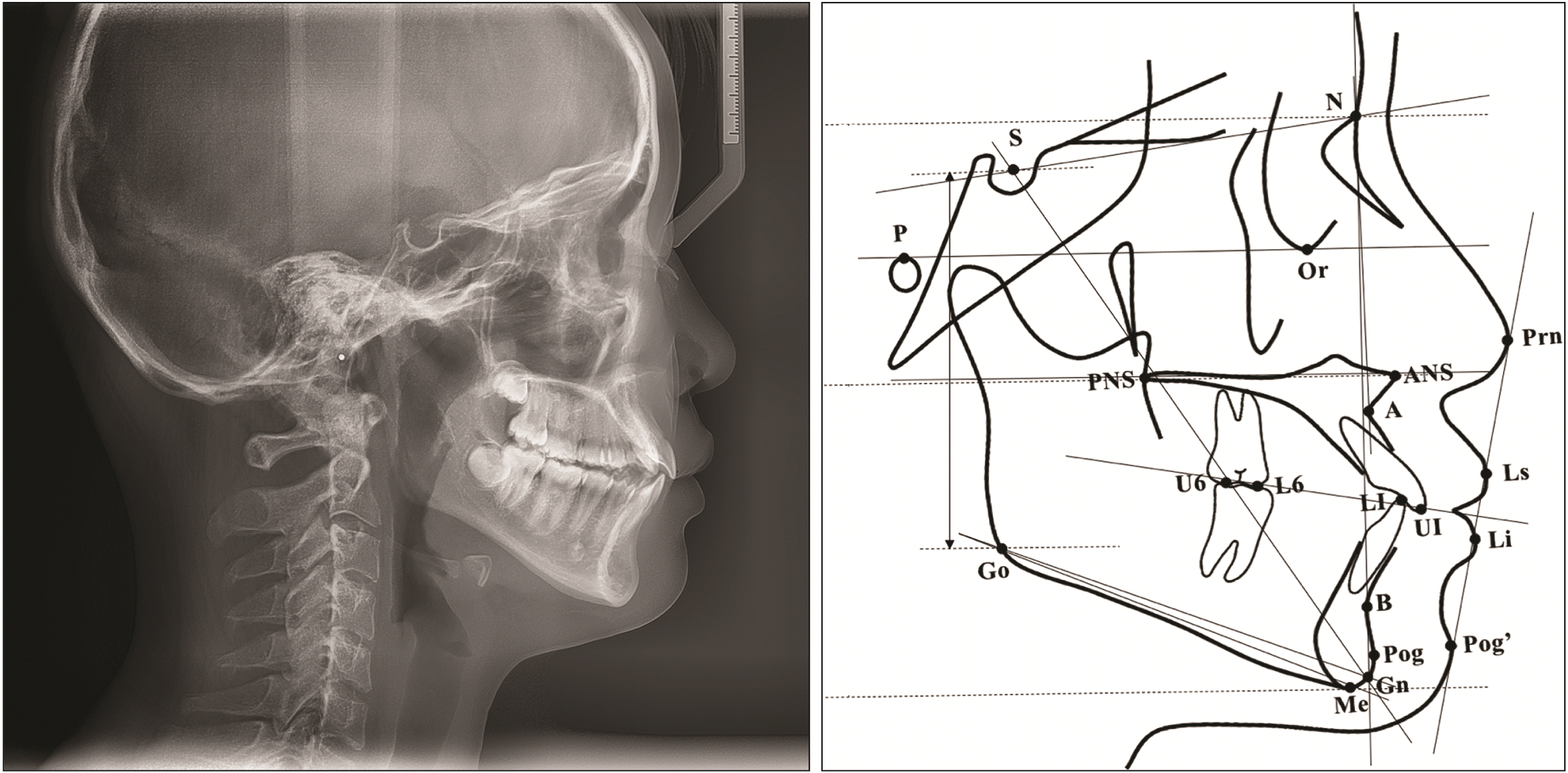
Clinical information, including age, sex, and number and type of missing teeth, was recorded. Craniofacial morphology was described by a customized cephalometric analysis that included the most frequently used cephalometric measurements from previous studies, comprising 10 angular and 7 linear measurements and 2 derived proportions.1,2,10,18,21,26-28 These measurements covered the maxillary, mandibular, and maxillomandibular skeletal characteristics, vertical relationship, and soft tissue morphology (Figure 1, Table 1). The cephalograms of the patients were taken with a natural head position and later analyzed using Uceph software (version 780; Uceph, Chengdu, China) by one independent operator. A month after the initial cephalometric analysis, 20 lateral cephalograms were randomly selected and repeatedly measured by the same operator to evaluate intra-operator reliability. The intraclass correlation coefficients of all measurements were > 0.9, indicating excellent agreement. Additionally, a Bland–Altman analysis was performed for all measurements, and the test-retest difference was plotted against the mean value for each measurement. Of the 19 measurements, 4 Bland–Altman plots comprising angular and linear measurements and proportions are shown in Figure 2. Test-retest bias ranged from –0.22 to 0.16, and all p-values were greater than 0.05, indicating no significant bias.

Statistical analysis

Statistical analyses were performed using the SPSS software (version 24.0; IBM Corp., Armonk, NY, USA), R software (http://www.R-project.org), and Empower (R) (www.empowerstats.com; X&Y Solutions, Inc., Boston, MA, USA). For all hypothesis tests, statistical significance was predetermined at α = 0.05. The Kolmogorov–Smirnov test was performed to analyze the normality of age and cephalometric measurements. Non-normally distributed data (age, NA-AP, Pog-NB, N-Me, N-ANS, and LL-EP) among groups were analyzed using the Mann–Whitney U test or Kruskal–Wallis test followed by multiple comparisons using the Dunn–Bonferroni approach if the difference was significant. Normally distributed data among groups were analyzed by independent samples t-test or one-way analysis of variance (ANOVA), followed by multiple comparisons using the least significant difference (LSD) test. The chi-square test was used to examine the differences in sex among the groups. A multivariate linear regression test was performed to evaluate the effect of the number of congenitally missing teeth on cephalometric measurements adjusted for sex and age. The number of congenitally missing teeth was the independent variable. Subsequently, six cephalometric measurements with high regression coefficients were selected to perform smooth curve fitting to determine the association between the number of congenitally missing teeth and cephalometric measurements.

RESULTS

Demographic characteristics

The number of patients of both sexes and the mean age of each group are presented in Tables 2 and 3. No significant difference was found between the prepubertal/circumpubertal and postpubertal groups.
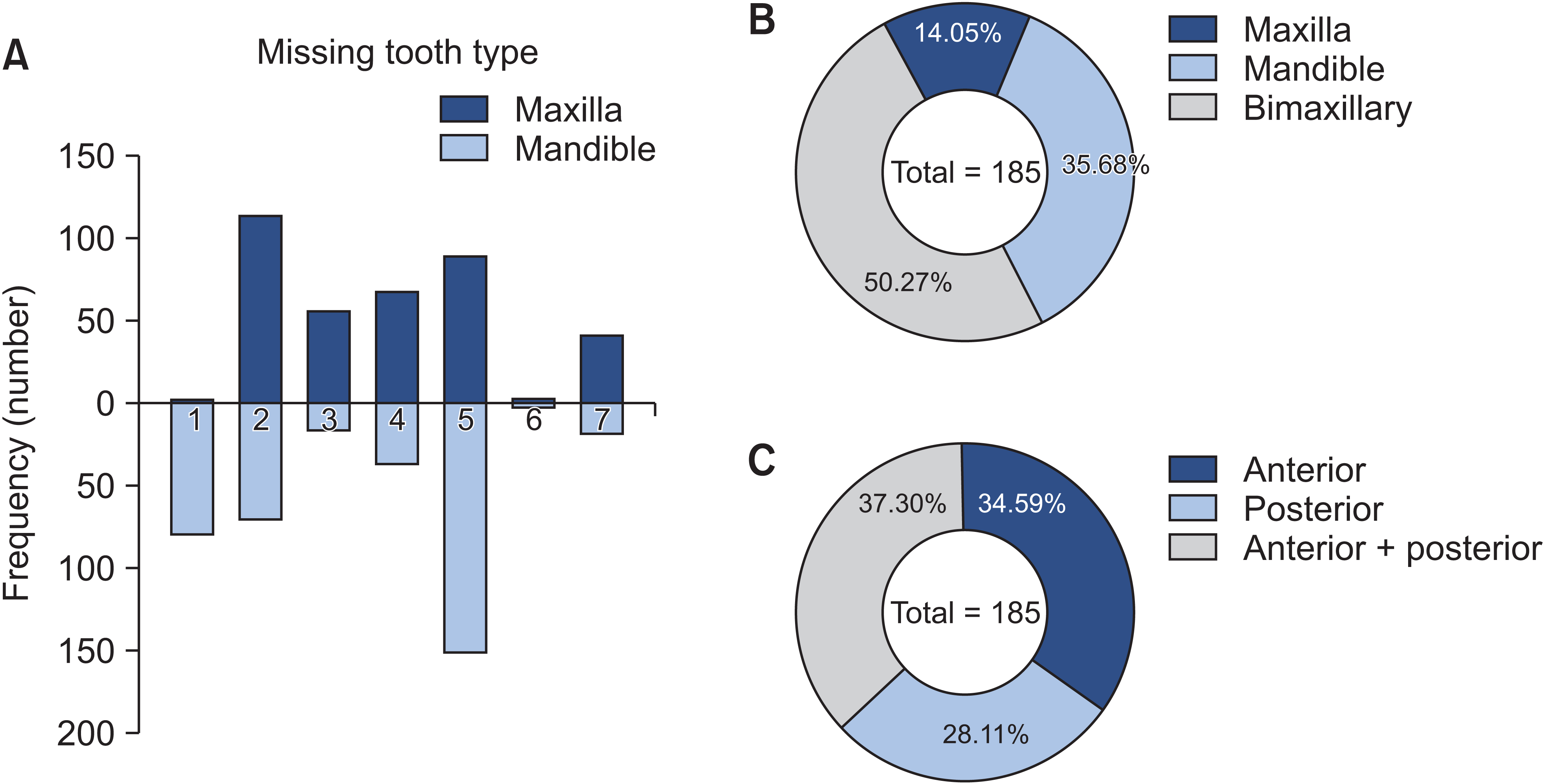
The frequencies of different types of permanent teeth (maxillary or mandibular incisors, canines, premolars, and molars) missing are shown (Figure 3A). Each type of permanent tooth has the potential to be congenitally absent, with the mandibular second premolar being the most common, followed by the maxillary lateral and mandibular incisors. Of the patients with hypodontia, 50.27% were missing both maxillary and mandibular teeth, 35.68% were missing only mandibular teeth, and 14.05% were missing only maxillary teeth (Figure 3B). Regarding patterns in different sites of dental arches, 37.30% of patients with hypodontia were missing both anterior and posterior teeth, 34.59% were missing only anterior teeth, and 28.11% were missing only posterior teeth (Figure 3C).

Cephalometric analysis

A comparison of cephalometric measurements between patients with and without hypodontia was conducted, with seven measurements showing statistically significant differences in the prepubertal/circumpubertal group and thirteen in the postpubertal group (Table 2).
Moreover, a comparison of cephalometric measurements among patients with different hypodontia severities (mild, moderate, severe) and individuals without hypodontia was conducted, with seven measurements showing statistically significant differences in the prepubertal/circumpubertal group and ten in the postpubertal group. Multiple comparisons were conducted using these parameters (Table 3).
The adjusted model showed that SNB, Pog-NB, and S-Go/N-Me were positively related to the number of congenitally missing teeth. Additionally, NA-AP, FH-NA, ANB, Wits, N-Me, ANS-Me, ANS-Me/N-Me, GoGn-SN, SGn-FH (Y-axis), UL-EP, and LL-EP were negatively correlated with the number of congenitally missing teeth in the multivariate linear regression analysis, with absolute values of regression coefficients ranging from 0.147 to 0.357 (Table 4).
The smooth curves between the number of congenitally missing teeth and cephalometric measurements with high regression coefficients, which were adjusted for age and stratified by sex, revealed that NA-AP, Pog-NB, S-Go/N-Me, and GoGn-SN showed the same tendency in both sexes, while UL-EP and LL-EP were different (Figure 4). Further, NA-AP, Pog-NB, and GoGn-SN changed more significantly in males with less than 10 congenitally missing teeth; however, no such trait was found in females. Pog-NB of males tended to be more affected by the number of congenitally missing teeth than females. With the increasing number of congenitally missing teeth, the UL-EP and LL-EP first decreased and then increased in male patients with 10 and 13 congenitally missing teeth as turning points, respectively.

DISCUSSION

Statistical analysis revealed significant differences in several measurements between the control group and the patients with mild, moderate, and severe hypodontia; hence, the null hypothesis was rejected.
This study found that the most frequently congenitally missing teeth were mandibular second premolars, followed by maxillary lateral incisors and mandibular incisors, as reported by several studies.7,18,26,29 It has been proposed that human dentition tends to be smaller with fewer teeth. The most distal tooth in each tooth type was suggested to be the least genetically stable tooth with the highest potential to be congenitally missing,6 which could partially explain the results found in this study.
Chan et al.18 reported that the maxilla was significantly retrognathic in patients with severe hypodontia by revealing a significant reduction in SNA and NA-FH, which partially agrees with the significantly reduced SNA and NA-FH found in the postpubertal group in the current study. In the prepubertal/circumpubertal, and postpubertal groups with varying hypodontia severity, a significantly reduced NA-AP was found, indicating that the maxilla position related to the face was more retrognathic in patients with hypodontia, consistent with the findings of Ben-Bassat and Brin21 and Ogaard and Krogstad.27 Notably, in the prepubertal/circumpubertal groups, such differences were not detected when comparing patients with and without hypodontia. ANB exhibited the same tendency, revealing that hypodontia severity plays an essential role in craniofacial morphology. Additionally, it was observed that as the hypodontia severity increased, ANB reduced; therefore, it was concluded that patients with hypodontia tended toward a Class III skeletal relationship.2,9,10,18,26 Meanwhile, Bassiouny et al.30 studied a sample of patients with congenitally missing maxillary lateral incisors and proved them to have a significant tendency to develop a Class III skeletal relationship with reduced ANB and Wits. In the present study, chin protrusion, measured by Pog-NB, was significantly increased in postpubertal patients with hypodontia, indicating that patients with hypodontia appeared to have a more prominent chin. This finding is supported by Chan et al.18 and Lisson and Scholtes,31 who found that patients with hypodontia generally had a thicker chin button.
Permanent teeth absence has been reported to result in underdevelopment of the maxilla or mandible.16 Therefore, retrognathism may occur in the maxilla, mandible, or a combination of both, depending on the hypodontia location. Another theory proposed by Ogaard and Krogstad27 stated that maxillary retrusion occurring in patients with hypodontia was due to anterior rotation of the mandible due to the lack of support from posterior teeth. This theory could also explain the tendency toward a Class III skeletal relationship and chin protrusion discovered in this study.
Regarding vertical relationship measurements, N-Me and ANS-Me were significantly smaller in prepubertal/circumpubertal patients with hypodontia. In contrast, ANS-Me/N-Me was significantly smaller in the postpubertal group, revealing either an absolute decrease or a relative decrease in lower anterior face height, which concurred with the results of previous studies.7,32,33
Several studies have reported a flatter mandibular plane in patients with hypodontia,18,20,27,34 which supports the finding of a significantly reduced GoGn-SN in prepubertal/circumpubertal, and postpubertal hypodontia patients. A significantly increased S-Go/N-Me was also observed in these groups, demonstrating a counterclockwise growth rotation, in agreement with Acharya et al.,10 who found that in severe hypodontia, the total posterior face height increased, the total anterior face height decreased, or both, leading to increased S-Go/N-Me. Conversely, Vucic et al.1 spotted a decrease in the lower posterior facial height in children with anterior hypodontia, suggesting a tendency to develop hyperdivergent craniofacial patterns.
The presence of sufficient teeth contributes to the vertical development of the alveolar process in both the maxilla and mandible. With the increasing number of absent teeth in patients with hypodontia, the deficiency of vertical development of the alveolar process might occur.16 Additionally, permanent teeth absence results in a lack of posterior occlusal support,35 which could lead to anterior mandible rotation. The significantly different vertical relationship measurements in patients with hypodontia found in this study, including a reduced lower anterior face height and a flatter mandibular plane, may be attributed to the deficiency of vertical development and anterior mandible rotation.
Further, UL-EP and LL-EP significantly decreased in postpubertal patients with hypodontia, indicating that they had more retrusive lower lips, in agreement with previous studies.20,27,30 It has been hypothesized that the difference in soft tissues in patients with hypodontia could be explained by an altered tongue-lip-pressure balance or an adaption of the tongue in the hypodontia region.7,28 Remarkably, UL-EP in the prepubertal/circumpubertal group with different hypodontia severities significantly decreased; however, such a difference was not discovered in the comparison between the two groups with or without hypodontia, which further emphasized the potential impact of hypodontia severity.
More importantly, sex may play a role in the relationship between hypodontia severity and craniofacial morphological characteristics. Sex differences in masticatory performance and occlusal force have been reported previously, revealing that males usually have greater occlusal force than females,36,37 mainly attributed to the greater muscular potential of males. In contrast, females might compensate for their low muscle strength with enhanced coordination of other motor and sensory functions.38,39 Therefore, based on the results of this study, it is speculated that males are more affected by the number of congenitally missing teeth because they are more dependent on occlusal force support. However, above a certain threshold (for example, more than 10 congenitally missing teeth), the influence of absent teeth decreases.
Although most of this study’s results are consistent with those of most previous studies, the association between hypodontia severity and craniofacial morphology was further elaborated in this study. Moreover, this study revealed that sex may play a role in craniofacial morphology in patients with hypodontia.
This study had some limitations. First, the standard deviations of a few measurements were relatively large, which could be attributed to the small sample size of the severe hypodontia group due to the low prevalence of severe hypodontia. However, according to the sample size calculation, 80% power could be achieved based on the current sample size. Second, only Chinese subjects who sought treatment in one center were included, which could have biased the findings. Therefore, the results should be extrapolated to patients with hypodontia of other ethnicities with extreme caution. In addition, the possible late formation of premolars or second molars could introduce bias since participants from 7 years of age were included. Finally, since this was a cross-sectional study, it cannot reflect any cause-and-effect relationship, and further longitudinal studies should be conducted to clarify the exact relationship.

CONCLUSIONS

Significant differences were observed in several cephalometric measurements of patients with hypodontia. Compared with individuals without hypodontia, patients with hypodontia tend toward a Class III skeletal relationship, a reduced lower anterior face height, a flatter mandibular plane, and more retrusive lips. Although the regression coefficients were weak, several cephalometric measurements were significantly associated with the number of congenitally missing teeth. Additionally, certain characteristics of male craniofacial morphology are more affected by the number of congenitally missing teeth than those of females.

Notes

AUTHOR CONTRIBUTIONS

Conceptualization: JW, XX. Data curation: JL, YW. Formal analysis: XX, CY. Funding acquisition: JW. Investigation: QZ, JL. Methodology: WY, YZ. Supervision: JW. Writing–original draft: JL, YW. Writing–review & editing: XX, JW.

CONFLICTS OF INTEREST

No potential conflict of interest relevant to this article was reported.

REFERENCES

1. Vucic S, Dhamo B, Kuijpers MA, Jaddoe VW, Hofman A, Wolvius EB, et al. 2016; Craniofacial characteristics of children with mild hypodontia. Am J Orthod Dentofacial Orthop. 150:611–9. https://doi.org/10.1016/j.ajodo.2016.03.021. Erratum in: Am J Orthod Dentofacial Orthop 2017;151:1023. https://doi.org/10.1016/j.ajodo.2017.03.014. DOI: 10.1016/j.ajodo.2017.03.014. PMID: 27692418.
crossref
2. Bajraktarova Miševska C, Kanurkova L, Bajraktarova Valjakova E, Georgieva S, Bajraktarova B, Georgiev Z, et al. 2016; Craniofacial morphology in individuals with increasing severity of hypodontia. South Eur J Orthod Dentofac Res. 3:12–7. https://doi.org/10.5937/sejodr3-15218. DOI: 10.5937/sejodr3-15218.
crossref
3. Tunç EŞ, Bayrak S, Koyutürk AE. 2011; Dental development in children with mild-to-moderate hypodontia. Am J Orthod Dentofacial Orthop. 139:334–8. https://doi.org/10.1016/j.ajodo.2009.04.024. DOI: 10.1016/j.ajodo.2009.04.024. PMID: 21392687.
crossref
4. Rodrigues AS, Antunes LS, Pinheiro LHM, Guimarães LS, Calansans-Maia JA, Küchler EC, et al. 2020; Is dental agenesis associated with craniofacial morphology pattern? A systematic review and meta-analysis. Eur J Orthod. 42:534–43. https://doi.org/10.1093/ejo/cjz087. DOI: 10.1093/ejo/cjz087. PMID: 31783403.
crossref
5. Polder BJ, Van't Hof MA, Van der Linden FP, Kuijpers-Jagtman AM. 2004; A meta-analysis of the prevalence of dental agenesis of permanent teeth. Community Dent Oral Epidemiol. 32:217–26. https://doi.org/10.1111/j.1600-0528.2004.00158.x. DOI: 10.1111/j.1600-0528.2004.00158.x. PMID: 15151692.
crossref
6. Al-Ani AH, Antoun JS, Thomson WM, Merriman TR, Farella M. 2017; Hypodontia: an update on its etiology, classification, and clinical management. Biomed Res Int. 2017:9378325. https://doi.org/10.1155/2017/9378325. DOI: 10.1155/2017/9378325. PMID: 28401166. PMCID: PMC5376450.
crossref
7. Kreczi A, Proff P, Reicheneder C, Faltermeier A. 2011; Effects of hypodontia on craniofacial structures and mandibular growth pattern. Head Face Med. 7:23. https://doi.org/10.1186/1746-160X-7-23. DOI: 10.1186/1746-160X-7-23. PMID: 22142280. PMCID: PMC3248361. PMID: f5d9cea90ca0440791ec8fe5f39e6128.
crossref
8. Johal A, Huang Y, Toledano S. 2022; Hypodontia and its impact on a young person's quality of life, esthetics, and self-esteem. Am J Orthod Dentofacial Orthop. 161:220–7. https://doi.org/10.1016/j.ajodo.2020.07.039. DOI: 10.1016/j.ajodo.2020.07.039. PMID: 34538709.
crossref
9. Costa AMG, Trevizan M, Matsumoto MAN, da Silva RAB, da Silva LAB, Horta KC, et al. 2017; Association between tooth agenesis and skeletal malocclusions. J Oral Maxillofac Res. 8:e3. https://doi.org/10.5037/jomr.2017.8203. DOI: 10.5037/jomr.2017.8203. PMID: 28791079. PMCID: PMC5541988. PMID: fa88637d22864192a97c9dd055a63253.
crossref
10. Acharya PN, Jones SP, Moles D, Gill D, Hunt NP. 2010; A cephalometric study to investigate the skeletal relationships in patients with increasing severity of hypodontia. Angle Orthod. 80:511–8. https://doi.org/10.2319/072309-411.1. DOI: 10.2319/072309-411.1. PMID: 20482356. PMCID: PMC8966445.
crossref
11. Dhamo B, Vucic S, Kuijpers MA, Jaddoe VW, Hofman A, Wolvius EB, et al. 2016; The association between hypodontia and dental development. Clin Oral Investig. 20:1347–54. https://doi.org/10.1007/s00784-015-1622-1. Erratum in: J Orthod 2017;44:74. https://doi.org/10.1080/14653125.2016.1275610. DOI: 10.1080/14653125.2016.1275610. PMID: 28079474.
crossref
12. Yu M, Fan Z, Wong SW, Sun K, Zhang L, Liu H, et al. 2021; Lrp6 dynamic expression in tooth development and mutations in oligodontia. J Dent Res. 100:415–22. https://doi.org/10.1177/0022034520970459. DOI: 10.1177/0022034520970459. PMID: 33164649.
crossref
13. Cunha AS, Dos Santos LV, Marañón-Vásquez GA, Kirschneck C, Gerber JT, Stuani MB, et al. 2021; Genetic variants in tooth agenesis-related genes might be also involved in tooth size variations. Clin Oral Investig. 25:1307–18. https://doi.org/10.1007/s00784-020-03437-8. DOI: 10.1007/s00784-020-03437-8. PMID: 32648061.
crossref
14. Rodrigues AS, Teixeira EC, Antunes LS, Nelson-Filho P, Cunha AS, Levy SC, et al. 2020; Association between craniofacial morphological patterns and tooth agenesis-related genes. Prog Orthod. 21:9. https://doi.org/10.1186/s40510-020-00309-5. DOI: 10.1186/s40510-020-00309-5. PMID: 32249341. PMCID: PMC7131971. PMID: dd9837aae09041de8e8121bb912d10d1.
crossref
15. Al-Ani AH, Antoun JS, Thomson WM, Merriman TR, Farella M. 2017; Maternal smoking during pregnancy is associated with offspring hypodontia. J Dent Res. 96:1014–9. https://doi.org/10.1177/0022034517711156. DOI: 10.1177/0022034517711156. PMID: 28535361.
crossref
16. Taju W, Sherriff M, Bister D, Shah S. 2018; Association between severity of hypodontia and cephalometric skeletal patterns: a retrospective study. Eur J Orthod. 40:200–5. https://doi.org/10.1093/ejo/cjx049. DOI: 10.1093/ejo/cjx049. PMID: 29016739.
crossref
17. Endo T, Yoshino S, Ozoe R, Kojima K, Shimooka S. 2004; Association of advanced hypodontia and craniofacial morphology in Japanese orthodontic patients. Odontology. 92:48–53. https://doi.org/10.1007/s10266-004-0034-5. DOI: 10.1007/s10266-004-0034-5. PMID: 15490305.
crossref
18. Chan DW, Samman N, McMillan AS. 2009; Craniofacial profile in Southern Chinese with hypodontia. Eur J Orthod. 31:300–5. https://doi.org/10.1093/ejo/cjn111. DOI: 10.1093/ejo/cjn111. PMID: 19193707.
crossref
19. Oeschger ES, Kanavakis G, Cocos A, Halazonetis DJ, Gkantidis N. 2022; Number of teeth is related to craniofacial morphology in humans. Biology (Basel). 11:544. https://doi.org/10.3390/biology11040544. DOI: 10.3390/biology11040544. PMID: 35453743. PMCID: PMC9029740. PMID: a0d2f4778979489cb80a89bcc0a41596.
crossref
20. Gungor AY, Turkkahraman H. 2013; Effects of severity and location of nonsyndromic hypodontia on craniofacial morphology. Angle Orthod. 83:584–90. https://doi.org/10.2319/091012-722.1. DOI: 10.2319/091012-722.1. PMID: 23311600. PMCID: PMC8754045.
crossref
21. Ben-Bassat Y, Brin I. 2003; Skeletodental patterns in patients with multiple congenitally missing teeth. Am J Orthod Dentofacial Orthop. 124:521–5. https://doi.org/10.1016/s0889-5406(03)00620-6. DOI: 10.1016/S0889-5406(03)00620-6. PMID: 14614419.
crossref
22. Dermaut LR, Goeffers KR, De Smit AA. 1986; Prevalence of tooth agenesis correlated with jaw relationship and dental crowding. Am J Orthod Dentofacial Orthop. 90:204–10. https://doi.org/10.1016/0889-5406(86)90067-3. DOI: 10.1016/0889-5406(86)90067-3. PMID: 3463196.
crossref
23. Yelmer ZA, Akbulut S. 2022; Evaluation of the effects of hypodontia on the morphology of craniofacial structures. Orthod Craniofac Res. 25:409–15. https://doi.org/10.1111/ocr.12550. DOI: 10.1111/ocr.12550. PMID: 34837458.
crossref
24. McNamara JA Jr, Franchi L. 2018; The cervical vertebral maturation method: a user's guide. Angle Orthod. 88:133–43. https://doi.org/10.2319/111517-787.1. DOI: 10.2319/111517-787.1. PMID: 29337631. PMCID: PMC8312535.
crossref
25. Lu W, Zhang X, Mei L, Wang P, He J, Li Y, et al. 2020; Orthodontic incisor retraction caused changes in the soft tissue chin area: a retrospective study. BMC Oral Health. 20:108. https://doi.org/10.1186/s12903-020-01099-2. DOI: 10.1186/s12903-020-01099-2. PMID: 32295586. PMCID: PMC7160892. PMID: fc4f4769b9134a62b239f59df8d574ff.
crossref
26. Chung LK, Hobson RS, Nunn JH, Gordon PH, Carter NE. 2000; An analysis of the skeletal relationships in a group of young people with hypodontia. J Orthod. 27:315–8. https://doi.org/10.1093/ortho/27.4.315. DOI: 10.1093/ortho/27.4.315. PMID: 11099569.
crossref
27. Ogaard B, Krogstad O. 1995; Craniofacial structure and soft tissue profile in patients with severe hypodontia. Am J Orthod Dentofacial Orthop. 108:472–7. https://doi.org/10.1016/s0889-5406(95)70047-1. DOI: 10.1016/S0889-5406(95)70047-1. PMID: 7484966.
crossref
28. Yüksel S, Uçem T. 1997; The effect of tooth agenesis on dentofacial structures. Eur J Orthod. 19:71–8. https://doi.org/10.1093/ejo/19.1.71. DOI: 10.1093/ejo/19.1.71. PMID: 9071047.
crossref
29. Higashihori N, Takada JI, Katayanagi M, Takahashi Y, Moriyama K. 2018; Frequency of missing teeth and reduction of mesiodistal tooth width in Japanese patients with tooth agenesis. Prog Orthod. 19:30. https://doi.org/10.1186/s40510-018-0222-4. DOI: 10.1186/s40510-018-0222-4. PMID: 30123921. PMCID: PMC6098995. PMID: e8fc9d6865314dc8a9954c5269008a1e.
crossref
30. Bassiouny DS, Afify AR, Baeshen HA, Birkhed D, Zawawi KH. 2016; Prevalence of maxillary lateral incisor agenesis and associated skeletal characteristics in an orthodontic patient population. Acta Odontol Scand. 74:456–9. https://doi.org/10.1080/00016357.2016.1193625. DOI: 10.1080/00016357.2016.1193625. PMID: 27306861.
crossref
31. Lisson JA, Scholtes S. 2005; Investigation of craniofacial morphology in patients with hypo- and oligodontia. J Orofac Orthop. 66:197–207. https://doi.org/10.1007/s00056-005-0437-0. DOI: 10.1007/s00056-005-0437-0. PMID: 15959633.
crossref
32. Bondarets N, McDonald F. 2000; Analysis of the vertical facial form in patients with severe hypodontia. Am J Phys Anthropol. 111:177–84. https://doi.org/10.1002/(SICI)1096-8644(200002)111:2<177::AID-AJPA4>3.0.CO;2-8. DOI: 10.1002/(SICI)1096-8644(200002)111:2<177::AID-AJPA4>3.0.CO;2-8. PMID: 10640945.
crossref
33. Bauer N, Heckmann K, Sand A, Lisson JA. 2009; Craniofacial growth patterns in patients with congenitally missing permanent teeth. J Orofac Orthop. 70:139–51. https://doi.org/10.1007/s00056-009-0744-y. DOI: 10.1007/s00056-009-0744-y. PMID: 19322532.
crossref
34. Takahashi Y, Higashihori N, Yasuda Y, Takada JI, Moriyama K. 2018; Examination of craniofacial morphology in Japanese patients with congenitally missing teeth: a cross-sectional study. Prog Orthod. 19:38. https://doi.org/10.1186/s40510-018-0238-9. DOI: 10.1186/s40510-018-0238-9. PMID: 30270414. PMCID: PMC6165831. PMID: 7244e6206a414cfaa0b9ab723af71417.
crossref
35. Nodal M, Kjaer I, Solow B. 1994; Craniofacial morphology in patients with multiple congenitally missing permanent teeth. Eur J Orthod. 16:104–9. https://doi.org/10.1093/ejo/16.2.104. DOI: 10.1093/ejo/16.2.104. PMID: 8005197.
crossref
36. Shiga H, Kobayashi Y, Katsuyama H, Yokoyama M, Arakawa I. 2012; Gender difference in masticatory performance in dentate adults. J Prosthodont Res. 56:166–9. https://doi.org/10.1016/j.jpor.2012.02.001. DOI: 10.1016/j.jpor.2012.02.001. PMID: 22613955.
crossref
37. Miwa S, Wada M, Murakami S, Suganami T, Ikebe K, Maeda Y. 2019; Gonial angle measured by orthopantomography as a predictor of maximum occlusal force. J Prosthodont. 28:e426–30. https://doi.org/10.1111/jopr.12598. DOI: 10.1111/jopr.12598. PMID: 28913893.
crossref
38. Koç D, Doğan A, Bek B. 2011; Effect of gender, facial dimensions, body mass index and type of functional occlusion on bite force. J Appl Oral Sci. 19:274–9. https://doi.org/10.1590/s1678-77572011000300017. DOI: 10.1590/S1678-77572011000300017. PMID: 21625746. PMCID: PMC4234342. PMID: 2d4fee5e633e4faa9dfc99d260fa4d78.
crossref
39. Ikebe K, Matsuda K, Kagawa R, Enoki K, Yoshida M, Maeda Y, et al. 2011; Association of masticatory performance with age, gender, number of teeth, occlusal force and salivary flow in Japanese older adults: is ageing a risk factor for masticatory dysfunction? Arch Oral Biol. 56:991–6. https://doi.org/10.1016/j.archoralbio.2011.03.019. DOI: 10.1016/j.archoralbio.2011.03.019. PMID: 21529776.
crossref

Figure 1
Cephalometric landmarks used in the customized analysis.
See Table 1 for definitions of each landmark.
kjod-53-3-150-f1.tif
Figure 2
Bland–Altman plots demonstrating the bias for cephalometric measurements. A, SNB. B, Pog-NB. C, S-Go/N-Me. D, LL-EP. Only four measurements are shown in this figure.
See Table 1 for definitions of measurement.
kjod-53-3-150-f2.tif
Figure 3
The patterns of permanent tooth missing within the sample. A, The frequency of different types of tooth missing. B, The proportion of different patterns of tooth missing in the maxilla and mandible. C, The proportion of different patterns of tooth missing in the anterior and posterior site of the dental arches.
kjod-53-3-150-f3.tif
Figure 4
Smooth curves between the number of congenitally missing teeth and (A) NA-AP, (B) Pog-NB, (C) S-Go/N-Me, (D) GoGn-SN, (E) UL-EP, and (F) LL-EP, stratified by sex.
See Table 1 for definitions of measurement.
kjod-53-3-150-f4.tif
Table 1
Cephalometric landmarks and measurements definitions
Landmark and measurement Definition
Landmark
Porion (P) The midpoint of the line connecting the most superior point of the radiopacity generated by
each of the two ear rods
Orbitale (Or) The lowest point on the inferior margin of the orbit
Nasion (N) The anterior point of the intersection between nasal and frontal bones
Sella (S) The midpoint of sella turcica cavity
Anterior nasal spine (ANS) The tip of anterior nasal spine
Posterior nasal spine (PNS) The tip of posterior nasal spine
Point A (A) The innermost point on the contour of the premaxilla between anterior nasal spine and the
incisor tooth
Upper incisor crown edge (UI) The most forward incisal point of the most prominent maxillary central incisor
Lower incisor crown edge (LI) The most forward incisal point of the most prominent mandibular central incisor
Point B (B) The innermost point on the contour of the mandible between the incisor tooth and the bony chin
Pogonion (Pog) The most anterior point on the contour of the chin
Gnathion (Gn) The center of the inferior point on the mandibular symphysis
Menton (Me) The most inferior point on the mandibular symphysis
Gonion (GO) The midpoint of the contour connecting the ramus and body of the mandible
Upper first molar (U6) Maxillary first molar distal cusp
Lower first molar (L6) Mandibular first molar mesial cusp
Pronasale (Prn) The most anterior point on the midsagittal profile of nose
Labiale superius (Ls) The most prominent point on the upper lip as measured from a perpendicular to nasal floor
Labiale inferius (Li) The most prominent point on the lower lip as determined by a perpendicular from nasal floor
Soft tissue pogonion (Pog’) The most prominent or anterior point on the soft tissue chin in the midsagittal plane
Measurements
SNA Angle between S, N, and point A
NA-AP Angle between N, point A, and Pog
FH-NA Angle formed by FH plane and N-A line
SNB Angle between S, N, and point B
FH-NP Angle formed by FH plane and N-Pog line
Pog-NB Distance from Pog to the N-B line
ANB Angle between point A, N, and point B
AB-NP Angle formed by A-B line and N-Pog line
Wits Distance between perpendiculars from point A and B onto the occlusal plane
N-Me Distance between N and Me
N-ANS Distance between N and ANS
ANS-Me Distance between ANS and Me
ANS-Me/N-Me Ratio of the distance between ANS and Me to the distance between N and Me
S-Go/N-Me Ratio of the distance between S and Go to the distance between N and Me
PP-MP Angle formed by palatal plane and mandibular plane
GoGn-SN Angle formed by Go-Gn line and S-N line
SGn-FH Angle formed by S-Gn line and FH plane
UL-EP Distance between Ls and Prn-Pog’ line
LL-EP Distance between Li and Prn-Pog’ line
Table 2
Comparison of demographic characteristics and cephalometric measurements of control and patients with hypodontia in prepubertal/circumpubertal, and postpubertal groups
Variable Prepubertal & circumpubertal Postpubertal
Control Hypodontia p-value Control Hypodontia p-value
Demographic characters
Sex 0.778 0.165
Male 25 (14.62) 56 (32.75) 9 (10.00) 34 (37.78)
Female 26 (15.20) 64 (37.43) 16 (17.78) 31 (34.44)
Age (yr) 13.31 ± 2.54 13.14 ± 3.82 0.160 16.68 ± 3.13 17.95 ± 3.08 0.079
Maxillary, mandibular and maxillomandibular skeletal measurements
SNA (°) 81.10 ± 3.32 81.27 ± 3.55 0.770 83.24 ± 4.28 81.06 ± 3.71 0.019*
NA-AP (°) 7.16 ± 5.58 5.07 ± 7.41 0.080 8.87 ± 6.42 1.73 ± 8.45 0.000*
FH-NA (°) 89.83 ± 3.38 89.68 ± 3.68 0.804 91.64 ± 3.77 88.97 ± 3.71 0.003*
SNB (°) 77.40 ± 3.36 78.28 ± 3.57 0.136 78.72 ± 4.10 79.16 ± 3.93 0.640
FH-NP (facial angle) (°) 86.55 ± 3.37 87.38 ± 3.82 0.179 87.35 ± 3.25 88.10 ± 4.02 0.402
Pog-NB (mm) 1.28 ± 0.99 1.75 ± 1.45 0.061 1.14 ± 0.73 2.39 ± 1.79 0.000*
ANB (°) 3.69 ± 2.41 2.98 ± 2.83 0.117 4.53 ± 2.74 1.91 ± 3.22 0.001*
AB-NP (°) –5.83 ± 4.36 –5.45 ± 4.88 0.636 –6.97 ± 3.72 –4.83 ± 4.42 0.035*
Wits (mm) –0.12 ± 4.17 –1.15 ± 3.89 0.122 –0.09 ± 3.15 –2.52 ± 4.56 0.016*
Vertical relationship measurements
N-Me (mm) 114.34 ± 7.30 110.80 ± 7.76 0.007* 116.92 ± 8.46 117.24 ± 7.05 0.630
N-ANS (mm) 51.40 ± 3.68 50.68 ± 3.90 0.349 51.10 ± 4.49 52.75 ± 3.49 0.012*
ANS-Me (mm) 61.87 ± 4.69 59.24 ± 5.48 0.003* 64.94 ± 4.96 63.70 ± 5.39 0.320
ANS-Me/N-Me (%) 54.62 ± 1.88 53.86 ± 2.46 0.030* 55.98 ± 1.91 54.66 ± 2.43 0.017*
S-Go/N-Me (%) 61.23 ± 4.75 64.91 ± 4.96 0.000* 62.77 ± 4.90 66.18 ± 5.55 0.008*
PP-MP (°) 27.39 ± 5.61 25.36 ± 7.19 0.050 27.03 ± 5.96 24.43 ± 7.11 0.109
GoGn-SN (°) 35.98 ± 5.29 32.56 ± 6.24 0.001* 35.47 ± 6.41 31.75 ± 6.82 0.021*
SGn-FH (Y-axis) (°) 62.73 ± 3.79 61.21 ± 3.97 0.022* 62.83 ± 3.85 61.68 ± 3.89 0.213
Soft tissue measurements
UL-EP (mm) 1.73 ± 2.41 0.89 ± 2.81 0.064 1.34 ± 2.01 –1.10 ± 3.07 0.000*
LL-EP (mm) 2.83 ± 2.48 1.60 ± 2.77 0.008* 2.79 ± 2.62 0.26 ± 3.29 0.001*

Values are presented as number (%) or mean ± standard deviation.

The chi-square test, independent samples t-test, and Mann–Whitney U test was used.

*p < 0.05.

See Table 1 for definitions of measurement.

Table 3
Comparison of demographic characters and cephalometric measurements of control, mild, moderate, and severe hypodontia in prepubertal/circumpubertal and postpubertal groups
Variable Prepubertal & circumpubertal Postpubertal
Control (1) Mild (2) Moderate (3) Severe (4) p-value Control (1) Mild (2) Moderate (3) Severe (4) p-value
Demographic characters
Sex 0.379 0.060
Male 25 (14.62) 19 (11.11) 28 (16.37) 9 (5.26) 9 (10.00) 11 (12.22) 8 (8.89) 15 (16.67)
Female 26 (15.20) 28 (16.37) 22 (12.87) 14 (8.19) 16 (17.78) 18 (20.00) 7 (7.78) 6 (6.67)
Age (yr) 13.31 ± 2.54 12.49 ± 2.28 13.56 ± 4.24 13.57 ± 5.13 0.488 16.68 ± 3.13 17.41 ± 3.29 17.40 ± 2.92 19.10 ± 2.70 0.068
Maxillary, mandibular, and maxillomandibular skeletal measurements
SNA (°) 81.10 ± 3.32 81.72 ± 3.72 81.28 ± 3.39 80.31 ± 3.48 0.454 83.24 ± 4.28 80.52 ± 3.95 81.43 ± 2.58 81.55 ± 4.08 0.091
NA-AP (°) 7.16 ± 5.85 7.17 ± 6.75 4.71 ± 7.33 1.55 ± 7.73 0.012*4<1 8.97 ± 6.42 3.99 ± 7.06 1.47 ± 8.40 –1.20 ± 9.64 0.000*(3,4) <1
FH-NA (°) 89.83 ± 3.38 90.72 ± 3.65 89.45 ± 3.77 88.98 ± 3.53 0.491 91.64 ± 3.77 89.66 ± 3.83 88.91 ± 3.98 88.07 ± 3.32 0.013*(3,4) <1
SNB (°) 77.40 ± 3.36 77.94 ± 3.52 78.46 ± 3.66 78.59 ± 3.61 0.400 78.72 ± 4.10 78.23 ± 3.41 79.80 ± 3.68 79.98 ± 4.62 0.379
FH-NP
(facial angle) (°)
86.55 ± 3.37 87.04 ± 3.49 87.31 ± 4.09 88.25 ± 3.88 0.323 87.35 ± 3.25 87.74 ± 4.24 88.21 ± 3.78 88.53 ± 4.01 0.749
Pog-NB (mm) 1.28 ± 0.99 1.57 ± 1.43 1.81 ± 1.55 2.00 ± 1.30 0.110 1.14 ± 0.73 1.56 ± 1.08 1.92 ± 1.07 3.88 ± 2.08 0.000*1 < (2,4)
ANB (°) 3.69 ± 2.41 3.77 ± 2.58 2.81 ± 2.75 1.72 ± 3.12 0.008*4 < (1,2) 4.53 ± 2.74 2.31 ± 3.16 1.64 ± 3.44 1.55 ± 3.25 0.005*(2,3,4) < 1
AB-NP (°) –5.83 ± 4.36 –6.09 ± 5.00 –5.01 ± 4.94 –5.12 ± 4.55 0.651 –6.97 ± 3.72 –3.77 ± 4.71 –4.83 ± 4.83 –6.29 ± 3.38 0.033*(1,4) <2
Wits (mm) –0.12 ± 4.17 –0.48 ± 3.39 –1.02 ± 3.85 –2.81 ± 4.58 0.050 –0.09 ± 3.15 –2.62 ± 4.23 –1.99 ± 5.17 –2.77 ± 4.73 0.110
Vertical relationship measurements
N-Me (mm) 114.34 ± 7.30 111.17 ± 7.12 111.48 ± 8.27 108.57 ± 7.81 0.013*4 <1 116.92 ± 8.46 119.09 ± 8.02 116.77 ± 6.08 115.03 ± 5.74 0.289
N-ANS (mm) 51.40 ± 3.68 50.88 ± 3.66 50.73 ± 3.97 50.15 ± 4.33 0.585 51.10 ± 4.49 52.76 ± 2.97 52.78 ± 3.08 52.72 ± 4.47 0.091
ANS-Me (mm) 61.87 ± 4.69 59.37 ± 4.82 59.84 ± 5.93 57.69 ± 5.67 0.010*(2,4) <1 64.94 ± 4.94 65.42 ± 6.36 63.33 ± 3.75 61.59 ± 4.17 0.054
ANS-Me/N-Me (%) 54.62 ± 1.88 53.83 ± 2.11 54.07 ± 2.55 53.45 ± 2.92 0.175 55.98 ± 1.91 55.26 ± 2.41 54.55 ± 1.32 53.90 ± 2.92 0.019*4< (1,2)
S-Go/N-Me (%) 61.23 ± 4.75 63.62 ± 3.64 66.72 ± 5.71 65.77 ± 5.20 0.000*1< (2,3,4) 62.77 ± 4.90 63.42 ± 4.68 67.84 ± 6.88 68.80 ± 3.82 0.000*(1,2) < (3,4)
PP-MP (°) 27.39 ± 5.61 26.12 ± 5.63 24.65 ± 8.01 25.34 ± 8.26 0.232 27.03 ± 5.96 26.08 ± 7.29 22.51 ± 8.05 23.54 ± 5.90 0.122
GoGn-SN (°) 35.98 ± 5.29 33.80 ± 4.14 31.88 ± 7.54 31.49 ± 6.51 0.002*(3,4) <1 35.47 ± 5.72 35.00 ± 5.72 29.32 ± 8.39 29.01 ± 5.14 0.000*(3,4) < (1,2)
SGn-FH (Y-axis) (°) 62.73 ± 3.79 61.76 ± 3.70 61.15 ± 4.28 60.24 ± 3.75 0.056 62.83 ± 3.85 62.45 ± 3.93 60.93 ± 3.90 61.16 ± 3.84 0.308
Soft tissue measurements
UL-EP (mm) 1.73 ± 2.41 1.45 ± 2.57 0.83 ± 2.86 –0.13 ± 2.98 0.032*4< (1,2) 1.34 ± 2.01 –0.17 ± 3.11 –1.33 ± 2.64 –2.20 ± 3.04 0.000*(2,3,4) <1
LL-EP (mm) 2.83 ± 2.48 1.60 ± 2.66 1.16 ± 3.00 1.56 ± 2.57 0.070 2.79 ± 2.62 1.85 ± 2.83 0.11 ± 2.24 –1.84 ± 3.42 0.000*4< (1,2)

Values are presented as number (%) or mean ± standard deviation.

The chi-square test and Kruskal–Wallis test, followed by multiple comparisons using the Dunn–Bonferroni approach, were used.

*p < 0.05.

See Table 1 for definitions of measurement.

Table 4
Multivariate linear regression analysis between the number of congenitally missing teeth and the cephalometric parameters, adjusted for age and sex
Cephalometric parameters Nonadjusted Adjusted model
B (95% CI) Adjusted p-value R-squared value B (95% CI) Adjusted p-value R-squared value
Maxillary, mandibular, and maxillomandibular skeletal measurements
SNA (°) –0.069 (–0.190, 0.052) 0.264 0.005 –0.080 (–0.203, 0.044) 0.207 0.009
NA-AP (°) –0.391 (–1.041, –0.576) 0.000* 0.153 –0.357 (–0.969, –0.506) 0.000* 0.194
FH-NA (°) –0.233 (–0.353, –0.114) 0.000* 0.054 –0.238 (–0.361, –0.117) 0.000* 0.056
SNB (°) 0.182 (0.062, 0.305) 0.003* 0.033 0.152 (0.031, 0.275) 0.014* 0.062
FH-NP (facial angle) (°) 0.130 (0.008, 0.256) 0.037* 0.017 0.094 (–0.027, 0.220) 0.126 0.063
Pog-NB (mm) 0.393 (0.113, 0.203) 0.000* 0.154 0.345 (0.095, 0.183) 0.000* 0.223
ANB (°) –0.316 (–0.346, –0.160) 0.000* 0.100 –0.291 (–0.327, –0.140) 0.000* 0.126
AB-NP (°) 0.050 (–0.090, 0.215) 0.419 0.003 0.052 (–0.089, 0.219) 0.405 0.029
Wits (mm) –0.247 (–0.411, –0.144) 0.000* 0.061 –0.236 (–0.400, –0.132) 0.000* 0.096
Vertical relationship measurements
N-Me (mm) –0.156 (–0.607, –0.077) 0.012* 0.024 –0.256 (–0.779, –0.345) 0.000* 0.372
N-ANS (mm) –0.013 (–0.143, 0.116) 0.833 0.000 –0.098 (–0.216, 0.009) 0.072 0.276
ANS-Me (mm) –0.196 (–0.488, –0.118) 0.001* 0.039 –0.285 (–0.602, 0.276) 0.000* 0.285
ANS-Me/N-Me (%) –0.183 (–0.196, –0.040) 0.003* 0.033 –0.200 (–0.208, –0.050) 0.001* 0.047
S-Go/N-Me (%) 0.375 (0.380, 0.710) 0.000* 0.140 0.318 (0.304, 0.619) 0.000* 0.248
PP-MP (°) –0.167 (–0.535, –0.086) 0.007* 0.028 –0.112 (–0.427, 0.009) 0.061 0.116
GoGn-SN (°) –0.324 (–0.768, –0.364) 0.000* 0.105 –0.278 (–0.683, –0.287) 0.000* 0.172
SGn-FH (Y-axis) (°) –0.157 (–0.297, –0.038) 0.011* 0.025 –0.147 (–0.289, –0.025) 0.020* 0.029
Soft tissue measurements
UL-EP (mm) –0.303 (–0.333, –0.148) 0.000* 0.092 –0.262 (–0.296, –0.120) 0.000* 0.214
LL-EP (mm) –0.294 (–0.333, –0.144) 0.000* 0.087 –0.264 (–0.308, –0.121) 0.000* 0.147

The model was adjusted for age and sex.

CI, confidence interval.

*p < 0.05.

See Table 1 for definitions of measurement.

TOOLS
Similar articles