INTRODUCTION
A considerable proportion of the attractiveness of a smiling face is attributed to the smile.
1 An attractive face with a beautiful smile leads to improved social communications.
2 Different tissue types are involved in the smile: teeth, lip, gingiva, and skin.
3 Considering the teeth, a generally accepted principle of smile design is that the maxillary dental midline should be aligned with the center of the face.
4 The gingival show is also a vital variable affecting smile esthetics.
5 The ideal amount and symmetry of the gingival display lead to a more beautiful smile.
5,6 Based on the etiology, different treatment modalities, including periodontal treatment, orthodontic therapy, orthognathic surgery, lip repositioning, and botulinum injection, are available to treat excessive and asymmetrical gingival display.
7-9 However, achieving an ideal symmetrical gingival show by orthodontic treatment can be particularly vexing. Virtually all mechanotherapies have limitations, and biomechanical side effects must be controlled. Furthermore, complete correction of the gingival display can result in a substantially protracted treatment, cumbersome mechanics, wire manipulations, and, in the case of elastic therapies, strict patient compliance. Besides, not every affected patient can or is willing to undergo invasive procedures such as surgical treatment.
10
Several studies have assessed the attractiveness or threshold of midline deviation.
3,11-25 Most of these investigations have been conducted on the smile or frontal facial images that are ideal and symmetrical except for the dental midline.
11-18,20-25 However, it has been shown that the smile and facial components can have interactions with each other, leading to different esthetic perceptions of the smile.
3,13,14,19,23,26 For instance, an asymmetrical nose, chin, or philtrum can affect the esthetic impact of upper dental midline deviation.
3,13,19 Facial attractiveness has been shown to influence the esthetic perception and preference of upper dental midline deviation, buccal corridor, gingival display, and smile arc.
14 The facial type has been demonstrated to affect the perception of upper dental midline deviation,
23 and gingival display can affect the perception of the smile arc
16 and maxillary incisor inclination.
27
To the best of our knowledge, no study has evaluated the perception of upper dental midline discrepancy in conjunction with the gingival display. In some orthodontic patients, the orthodontist might have limitations in providing an ideal upper dental midline relative to the face because of the prolonged treatment duration, increased risk of root resorption, alveolar defects such as cleft area, and the need for single or multiple tooth extraction, etc.
13 Particularly in these patients who have a concomitant unsatisfactory gingival display, it seems essential to know the threshold of midline deviation, considering difficulties encountered in their gummy smile treatment.
10 Therefore, this study aimed to quantitatively evaluate the influence of the upper dental midline discrepancy in conjunction with the amount and asymmetry of maxillary gingival display on the perception of smile attractiveness by different professional groups and laypersons using altered images of a male subject.
MATERIALS AND METHODS
The study protocol has been approved by ethical committee of Shiraz University of Medical Sciences (IR.SUMS.REC.1396.S995).
Individual photographs
A male subject having finished orthodontic treatment with high degree of facial attractiveness was selected, according to the smile characteristics close to textbook norms
28,29:
- Macro-esthetics: normal facial proportions in all three planes of space
- Mini-esthetics: normal buccal corridor and teeth display at rest, during speech and on smiling
- Micro-esthetics: light and bright tooth shade, normal tooth proportions in height and width, normal gingival shape and contour, connectors and embrasures, without black triangular holes
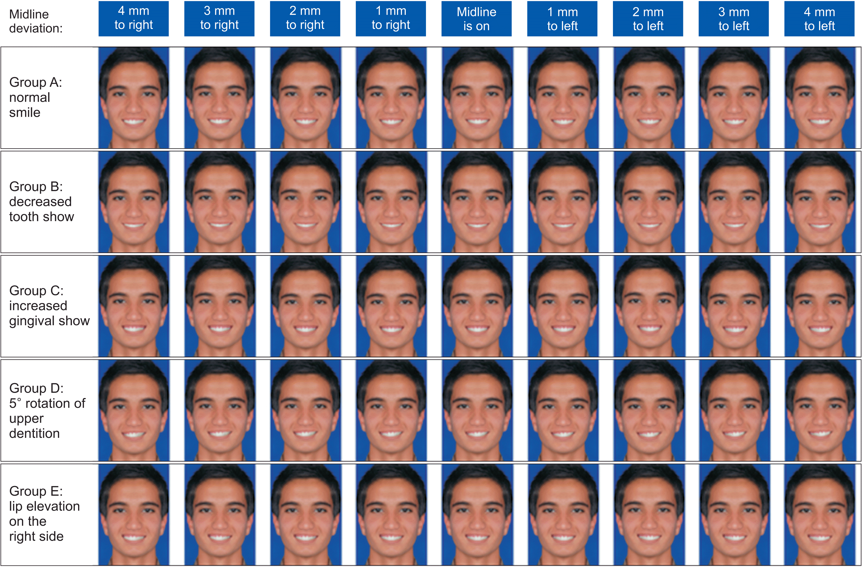
Informed consent was obtained from the subject, and his photograph was taken using a digital flash-on camera (c-2000; Olympus America, Melville, NY, USA) with a frontal smile under standard conditions. The smile served as a control and gold model (Series A) for other photographs. This image was altered using a software program (Adobe Photoshop CS, version 8.0; Adobe Systems, San Jose, CA, USA) according to the following measurements to create other picture series. Minimum distinguishable
5,6,11,30,31 modifications in the gingival show were applied in the following series so that all the raterscould recognize the modifications of the gingival show (without manipulatingthe lower facial third and buccal corridor size):
- Series A (normal): The ideal control smile in which the whole crowns of the upper anterior teeth were visible without gingival show.
- Series B (decreased toothshow): Gingival show decreased 4 mm symmetrically, compared to the normal status.
- Series C (increased gumminess): Gingival show was increased symmetrically for all the teeth representing the smile, with a maximal gingival show of 3.5 mm in the upper central incisor area.
- Series D (maxillary cant): The gingival show was increased asymmetrically by rotating the upper dentition 5º around the most incisal contact point of central incisors on the upper dental midline. The right side was moved downward, and the left side was elevated.
- Series E (asymmetric lip elevation): The gingival show was increased asymmetrically by moving the right commissure 2 mm upwards.
In each series, the upper dentition was displaced along the horizontal plane from –4 (left) to +4 (right) mm in 1-mm increments.
Each series of images was printed separately in the dimension of a typical human head and placed randomly in a binder.
Evaluation of the photographs
The sample size was calculated by Open-Epi software according to Johnston et al.,
16 who reported mean visual analog scale (VAS) scores of 5.5 ± 1.2 and 6.4 ± 1.7 for 2 mm of midline discrepancy by orthodontists and laypersons, respectively. The power of the study was set at 80% with a 95% confidence interval. The software showed that 42 raters should be included in each group. The 210 Iranian raters consisted of orthodontists, maxillofacial surgeons, prosthodontists, operative dentists, and laypeople (n = 42 in each group).
The laypeople’s selection criteria consisted of no previous orthodontic or facial surgical treatment and esthetic dental treatment, no facial deformities, and no healthcare employee as these factors might affect the perception of smile attractiveness.
12,32-36
Each rater received the frontal photographs in 5 series (with nine images) separately (series A, B, C, D, and E) and was asked to rate the subject’s attractiveness by selecting a point along a VAS from 1 to 100.
23 Furthermore, they were asked whether the subject required treatment via “Yes” or “No” response. The acceptance threshold for each evaluator on each side was based on their highest midline deviation that did not need orthodontic treatment.
The same researcher (MA) instructed all the 210 raters how to use the scale. Each rater was asked to rate each photograph’s attractiveness on whatever criteria they deemed satisfactory. The smiling frontal photographs in each set were randomized before rating using random numbers. Each questionnaire, with questions about the raters’ demographic characteristics, was marked by a numeric code to guarantee anonymity.
30% of the raters in each group were asked to re-rate the images and complete the questionnaires after two weeks to determine intra-examiner reliability.
Statistical analysis
All the statistical analyses were carried out using SPSS 22.0 (IBM Corp., Armonk, NY, USA).
The threshold data were not distributed normally based on the Kolmogorov–Smirnov test. Friedman test was used to compare the differences in acceptance thresholds of photos among each image series. Wilcoxon signed-ranks test was used to compare the right and left acceptance threshold of each image series and for pairwise comparisons. Thresholds of each image series were compared among the five groups of raters and the two sexes using Kruskal–Wallis and Mann–Whitney tests, respectively.
Repeated-measures analysis of variance (ANOVA) was used to compare the differences in attractiveness ratings of photos among each image series. Post hoc Sidak’s t-test was applied for pairwise comparisons.
One-way ANOVA and post hoc Duncan’s multiple range test were used to compare differences in the average attractiveness ratings among the five groups of raters. Student’s t-test was used to compare differences in the average attractiveness ratings among the two sexes.
The results were evaluated at the p < 0.05 significance level.
RESULTS
The 210 raters consisted of 114 females (mean age = 33.73) and 96 males (mean age = 34.23), with an age range of 18–56 (overall mean = 33.98, orthodontists’ mean = 34.11, prosthodontists’ mean = 33.71, oral and maxillofacial surgeons’ mean = 34.43, operative dentists’ mean = 33.67, laypeople’s mean = 33.98) with no significant difference in the mean age of the raters and the two sexes (p > 0.05).
The intraclass correlation coefficients were 0.75 (lower bound, 0.68; upper bound, 0.80; with 95% confidence interval) and 0.83 (lower bound, 0.71; upper bound, 0.95; with 95% confidence interval) for the thresholds and attractiveness scores, respectively, indicating moderate to good intra-rater reliability.
There was no significant difference between the male and female raters in the midline threshold and image attractiveness perception (
p > 0.05). Also, there were no significant differences between the groups of raters in the perception of the midline threshold and image attractiveness (
p > 0.05), except for 5 images (L3A,
p = 0.014; L4A,
p = 0.001; R3A,
p = 0.021; L4B,
p = 0.003; and L4D,
p = 0.029) regarding the attractiveness, with laypeople rating these images as significantly more attractive than the other raters (
Tables 2 and
3).
When the right and left thresholds were not significantly different, the two sides’ mean threshold was calculated as the mean threshold. Friedman test showed significant differences in acceptance thresholds of photos in each image series (p = 0.0001). In symmetrical image series, the right and left thresholds were statistically the same in all the rater groups. However, the right threshold of series D was significantly lower than the left threshold in groups of surgeons, operative dentists, and laypeople (p = 0.034, p = 0.038 and p = 0.007, respectively). Also, the right threshold of series E was significantly higher than the left threshold in prosthodontist and laypeople groups (p = 0.032 and p = 0.043, respectively).
In most rater groups, series B exhibited the highest mean threshold. The mean threshold of image series A was more than image series C in all the rater groups (orthodontists,
p = 0.0001; prosthodontists,
p = 0.014; oraland maxillofacial surgeons,
p = 0.0001; operative dentists,
p = 0.001; lay people,
p = 0.0001). Image series D exhibited the lowest mean threshold in all the rater groups (
Table 2).
The mean attractiveness of each image (midline deviation) in each image series, as rated by the groups, is presented in
Figure 2. The results showed significant differences in each image’s attractiveness within each image series (
p < 0.05) (
Table 4). In symmetrical image series, all the raters selected undeviated midline as the most attractive image of their series except for the laypeople. In these image series, the average attractiveness of the right and left midline deviation was not significantly different in all the rater groups (
p > 0.05). All the rater groups selected 4-mm midline discrepancy as the least attractive image in each image series. In image series E, all the rater groups selected E0 as the most attractive and L4E as the least attractive image. Most rater groups considered the right midline deviations more attractive than the left midline deviation. In image series D, most groups selected L2D as the most attractive image of its series. In this image series, the mean attractiveness of the left midline deviations was significantly higher than the right in all the rater groups (
p = 0.0001). All the rater groups chose R4D as the least attractive image of this image series (
Tables 3–
5 and
Figure 2).
DISCUSSION
A patient with a normal gingival show in an orthodontic office is more the exception than the rule.
7-9 In the orthodontic finishing stage, the range of acceptable position of the upper dental midline relative to the face becomes a question for an orthodontist in an individual with a disproportionate gingival show if there are limitations to follow the strict rule of the undeviated midline position.
It can be inferred from the result of the present study that as the gingival show increases, the threshold for midline deviation becomes more limited, and the strict rule of undeviated midline position is still applicable for symmetrical gingival show.
Most previous studies have reported an acceptance threshold of approximately 2 mm, consistent with our findings in the normal and decreased tooth show group.
5,13,14,16,21,23,24
Some studies have reported a higher threshold than ours,
12,18,22,25 with others reporting lower thresholds
17,20,26,37 of 1–5 mm.
In studies on attractiveness, the most attractive image was the one with undeviated dental midline, and the images were scored as less attractive as the midline discrepancy increased.
15,16,21 However, in some studies, some raters considered the images with midline deviations up to 3–5 mm as attractive as the undeviated status,
17,18,25 confirming our findings in symmetrical image series where the undeviated midline status was scored as the most attractive; however, in our study, up to 1 mm of midline deviation was rated as attractive as the coincident midline in most situations.
One reason for the different results in previous studies might be the raters’ ethnicity. A previous study
20 showed that the raters’ ethnicity affected upper dental midline deviation perception. Another factor might be the sex of the subject being rated. It has been shown that evaluators are somewhat less tolerant of deviations in female subjects.
13,15,24
Another variable affecting the rater’s judgment on the midline deviation is the lower anterior tooth show, with some studies reporting a higher midline threshold than the present study that included the lower anterior teeth in the images of upper dental midline deviation.
12,22,25
Moreover, the esthetic assessment method might play a role, with one study exhibiting a lower acceptable midline threshold using a digital esthetic assessment protocol.
20
One interesting finding is that in most studies reporting a lower midline threshold
15,17,20,37 than the present study, the extent of the image presented to the rater was the lips-only
15,17,37 or the lower face.
15,20 The broader perspective can dilute the attention to the smiles’ details, such as the acceptable upper midline position.
15,22
Regarding the asymmetrical image series, in the presence of maxillary cant, the present study indicated that the midline threshold for the side where the maxilla was relatively low with a higher gingival show was lower in almost all the groups. Also, in this image series, almost all the groups selected a 1–2-mm deviation to the left from the midline as the most attractive image of its series. In this image series, the mean attractiveness of the left midline deviations was higher than the right in all the rater groups.
In asymmetrical lip elevation, all the groups selected the undeviated position as the most attractive image of its series. Most rater groups considered right midline deviations more attractive than left midline deviations in this image series.
The present study showed that the strict predominance of undeviated midline position is probably less applicable for the asymmetrical gingival show, and in maxillary cant, the dental midline might be allowed to deviate to the side where the maxilla is relatively higher. In cases of asymmetrical lip elevation, although the most attractive position of the dental midline is the undeviated status, the dental midline might be permitted to deviate to the side with the increased gingival show.
A few studies have investigated the perception of upper dental midline deviation concerning facial asymmetry.
3,13
Silva et al.
3 concluded that facial asymmetries in the chin and nose affected the upper dental midline shift perception, consistent with the present study to some extent. It seems that the human eye is sensitive to facial asymmetries, such as asymmetrical nose and chin in the study above and maxillary cant in the current study, indicating that midline deviations to the side where the maxilla was low were rated less esthetic.
The present study showed that the raters’ sex did not affect the perception of midline threshold or image attractiveness. In most previous studies, male and female raters have not exhibited a statistically different perception of upper midline deviations, consistent with the present study.
3-5,13,18,20-23
In the present study, there was no significant difference between the groups of raters in the midline threshold perception. Moreover, no difference was observed between the rater groups in the attractiveness perception of most images. Our evaluators were young Iranian adults with similar mean age among the groups of raters. Since raters’ age and ethnicity have previously been found to result in significant differences in the perception of upper dental midline deviations in a few studies,
18,20 the results of this research in other age groups or races might be different. There is controversy over the effect of profession in previous studies. In some studies, dental professionals and laypersons had the same perception of midline deviation, consistent with the present study, where profession did not affect the perception of the threshold of upper midline deviation.
25,37 However, some studies have shown that professionals, especially orthodontists, are significantly less tolerant of midline deviation.
13,16,18
Finally, during the orthodontic treatment of a patient without an ideal gingival show, the following can be suggested about the position of the upper dental midline with caution as limitations of the present study:
Increasing the gingival show for asymmetry limits the amount of acceptable midline deviation.
In symmetrical gingival discrepancy, the coincident midline position should be strictly followed, especially in gummy smile patients.
In the maxillary cant, the undeviated midline position predominance is possibly less applicable, and the dental midline can be deviated to the side where the maxilla is relatively higher, or there is less gingival show.


 PDF
PDF Citation
 Print
Print


 XML Download