

 PDF
PDF Citation
 Print
Print


INTRODUCTION
Unconventional biliary anatomy is encountered in less than half of patients. It is mostly related to the intrahepatic biliary system [1]. However, duplication of the extrahepatic bile duct (EBD) is one of the rare biliary variations reported uncommonly. In this anomaly, there is a presence of two EBDs, one of which has normal drainage into the duodenum and the other (usually called accessory common bile duct [CBD]) opens in different parts of the upper gastrointestinal tract like the stomach, duodenum, or pancreatic duct [2,3]. This intriguing anomaly has been reported in association with pancreaticobiliary maljunction (PBM) & choledochal cysts. It can predispose to choledocholithiasis, cholangitis, pancreatitis, and biliary malignancies [2,4]. Among various types of duplicated extrahepatic bile duct (DEBD), type V has a single biliary drainage in the duodenum with or without a communicating channel. It is considered the rarest variant [2].
In this report, we present a case of type Vb DEBD associated with cholelithiasis and multiple calculi in the right hepatic duct managed successfully by surgical intervention. It is essential to discuss and report such rare biliary anomalies to gain knowledge about relevant investigations and treatment options for them.
CASE
A 38-year-old lady presented with right upper abdominal pain for the past week with a low-grade fever. Evaluation by ultrasound elsewhere showed cholelithiasis with a dilated bile duct. In initial laboratory tests, she had a total bilirubin level of 2.2 mg/dL (normal range, 0.3–1.2 mg/dL), an aspartate aminotransferase level of 142 IU/L (normal: < 35 IU/L), an alanine-aminotransferase level of 104 IU/L (normal: < 35 U/L), and an alkaline phosphatase level of 191 IU/L (normal range, 30–120 IU/L).
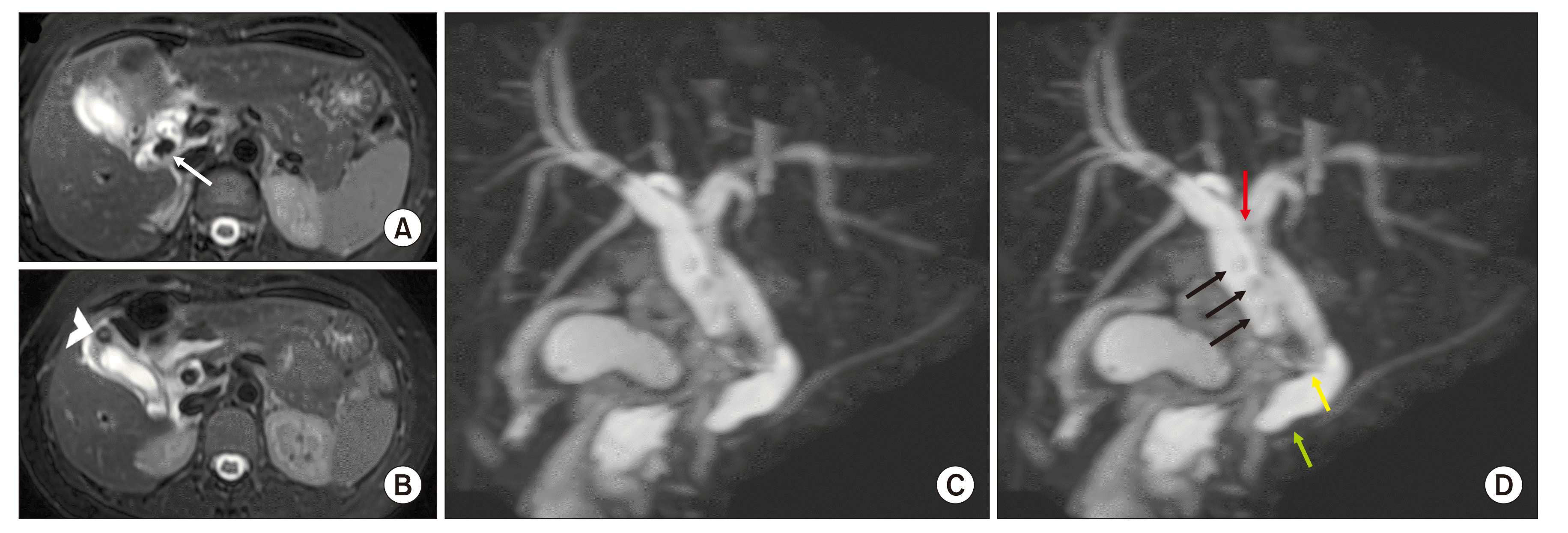
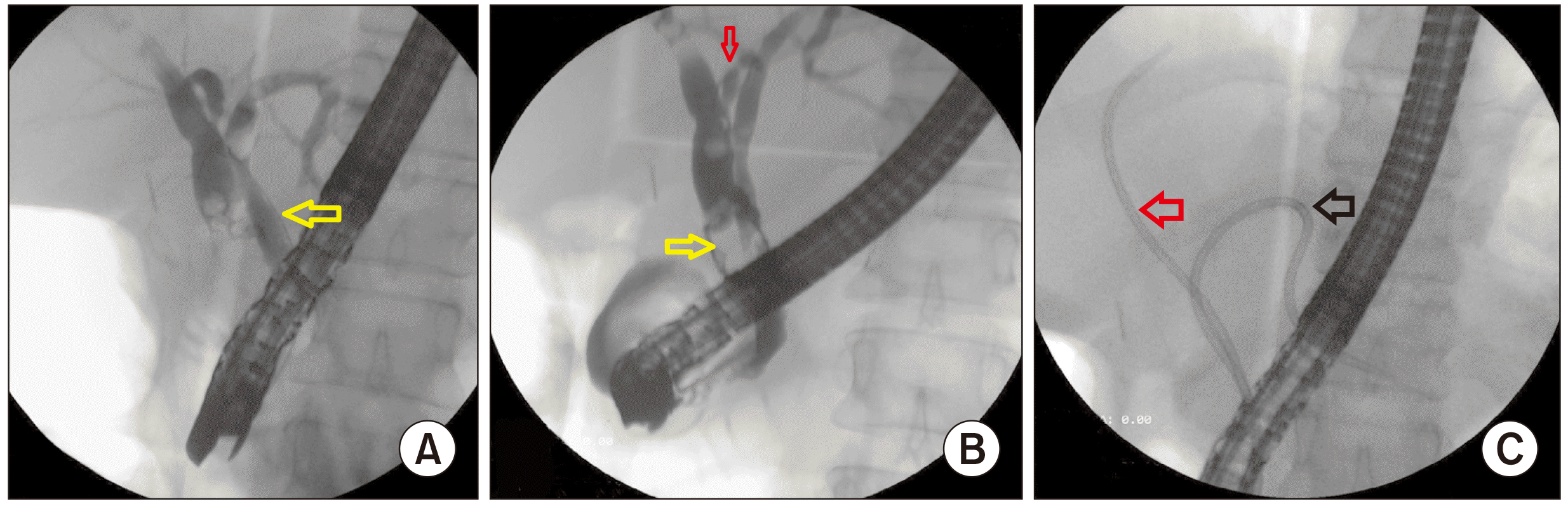
As the patient had an intermediate risk of choledocholithiasis, magnetic resonance cholangiopancreatography (MRCP) was performed, which revealed duplication of EBD with multiple calculi in the right EBD (ductolithiasis). Right and left EBDs had a small communication at the location of primary confluence, with the right EBD joining the left EBD in the intrapancreatic region. Cholelithiasis was also present with cystic duct opening into the right EBD (Fig. 1). Endoscopic retrograde cholangiography (ERC) was done to clear the right EBD calculi. Although the right EBD was visualized via contrast passage, the actual junction of the right EBD with the left EBD could not be cannulated. Biliary stents were placed, one in the left EBD and the other in the right anterior sectoral duct, via the cranial communication between the two ducts. She was planned for surgical exploration (Fig. 2).
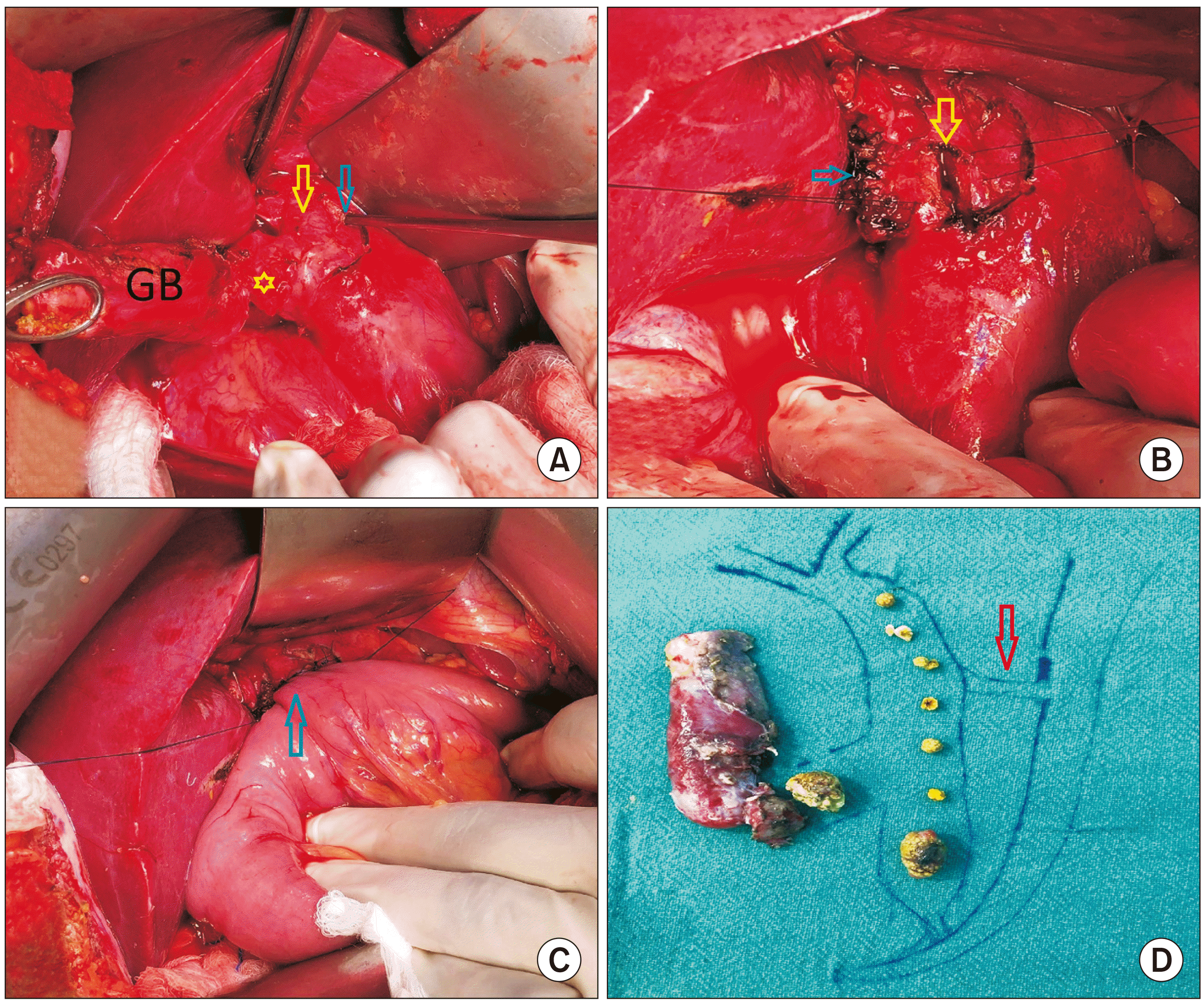
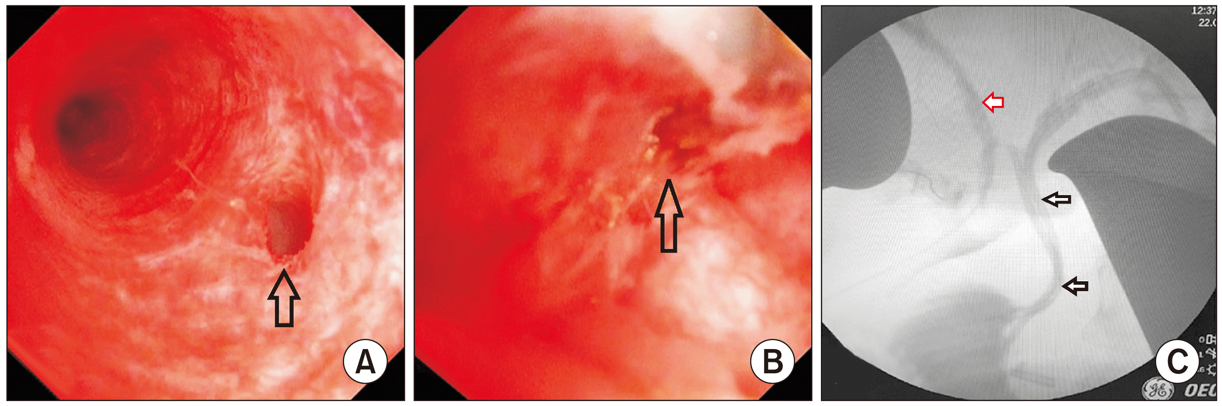
Intraoperatively, the gall bladder was distended and thick-walled with an impacted stone in the cystic duct. Cholecystectomy was completed and the cystic duct was ligated at its junction with the right EBD (Fig. 3A, 3B). The right EBD was dilated up to 15 mm with pericholedochitis, while stents were felt in the left EBD. Stone clearance was achieved via a longitudinal choledochotomy in the right EBD. On video choledochoscopy (Olympus Medical System), the proximal communication between left and right EBDs was visualized as well as the intrapancreatic joining of the right EBD into the left. However, none was negotiable (Fig. 4A, 4B). Intraoperative cholangiography confirmed these findings, with both right and left EBDs being opacified without filling defects. The contrast was reaching the duodenum via the left EBD, with a hold-up of contrast in the right EBD (Fig. 4C). Two stents were noted in situ in the left EBD. Side to Side Roux-en-Y hepaticojejunostomy (HJ) was done with the right EBD at the site of the previous choledochotomy (Fig. 3C, 3D). Hand-sewn side-to-side jejunojejunostomy was done in two layers ~40 cm distal to HJ.
Her immediate postoperative course was uneventful. She was allowed oral liquids on 1st postoperative day (POD) and a soft diet by 2nd POD. Her drain was removed on the 3rd POD. She had a mild superficial surgical site infection which was managed with daily dressings. She was discharged in a stable condition on the 6th POD. After six weeks, both her biliary stents were removed. She is currently doing well with normal liver function tests at three-month follow-up.
DISCUSSION
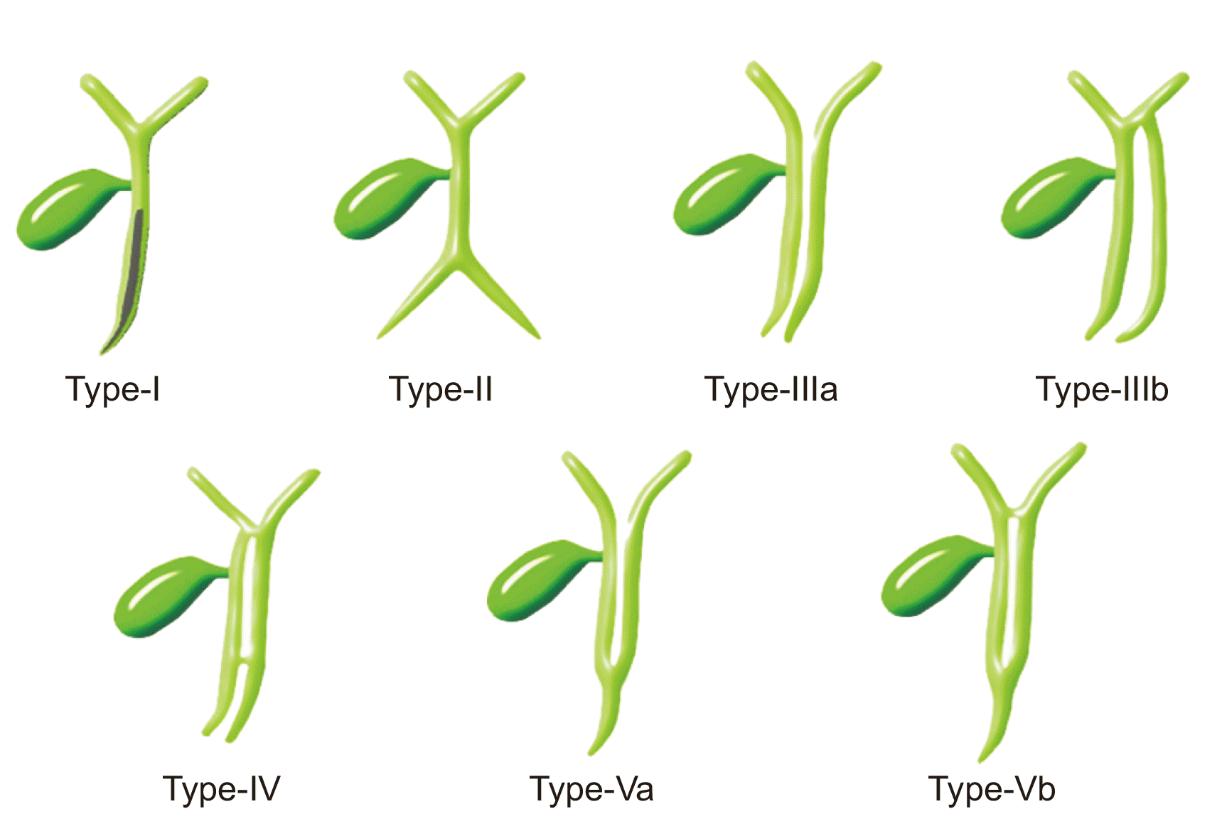
DEBD is a rare congenital anomaly of the biliary system. It is characterized by a septum within the CBD or an accessory CBD. Although the first description of this anomaly dates back to the 16th century, the most recent classification for these anomalies has been presented by Choi et al., [2] with DEBD classified into five different types: type I, a septum within the CBD lumen; type II, the distal bile duct bifurcates and each channel independently drains into the different sites; type III, duplicated extrahepatic bile ducts without (type IIIa) or with (type IIIb) intrahepatic communicating duct; type IV, duplicated extrahepatic bile ducts with the extrahepatic communicating channel or both intrahepatic and extrahepatic communicating channels; and type V, single biliary drainage of double extrahepatic bile ducts without (a) or with communicating channels (b) (Fig. 5). The most commonly reported anomalies are types III and IV, with one duct opening into the major duodenal papilla and a second duct (often referred to as accessory bile duct) opening into the stomach, duodenum, or pancreatic duct [5,6]. Type V anomaly is the rarest. It is characterized by DEBD joining as a single duct and draining into the duodenum. It is subdivided based on the absence (type Va) or presence (type Vb) of communicating channel between left and right EBDs. Type Vb is an extremely rare anomaly, with only two cases reported till now [7,8]. Our case, the third one in this category, presented with cholelithiasis and multiple calculi in the right EBD (Table 1). The term choledocholithiasis is used for calculi in the CBD and the term hepatolithiasis is used for intrahepatic calculi proximal to biliary confluence. However, both terms did not concur in the present case. Thus, we labelled them as ductolithaisis (calculi in the right EBD).
Despite being rare, it is necessary to understand this anomaly due to its important clinical implication. The development of DEBD occurs due to interference in the hepatic primordium recanalization, which involves a division of the hepatic diverticulum in the first week of embryogenesis [6]. It results in persistence of an extrahepatic accessory duct, which usually regresses during a normal course [2]. This anomaly is predominantly found in middle-aged females, showing symptoms such as right upper abdominal pain, fever with chills, and jaundice [6]. Although the presence of multiple stones is common in DEBD, there is usually no evidence of significant cholestatic features or profound dilatation of intrahepatic biliary radicals as shown in our case. This is attributed to a communication channel between the two EBDs which tends to decompress the obstructed duct.
The actual incidence of this anomaly is difficult to know as only symptomatic patients come under the scanner or are encountered after complication appears during surgical intervention [9]. Hence, an attempt to diagnose these anomalies correctly before any intervention is essential. Ultrasound is usually performed as the first investigation in hepatobiliary disorders. However, ultrasound is known to be operator-dependent. In addition, its sensitivity and specificity to identify complicated biliary anomalies are lower than cross-sectional imaging, which is usually required to diagnose them and their associated complications. MRCP is considered the foremost noninvasive imaging technique that provides adequate diagnostic information [2,5]. It can help us classify the type of DEBD and determine the presence of PBM. It can also help us look for calculi in EBDs. MR angiography can also be added to obtain information about any vascular anomalies. In a few instances, information from the MRCP is limited due to the presence of multiple impacted calculi. In those cases, an abdominal contrast-enhanced computed tomography (CECT) with a MinIP mode might be helpful for depicting complex anatomical details of this anomaly, especially when there is doubt of associated malignancy [2,4,7].
ERC is an invasive procedure with associated complications. Thus, it is used predominantly as a therapeutic modality. Endoscopic sphincterotomy with successful stone extraction can help us avoid further CBD exploration. However, due to the complex anatomy and difficult cannulation access, it may not be possible to access both EBDs [10]. In our case, although calculi were present in the right EBD, only those in the left EBD could be cannulated during ERC. We could not enter the right EBD as its intrapancreatic communication with the left EBD was small and probably strictured, which could have lead to bile stasis and stone formation in the first place.
Cholecystectomy, along with bile duct exploration, is required to remove all calculi. It is important to avoid any inadvertent bile duct injury. In addition, a choledochoscope can ensure complete clearance of ductolithiasis in the proximal and distal duct, as we did for our case. Intraoperative cholangiography is required to precisely delineate the biliary anomaly with the presence of communication between the two ducts and localization of the ectopic opening of the accessory duct, if any. It also helps us avoid injuring the undilated normal EBD during surgery (the left EBD in our case). In the presence of PBM, cholecystectomy with EBD resection is required as this malformation predisposes to malignancy. However, it is not necessary in type V DEBD. In our case, due to a distal stricture in the right EBD, we added biliary drainage after stone clearance to prevent recurrence of symptoms, EBD stones, and future cholangitis. Furthermore, the left biliary system was neither dilated nor obstructed on ERCP or MRCP. Hence, no intervention was offered for it.
In conclusion, DEBDs are rare anomalies and often present with cholelithiasis, ductolithiasis, and cholangitis. MRCP is the most important investigation for diagnosis. ERC is both diagnostic and therapeutic in certain scenarios. When indicated, CBD exploration and clearance with biliary drainage is the optimum treatment. It is crucial to identify these anomalies to avoid intraoperative injuries to extrahepatic ducts. However, regular follow-up is required to prevent any future complications.
 XML Download