Journal List > Ann Hepatobiliary Pancreat Surg > v.27(2) > 1516082793

Mownah and Aroori: The Pringle maneuver in the modern era: A review of techniques for hepatic inflow occlusion in minimally invasive liver resection

Abstract

During minimally invasive liver resection (MILR), the Pringle maneuver aims to minimize blood loss and provide a clear operative field, thereby identifying intrahepatic structures and facilitating safe parenchymal transection. Several techniques for using the Pringle maneuver in MILR have been described. This review presents various methods which have been reported in the literature. A systematic literature search used the MEDLINE/PubMed database from its earliest records to August 2022 using appropriate search headings and keywords. The primary outcome was identifying techniques for performing hepatic inflow occlusion during laparoscopic/robotic hepatectomy. Inclusion criteria consisted of publications describing technical steps to obtain hepatic inflow occlusion during minimally invasive hepatectomy. A literature search identified 23 relevant publications, and the full texts were examined. The techniques described in the reports can be broadly categorized into three groups: (1) the Rummel-tourniquet technique, (2) vascular clamp use, and (3) the Huang Loop technique. Various techniques have been used in MILR to achieve inflow confinement successfully. The authors prefer the modified Huang Loop technique because it is inexpensive, reliable, and quick to apply or release. Hepatobiliary surgeons are advised to familiarize themselves with these MILR techniques, which have proven effective and safe inflow occlusion.

INTRODUCTION

Liver resection performed by a minimally invasive technique (laparoscopic, robotic or hybrid approaches) is an increasingly used treatment strategy for patients requiring hepatectomy for various benign and malignant diseases [1,2]. Minimally invasive liver resection (MILR) has demonstrated oncological safety and is associated with low mortality and morbidity rates [3]. However, the procedure requires experienced surgical teams because the operative aspects are complex. One of the significant challenges is maintaining haemostasis at the transection plane.
The Pringle maneuver has been successfully used in open hepatectomy to address this challenge and facilitate safe hepatectomy [4]. The concept of inflow occlusion to control hepatic bleeding is named after James Pringle, a 19th century surgeon from Glasgow. It is a valuable procedure to reduce hemorrhage in the setting of hepatic trauma [5]. When applied to MILR, the Pringle maneuver may provide several advantages. Primarily the Pringle maneuver helps reduce blood loss and reduces need for perioperative blood transfusions. Additional advantages include providing a more apparent operative field and an enhanced ability for the surgeon to visualize intrahepatic vascular and biliary structures during the transaction. This could theoretically reduce bleeding or damage to major vascular/biliary radicles.
Several techniques describe performance of the Pringle maneuver in minimally invasive surgery. However, an optimal method has yet to be identified. This review article aims to set out the various techniques described in the literature with their respective advantages and disadvantages.

MATERIALS AND METHODS

A systematic literature search on MEDLINE/PubMed was performed from its earliest records to June 2022. The following medical search headings and keywords were used: "liver resection" OR "hepatectomy" AND "Pringle maneuver" OR "Pringle maneuver" OR "inflow occlusion" OR "hepatic inflow occlusion" AND "minimally invasive" OR "laparoscopic" OR "robotic" in non-MeSH terms.
The primary outcome was identifying techniques for performing hepatic inflow occlusion during laparoscopic/robotic hepatectomy. Inclusion criteria consisted of publications describing technical steps to obtain hepatic inflow occlusion during minimally invasive hepatectomy. Includes only articles reporting studies using human subjects written in English. The full-text articles were acquired and screened for eligibility. Publications which did not include laparoscopic or robotic surgical approaches to hepatectomy were excluded.
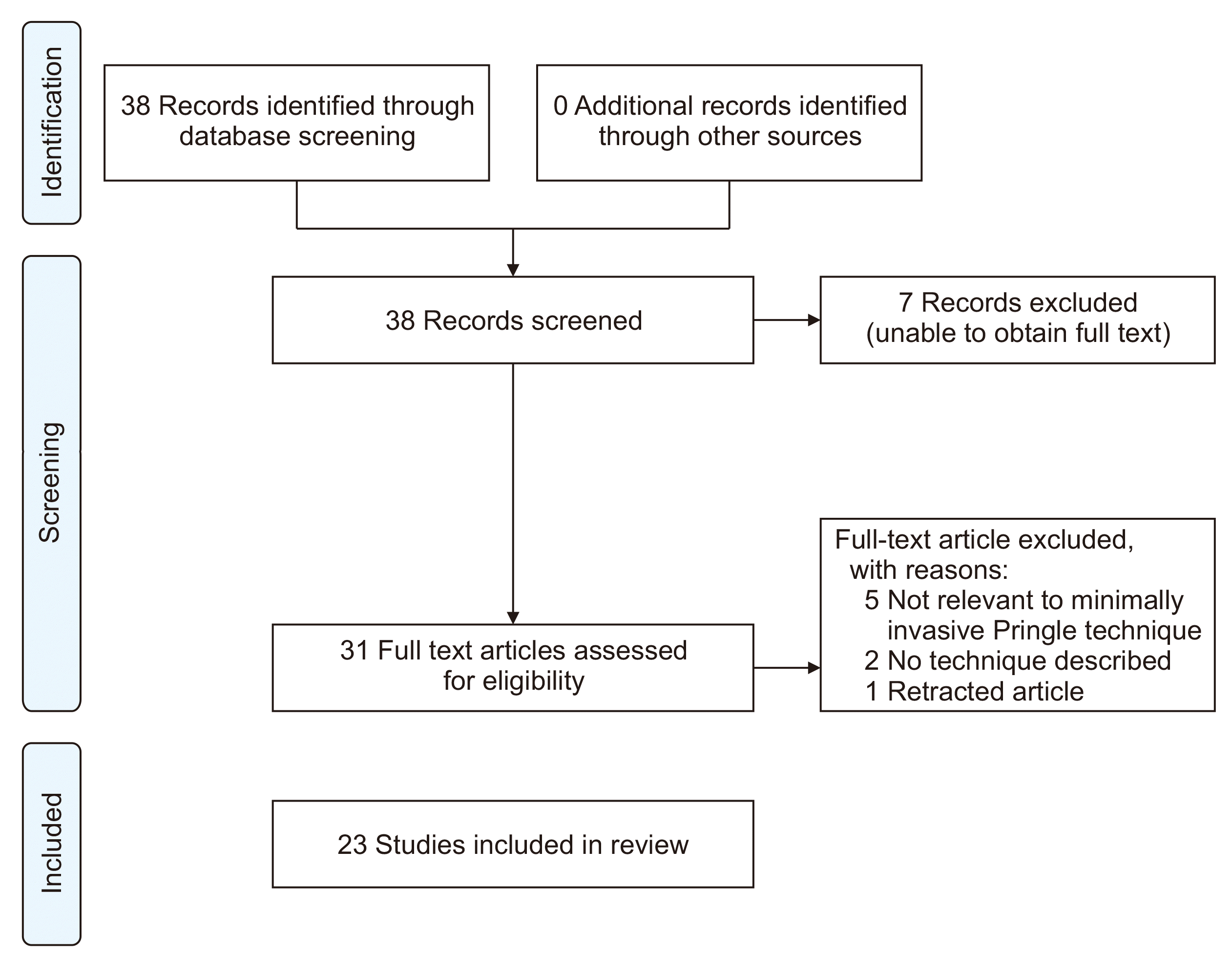
Descriptive technical reports were requested rather than statistical analyses of outcomes and therefore no validated scale was used. Author assessment (OAM and SA) of the quality of individual articles was performed. Report of the literature search results according to PRISMA guidelines (Fig. 1).

RESULTS

After the initial search, 38 records were found. Seven studies were excluded because of inability to retrieve the full-text article. Five of the 31 full-text articles screened were excluded because they did not comply with the study topic. Furthermore, two were excluded because no description of the technique for using the Pringle maneuver was provided or the technique did not pertain to minimally invasive surgery. One article was subsequently withdrawn by the authors and was therefore excluded. Finally, 23 publications were identified and the full texts were examined. Table 1 presents the records [6-28]. Table 2 presents the reported advantages and disadvantages of the techniques.
The techniques described in the 23 articles included in the review can be classified as intracorporeal or extracorporeal. Extracorporeal usually requires an additional laparoscopic port. The techniques can be placed into 3 groups as similar principles were followed, albeit using slight modifications in technique. The three types of technique which emerged were:
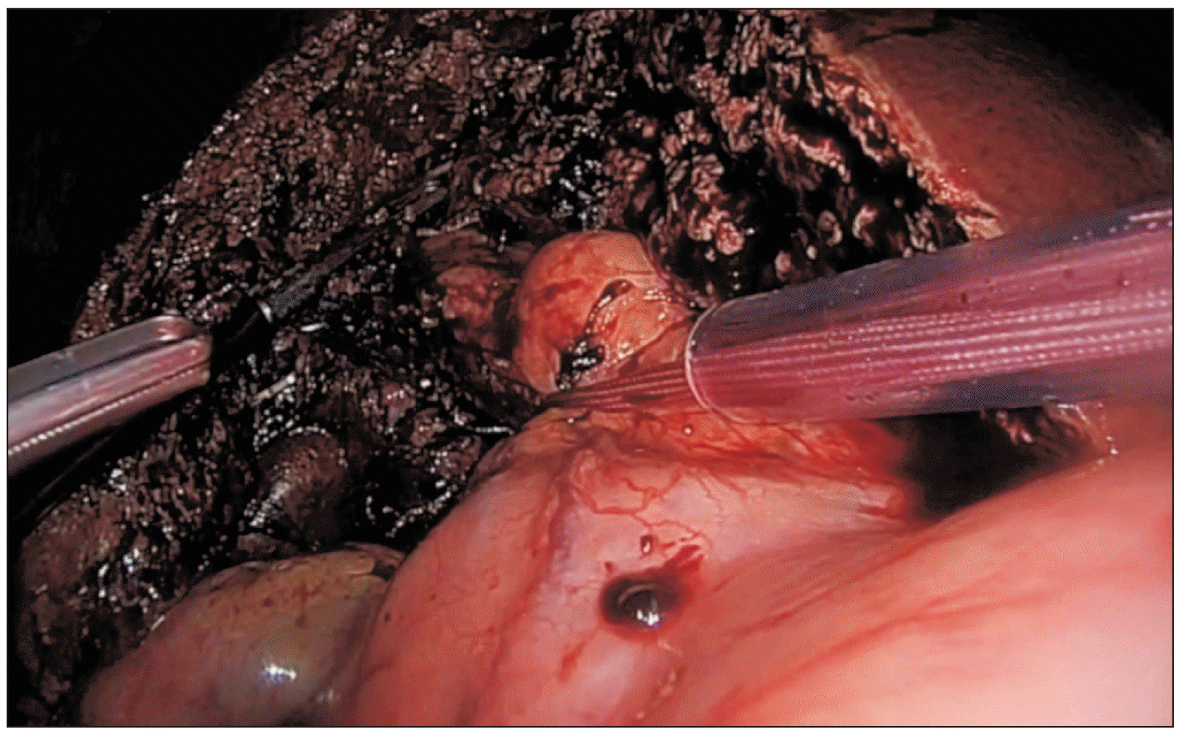
1. The Rummel tourniquet: wrapping around the hilar structures using tape (cotton, nylon, or umbilical), which is then passed through a rigid tube (Fig. 2).
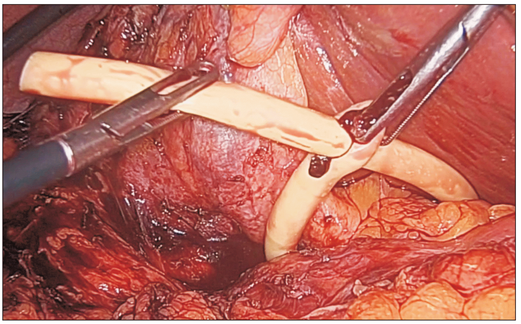
2. Direct compression to occlude the inflow structures using a vascular clamp (Fig. 3).
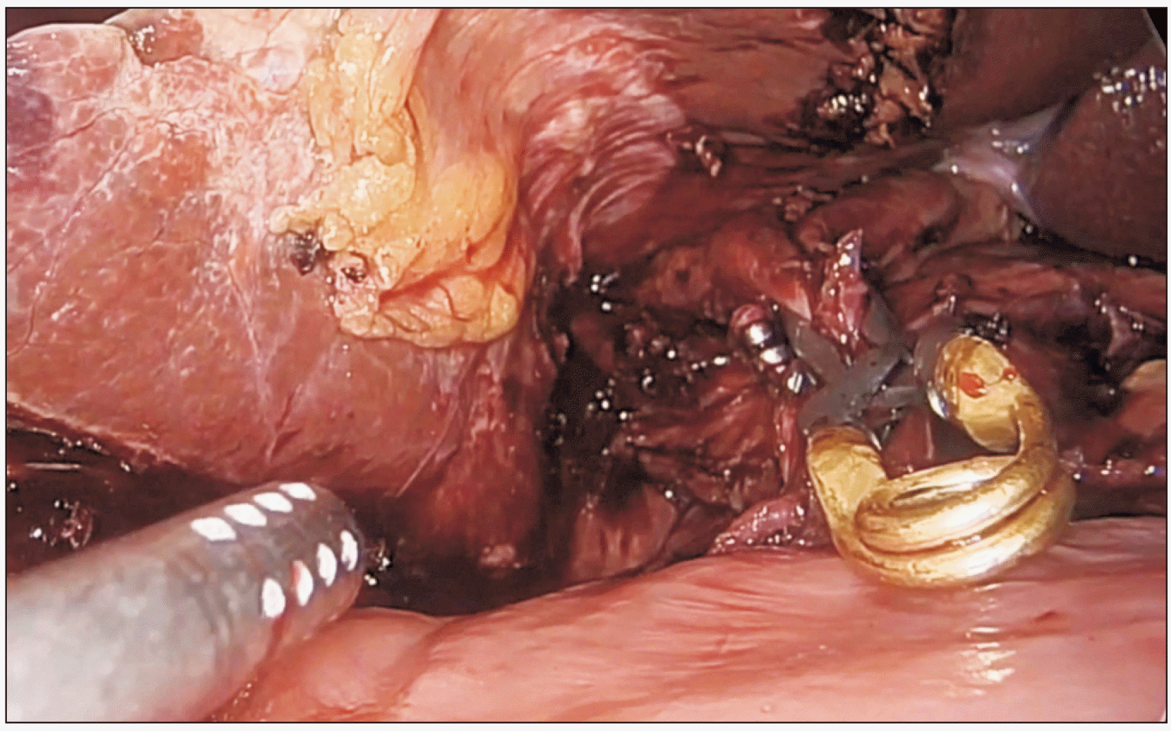
3. Constriction of hilar structures using Foley catheter—Huang Loop technique (Fig. 4).

CONCLUSIONS

Laparoscopic liver resection is a complex procedure that requires extensive training and experience [29]. Intra-operative hemorrhage is a primary challenge facing the surgeon, with ramifications including the requirement for a blood transfusion associated with increased postoperative complications, mortality, and reduced disease-free survival [30,31]. Theoretically, bleeding during the transection may increase operative times and impair the operative field, resulting in difficulty identifying intrahepatic biliary and vascular structures. The Pringle maneuver is an established strategy to facilitate hepatectomy whilst maintaining haemostasis in the transection plane.
The early descriptions of the laparoscopic Pringle maneuver tended to describe the Rummel tourniquet technique, widely used by liver surgeons in open hepatectomy. The clear advantage is that the surgeon is familiar with the technique and uses the same apparatus in their open practice. The Rummel tourniquet method is safe and can be used efficiently. Okuda et al. [6] reported that preparing this technique would take 354 seconds. A significant advantage offered by this technique during right hepatectomy is that extracorporeal retraction can be used to retract the hilum to the left, which improves the view of the inflow structures during hilar dissection.
The option of using a vascular clamp to achieve inflow occlusion can be undertaken using an extracorporeal vascular clamp [7]. However, this can be problematic depending on the type of hepatectomy which is being performed, as the long handle of the instrument may obstruct the surgeon’s view or clash with other instruments. The intracorporeal vascular clamps (bulldog clamps) are particularly useful in a recent study demonstrating comparable short-term outcomes to laparoscopic hepatectomy versus cotton tourniquet technique [32]. The bulldog technique is particularly applicable in instances where there is concern that the umbilical tape may damage hilar structures, for example, after hilar lymphadenectomy, where the structures are devoid of protective lymphatics and connective tissue.
The Huang Loop is a relatively recent innovation and is our institution’s current method of choice [8]. It has the benefit of being intracorporeal and relatively cost-effective. The silastic properties of the Foley catheter provide uniform constriction of the inflow structures whilst exerting minimal trauma. A particularly useful property of the 16- or 18-French Foley catheter is its relative rigidity which allows a passage through the Foramen of Winslow to be safely negotiated as it passes posterior from the pars flaccida to the hepatoduodenal ligament. This passage is performed without direct visualization of Winslow’s Foramen, but the Foley catheter’s atraumatic structure is unlikely to cause caval or other injury. The hepatoduodenal ligament may be closed following previous upper abdominal surgery due to adhesions. In such cases, a small amount of adhesiolysis is required before passing the Foley catheter through a window in the pars flaccida. The Foley then enters the safe window and emerges to the right of the ligament. Pringle may be released inadvertently under certain circumstances. This has been noted in our experience when the surface of the Foley is very wet/lubricated, and we advise keeping the Foley as dry as possible.
Intracorporeal techniques have the benefit of not requiring an additional port. Cai et al. [9] report that a straightforward technique successfully uses an inexpensive instrument to apply laparoscopic Pringle in 34 patients. The authors used the elastic rim of a size seven surgical glove to encircle the inflow structures and maintained tension using Hem-o-Lok. Although they reported that Pringle release using loose clip forceps is straightforward, this instrument may be have limited availability in laparoscopic centers, and removal of Hem-o-Loks once applied is associated with difficulty.
The patient’s left lateral decubitus positioning during laparoscopic liver surgery facilitates proper liver mobilization and provides access to the right superior segments (VII and VIII) but increases the technical difficulty of achieving hepatoduodenal ligament encirclement. In particular, the pars flaccida is challenging to access in this position. A preferred option is to use curved retractors, such as the Endo Retract Maxi (Medtronic) to pass the nylon tape around the hilar structures [10]. Using a vascular clamp (intra- or extracorporeal) is another option, and in recent cases, our center has had success using the Huang Loop technique.
Recent years have seen much progress in the robotic approach to MILR. Only two publications identified in this review reported the use of their technique for robotic hepatectomy [6,14]. It is appropriate to appreciate that Pringle technique used an extracorporeal method for both publications. Therefore, the assistant uses Pringle extracorporeally whilst the lead surgeon operates from the robotic console.
In conclusion, the optimal method of performing the Pringle maneuver should be based on simplicity, safety, reproducibility and cost-effectiveness. There are several techniques for using Pringle in both laparoscopic and robotic approaches without evidence of superiority. The surgeon or institutional preference dictates the technique chosen. It is recommended that surgical teams become familiar with more than one technique because of many factors, including adhesions from previous surgery or patient positioning. However, we recommend the Huang loop technique using a Foley catheter because it is inexpensive, straightforward, rapid and a safe way of applying Pringle in both open and laparoscopic cases.

Notes

CONFLICT OF INTEREST

No potential conflict of interest relevant to this article was reported.

AUTHOR CONTRIBUTIONS

Conceptualization: All authors. Data curation: All authors. Methodology: All authors. Visualization: All authors. Writing - original draft: All authors. Writing - review & editing: All authors.

REFERENCES

1. Haney CM, Studier-Fischer A, Probst P, Fan C, Müller PC, Golriz M, et al. 2021; A systematic review and meta-analysis of randomized controlled trials comparing laparoscopic and open liver resection. HPB (Oxford). 23:1467–1481. DOI: 10.1016/j.hpb.2021.03.006. PMID: 33820689.
crossref
2. Kamarajah SK, Bundred J, Manas D, Jiao L, Hilal MA, White SA. 2021; Robotic versus conventional laparoscopic liver resections: a systematic review and meta-analysis. Scand J Surg. 110:290–300. DOI: 10.1177/1457496920925637. PMID: 32762406.
crossref
3. Tsilimigras DI, Moris D, Vagios S, Merath K, Pawlik TM. 2018; Safety and oncologic outcomes of robotic liver resections: a systematic review. J Surg Oncol. 117:1517–1530. DOI: 10.1002/jso.25018. PMID: 29473968.
crossref
4. Belghiti J, Noun R, Zante E, Ballet T, Sauvanet A. 1996; Portal triad clamping or hepatic vascular exclusion for major liver resection. A controlled study. Ann Surg. 224:155–161. DOI: 10.1097/00000658-199608000-00007. PMID: 8757378. PMCID: PMC1235336.
crossref
5. Pringle JH. 1908; V. Notes on the arrest of hepatic hemorrhage due to trauma. Ann Surg. 48:541–549. DOI: 10.1097/00000658-190810000-00005. PMID: 17862242. PMCID: PMC1406963.
6. Okuda Y, Honda G, Kurata M, Kobayashi S. 2013; Useful and convenient procedure for intermittent vascular occlusion in laparoscopic hepatectomy. Asian J Endosc Surg. 6:100–103. DOI: 10.1111/ases.12003. PMID: 23126444.
crossref
7. Onda S, Haruki K, Furukawa K, Yasuda J, Shirai Y, Sakamoto T, et al. 2021; Newly-revised Pringle maneuver using laparoscopic Satinsky vascular clamp for repeat laparoscopic hepatectomy. Surg Endosc. 35:5375–5380. DOI: 10.1007/s00464-021-08516-9. PMID: 33913029.
crossref
8. Huang JW, Su WL, Wang SN. 2018; Alternative laparoscopic intracorporeal Pringle maneuver by Huang's loop. World J Surg. 42:3312–3315. DOI: 10.1007/s00268-018-4584-z. PMID: 29589115.
crossref
9. Cai J, Zheng J, Xie Y, Kirih MA, Jiang G, Liang Y, et al. 2020; A novel simple intra-corporeal Pringle maneuver for laparoscopic hemihepatectomy: how we do it. Surg Endosc. 34:2807–2813. DOI: 10.1007/s00464-020-07513-8. PMID: 32206920.
crossref
10. Belli G, Fantini C, D'Agostino A, Cioffi L, Limongelli P, Russo G, et al. 2008; Laparoscopic segment VI liver resection using a left lateral decubitus position: a personal modified technique. J Gastrointest Surg. 12:2221–2226. DOI: 10.1007/s11605-008-0537-4. PMID: 18473147.
crossref
11. Zhang C, Yu J, Ma J. 2021; How to perform laparoscopic intracorporeal Pringle manoeuvre: Zhang's modified method. ANZ J Surg. 91:742–743. DOI: 10.1111/ans.16710. PMID: 33665920.
crossref
12. SteinbrÜck K, Fernandes R, D'Oliveira M, Capelli R, Cano R, Vasconcelos H, et al. 2021; External pringle maneuver in laparoscopic liver resection: a safe, cheap and reproducible way to perform it. Arq Bras Cir Dig. 33:e1555. DOI: 10.1590/0102-672020200004e1555. PMID: 33503115. PMCID: PMC7836078.
crossref
13. Gao Z, Li Z, Zhou B, Chen L, Shen Z, Jiang Y, et al. 2021; A self-designed liver circle for on-demand Pringle's manoeuver in laparoscopic liver resection. J Minim Access Surg. 17:120–126. DOI: 10.4103/jmas.JMAS_130_19. PMID: 33353898. PMCID: PMC7945630.
crossref
14. Choi YI. 2020; The usefulness of the totally intra-corporeal pringle maneuver with Penrose drain tube during laparoscopic left side liver resection. Ann Hepatobiliary Pancreat Surg. 24:252–258. DOI: 10.14701/ahbps.2020.24.3.252. PMID: 32843589. PMCID: PMC7452809.
crossref
15. Peng Y, Wang Z, Wang X, Chen F, Zhou J, Fan J, et al. 2019; A novel very simple laparoscopic hepatic inflow occlusion apparatus for laparoscopic liver surgery. Surg Endosc. 33:145–152. DOI: 10.1007/s00464-018-6285-y. PMID: 29943053.
crossref
16. Lim C, Osseis M, Lahat E, Azoulay D, Salloum C. 2018; Extracorporeal Pringle maneuver during laparoscopic and robotic hepatectomy: detailed technique and first comparison with intracorporeal maneuver. J Am Coll Surg. 226:e19–e25. DOI: 10.1016/j.jamcollsurg.2018.02.003. PMID: 29501783.
crossref
17. Laurenzi A, Cherqui D, Figueroa R, Adam R, Vibert E, Sa Cunha A. 2018; Totally intra-corporeal Pringle maneuver during laparoscopic liver resection. HPB (Oxford). 20:128–131. DOI: 10.1016/j.hpb.2017.05.013. PMID: 29239841.
crossref
18. Piardi T, Lhuaire M, Memeo R, Pessaux P, Kianmanesh R, Sommacale D. 2016; Laparoscopic Pringle maneuver: how we do it? Hepatobiliary Surg Nutr. 5:345–349. DOI: 10.21037/hbsn.2015.11.01. PMID: 27500146. PMCID: PMC4960419.
crossref
19. Mizuguchi T, Kawamoto M, Nakamura Y, Meguro M, Hui TT, Hirata K. 2015; New technique of extracorporeal hepatic inflow control for pure laparoscopic liver resection. Surg Laparosc Endosc Percutan Tech. 25:e16–e20. DOI: 10.1097/SLE.0b013e3182a4c0f4. PMID: 25533749.
crossref
20. Nomi T, Fuks D, Agrawal A, Govindasamy M, Araki K, Gayet B. 2015; Modified Pringle maneuver for laparoscopic liver resection. Ann Surg Oncol. 22:852. DOI: 10.1245/s10434-014-4088-5. PMID: 25223928.
crossref
21. Dua MM, Worhunsky DJ, Hwa K, Poultsides GA, Norton JA, Visser BC. 2015; Extracorporeal Pringle for laparoscopic liver resection. Surg Endosc. 29:1348–1355. DOI: 10.1007/s00464-014-3801-6. PMID: 25159645.
crossref
22. Chao YJ, Wang CJ, Shan YS. 2012; Technical notes: a self-designed, simple, secure, and safe six-loop intracorporeal Pringle's maneuver for laparoscopic liver resection. Surg Endosc. 26:2681–2686. DOI: 10.1007/s00464-012-2210-y. PMID: 22437948.
crossref
23. Rotellar F, Pardo F, Bueno A, Martí-Cruchaga P, Zozaya G. 2012; Extracorporeal tourniquet method for intermittent hepatic pedicle clamping during laparoscopic liver surgery: an easy, cheap, and effective technique. Langenbecks Arch Surg. 397:481–485. DOI: 10.1007/s00423-011-0887-3. PMID: 22183106.
crossref
24. Patriti A, Ceccarelli G, Bartoli A, Casciola L. 2011; Extracorporeal Pringle maneuver in robot-assisted liver surgery. Surg Laparosc Endosc Percutan Tech. 21:e242–e244. DOI: 10.1097/SLE.0b013e31822d7fb4. PMID: 22002285.
crossref
25. Herman P, Perini MV, Coelho F, Saad W, D'Albuquerque LA. 2010; Half-Pringle maneuver: a useful tool in laparoscopic liver resection. J Laparoendosc Adv Surg Tech A. 20:35–37. DOI: 10.1089/lap.2009.0215. PMID: 20059322.
crossref
26. Cho A, Yamamoto H, Nagata M, Takiguchi N, Shimada H, Kainuma O, et al. 2009; Safe and feasible inflow occlusion in laparoscopic liver resection. Surg Endosc. 23:906–908. DOI: 10.1007/s00464-008-0257-6. PMID: 19116742.
crossref
27. Belli G, Fantini C, D'Agostino A, Belli A, Langella S. 2005; Laparoscopic hepatic resection for completely exophytic hepatocellular carcinoma on cirrhosis. J Hepatobiliary Pancreat Surg. 12:488–493. DOI: 10.1007/s00534-005-1006-z. PMID: 16365825.
crossref
28. Kurokawa T, Inagaki H, Sakamoto J, Nonami T. 2002; Hand-assisted laparoscopic anatomical left lobectomy using hemihepatic vascular control technique. Surg Endosc. 16:1637–1638. DOI: 10.1007/s00464-002-4212-7. PMID: 12085137.
crossref
29. Nomi T, Fuks D, Kawaguchi Y, Mal F, Nakajima Y, Gayet B. 2015; Learning curve for laparoscopic major hepatectomy. Br J Surg. 102:796–804. DOI: 10.1002/bjs.9798. PMID: 25873161.
crossref
30. Lyu X, Qiao W, Li D, Leng Y. 2017; Impact of perioperative blood transfusion on clinical outcomes in patients with colorectal liver metastasis after hepatectomy: a meta-analysis. Oncotarget. 8:41740–41748. DOI: 10.18632/oncotarget.16771. PMID: 28410243. PMCID: PMC5522331.
crossref
31. Martin AN, Kerwin MJ, Turrentine FE, Bauer TW, Adams RB, Stukenborg GJ, et al. 2016; Blood transfusion is an independent predictor of morbidity and mortality after hepatectomy. J Surg Res. 206:106–112. DOI: 10.1016/j.jss.2016.07.013. PMID: 27916348. PMCID: PMC5142215.
crossref
32. He L, Li W, Zhou D, Wang L, Hou H, Geng X. 2021; Comparative analysis of vascular bulldog clamps used in laparoscopic liver resection. Medicine (Baltimore). 100:e26074. DOI: 10.1097/MD.0000000000026074. PMID: 34114991. PMCID: PMC8202581.
crossref

Fig. 1
PRISMA diagram of study inclusion.
ahbps-27-2-131-f1.tif
Fig. 2
Rummel tourniquet technique.
ahbps-27-2-131-f2.tif
Fig. 3
The Bulldog technique.
ahbps-27-2-131-f3.tif
Fig. 4
The Huang Loop technique.
ahbps-27-2-131-f4.tif
Table 1
Summary of published articles describing the Pringle technique in minimally invasive liver resection
Authors Year of publication Publication type Intra/extra-corporeal Laparoscopic/robotic/both Technical details
1 Onda et al. [7] 2021 Case series (n = 14) Extracorporeal Laparoscopic A laparoscopic Satinksy vascular clamp was used to occlude portal structures.
2 Zhang et al. [11] 2021 Technical report Intracorporeal Laparoscopic This technique described a modification of the Huang Loop technique.
Encirclement of the hepatoduodenal ligament was achieved using a Foley catheter with a suture attached to the distal end.
The Goldfinger dissector was utilised from the patient's right side to grasp the suture and guide the Foley behind the portal structures.
3 SteinbrÜck et al. [12] 2021 Case series (n = 35) Extracorporeal Laparoscopic This technique used a cotton tape as a tourniquet threaded through a 24-French chest tube.
The chest tube was controlled extracorporeally to apply Pringle.
4 Gao et al. [13] 2021 Technical report Intracorporeal Laparoscopic This technique described a modification of the Huang Loop technique.
A custom-made tourniquet (silica gel-based) was used with a Hem-o-Lok used to prevent slippage when not in occlusive mode.
5 Choi et al. [14] 2020 Case series (n = 25) Intracorporeal Laparoscopic This technique described use of a Penrose drain to perform occlusion.
The Goldfinger dissector was used to facilitate encirclement of the hepatoduodenal ligament.
6 Cai et al. [9] 2020 Case series (n = 34) Intracorporeal Laparoscopic This intracorporeal technique used the elasticated cuff from surgical gloves.
The elasticated cuff was passed behind the hepatoduodenal ligament with Desjardins forceps.
The cuff was secured with a Hem-o-Lok at the time of occlusion and removed using loose clip forceps.
7 Huang et al. [8] 2018 Technical report Intracorporeal Laparoscopic This technique used a 14-French Foley catheter to encircle portal structures.
8 Peng et al. [15] 2019 Propensity case-matched analysis (n = 92) Extracorporeal Laparoscopic Encirclement of the portal structures was performed with intravenous infusion tubing (cut to 70 cm).
The infusion tubing was passed through a size 7.5 endotracheal tube (cut to 30 cm).
Pringle was applied extracorporeally.
9 Lim et al. [16] 2018 Case series (n = 48) Intracorporeal and extra­corporeal Both (Laparo­scopic and Robotic) This technique used a cotton tape passed around the portal structures and through a catheter.
10 Laurenzi et al. [17] 2018 Case series (n = 400) Intracorporeal Laparoscopic In this technique a Teflon tape and drain tubing was used (when applying occlusion a heavy duty clip was applied to maintain compression).
11 Piardi et al. [18] 2016 Technical report Both intra­corporeal and extra­corporeal Laparoscopic This publication describes an intracorporeal and extracorporeal technique, which were similar in using a cotton tape passed through a tube.
12 Mizuguchi et al. [19] 2015 Case series (n = 11) Extracorporeal Laparoscopic In this technique clamping took place outside the abdominal wall.
The authors demonstrated feasibility in 11 patients.
13 Nomi et al. [20] 2015 Case series (n = 6) Intracorporeal Laparoscopic This publication reported 6 cases using "modified laparoscopic Pringle" in which only the hepatic artery was clamped—as an alternative to total inflow occlusion which is postulated may result in greater ischaemia-reperfusion injury.
14 Dua et al. [21] 2015 Case series (n = 88) Extracorporeal Laparoscopic In this technique encirclement of the hepatoduodenal ligament is performed with umbilical tape which is threaded through a flexible Rummel tourniquet (7 inch, 10 French).
One end is intracorporeal and in contact with the hepatoduodenal ligament and the other end is passed extracorporeally alongside a working port.
A Mayo clamp is extracorporeally used to maintain constriction of the hepatoduodenal ligament.
15 Okuda et al. [6] 2013 Case series (n = 23) Extracorporeal Laparoscopic In this technique cotton tape is threaded through a tube which is partly intracorporeal and partly extracorporeal (passes through an abdominal wall defect created by 5 mm trocar/port).
16 Chao et al. [22] 2012 Case series (n = 20) Intracorporeal Laparoscopic This technique described the use of a T-tube and 10-French urethral catheter to fashion a "6-loop" with a stitch inserted to maintain occlusion.
17 Rotellar et al. [23] 2012 Case series (n = 39) Extracorporeal Laparoscopic Tourniquet technique: this technique uses a cotton tape (with ink to mark the midpoint) to encircle hepatoduodenal ligament, which is threaded through a Tiemanns catheter (22 Fr) with the internal tip at the hepatoduodenal ligament and the other end passed externally.
18 Patriti et al. [24] 2011 Case series (n = 10) Extracorporeal Robotic This technique is used in robotic hepatectomy.
Exteriorisation takes place at the epigastrium allowing the assistant to apply Pringle whilst the operating surgeon remains at the console.
19 Herman et al. [25] 2010 Case series (n = 9) Extracorporeal Laparoscopic This technique describes use of a laparoscopic vascular clamp to occlude the pedicle to the hemi-liver only—as the authors cited concerns about the possibility of ischaemia-reperfusion injury with total pedicle clamping.
Hence this technique was described by the authors as a "half-Pringle" manoeuvre.
20 Cho et al. [26] 2009 Case series (n = 32) Extracorporeal Laparoscopic Use of the Endo Retract Maxi to pass behind the hepatoduodenal ligament and encircle with vascular tape with use of a Nelaton catheter for a Rummel tourniquet-type technique.
21 Belli et al. [10] 2008 Case report Extracorporeal Laparoscopic Use of the Endo Retract Maxi to pass behind the hepatoduodenal ligament and encircle with vascular tape with use of a Nelaton catheter for a Rummel tourniquet-type technique.
22 Belli et al. [27] 2005 Case report Not stated Laparoscopic The porta hepatis was surrounded by a tape that was passed through a 16-French rubber catheter for use as a tourniquet to enable rapid and nontraumatic clamping of the pedicle.
This precaution was only preventive; the Pringle manoeuvre was never performed.
23 Kurokawa et al. [28] 2002 Case report Intracorporeal Laparoscopic Short report where the technique not described but mentions that it is the Glisson's left hemi-liver pedicle which is selectively clamped.
Table 2
Reported results from each publication with stated advantages and disadvantages
Author Technique detail Time to prepare Pringle (sec) Failure to achieve vascular occlusion (rate %) Complication rate from placement and application of Pringle (rate %) Advantages and disadvantages of the described technique
1 Onda et al. [7] Laparoscopic
Extracorporeal
Satinksy clamp
n = 14
Not stated 0% (reported) 0% (reported) This technique is particularly advantageous in cases of repeat laparoscopic hepatectomy where adhesions may be present at the hepatoduodenal ligament. The Satinksy clamp is applied effectively without need for adhesiolysis which may be challenging, time-consuming and risk inadvertent vascular/biliary injury at the hepatoduodenal ligament.
No disadvantages were stated.
2 Zhang et al. [11] Laparoscopic
Intracorporeal
Modified Huang Loop technique
Not stated Not stated Not stated No advantages or disadvantages to this technique are stated in the report.
3 SteinbrÜck et al. [12] Laparoscopic
Extracorporeal
Rummel tourniquet technique
n = 35
Not stated 0% (reported) 0% (reported) Key advantage reported is safety and effectiveness.
The authors also cite cost-effectiveness, reporting that the cost for each case is < US$1.
4 Gao et al. [13] Laparoscopic
Intracorporeal
Modified Huang Loop technique
n = 18
41
(versus 60 seconds when using a tourniquet technique)
Not stated Not stated The key advantage of their technique is the speed of preparing, applying and releasing Pringle. The time to complete these steps was faster compared with tourniquet technique.
5 Choi et al. [14] Laparoscopic
Intracorporeal
Tourniquet technique using Penrose drain
n = 25
Not stated Not stated Not stated Advantages stated are that the technique is simple, reliable, cost-effective and reproducible. Furthermore, it does not require an additional working port to be inserted.
Due to the flexibility of the Penrose drain it does not obscure the surgeon’s view.
A limitation may occur in repeat hepatectomy cases where adhesions hinder encirclement of the hepatoduodenal ligament.
6 Cai et al. [9] Laparoscopic
Intracorporeal
Tourniquet technique using elasticated cuff from surgical glove
n = 34
Not stated Not stated Not stated Advantages stated for this technique are cost-effectiveness and safety. The elasticated cuff is postulated by the authors to be less traumatic to structures of the hepatoduodenal ligament compared with rape/ribbon.
Furthermore, the technique does not require an additional working port.
No disadvantages specific to the technique are stated.
7 Huang et al. [8] Laparoscopic
Intracorporeal
Original description of the Huang Loop
Not stated Not stated Not stated Advantages stated are ease of use and no requirement for additional port. In addition, the soft nature of the Foley catheter is deemed less likely to injury portal structures during passage behind the hepatoduodenal ligament and during inflow occlusion.
8 Peng et al. [15] Laparoscopic
Extracorporeal
Tourniquet technique using intravenous infusion tubing
n = 92
408 Not stated Not stated The advantage of this technique is the ease of application and ready availability of the apparatus.
The disadvantage is the need for an extra working port due to the extracorporeal nature of the described technique.
9 Lim et al. [16] Laparoscopic and robotic
Intracorporeal and extracorporeal
Tourniquet technique using cotton tape
n = 48
Not stated 0% (reported) Not stated The authors postulate that the ability of the surgeon to obtain tactile feedback when applying Pringle is greater with the extracorporeal technique, hence a less theoretical risk of causing injury.
Furthermore the intracorporeal technique may have a higher rate of incomplete occlusion.
Although specific time duration is not stated the authors state that locking/unlocking of Pringle is slower with the intracorporeal method.
10 Laurenzi et al. [17] Laparoscopic
Intracorporeal
Tourniquet technique using Teflon tape and clip
n = 400
Not stated 3% (failure occurred in cases where adhesions at the hepatoduodenal ligament were present) 0% (reported) A key advantage reported is disengaging of the Pringle using this technique is fast (by releasing the clip).
A disadvantage is the difficulty of an effective Pringle in cases of adhesions at the hepatoduodenal ligament.
11 Piardi et al. [18] Laparoscopic
Intracorporeal and extracorporeal
Tourniquet technique using cotton tape
Not stated Not stated Not stated The advantage of the intracorporeal technique is not requiring an additional port but it may be challenging to apply in “critical moments”.
The advantage of the extracorporeal technique is cost-effectiveness and ease of application. It allows quick clamping and unclamping. However it requires an extra port and may be difficult to manoeuvre in the left side/lateral decubitus positions.
12 Mizuguchi et al. [19] Laparoscopic
Extracorporeal
Tourniquet technique using Teflon tape and clip
n = 11
Not stated Not stated 0% (reported) A cheap, reproducible method for laparoscopic Pringle without specific limitations described.
13 Nomi et al. [20] Laparoscopic
Intracorporeal
Hepatic artery occlusion only
n = 6
Not stated Not stated Not stated The authors postulate that their method of selective arterial clamping may be preferable in cases of major liver resection to avoid ischaemia-reperfusion effects in small liver remnants.
14 Dua et al. [21] Laparoscopic
Extracorporeal
Tourniquet technique using umbilical tape and Mayo clamp
n = 88
Not stated Not stated Not stated Advantages stated are ease of placement, effectiveness and reproducibility.
The tourniquet is placed pre-emptively and thus Pringle can be applied quickly in cases of bleeding. This is in contrary to vascular clamps which may cause injury during placement if the view of the hepatoduodenal ligament is obscured by bleeding.
15 Okuda et al. [6] Laparoscopic
Extracorporeal
Tourniquet technique using cotton tape
n = 23
354 Not stated 0% (reported) Advantages stated are ease and speed of application. There were no complications associated with use of Pringle using this technique.
No limitations are stated.
16 Chao et al. [22] Laparoscopic
Intracorporeal
Tourniquet technique using T-tube and catheter
n = 20
Not stated Not stated 0% (reported) Advantages are safety of the technique, ease of assembly with materials readily available in operating room. No specific disadvantages stated.
17 Rotellar et al. [23] Laparoscopic
Extracorporeal
Tourniquet technique using cotton tape
n = 39
Not stated Not stated 0% (reported) Advantages include that the technique is fast, simple and safe. No injuries were identified related to setup and application of Pringle.No specific disadvantages stated.
18 Patriti et al. [24] Robotic
Extracorporeal
Tourniquet technique
n = 10
Not stated 0% (reported) 0% (reported) A safe, fast and reproducible technique described for use in robotic hepatectomy. No complications or failure to achieve inflow occlusion were reported.
19 Herman et al. [25] Laparoscopic
Extracorporeal
Use of vascular clamp
n = 9
Not stated 0% (reported) 0% (reported) The authors describe a technique for hemi-liver Pringle which they state is advantageous to reduce ischaemia/reperfusion injury without excessive splanchnic venous congestion. The other advantages are safety and ease of application.
20 Cho et al. [26] Laparoscopic
Extracorporeal
Tourniquet technique using vascular tape
n = 32
Not stated 0% (reported) 0% (reported) The main advantages are safety and effectiveness of the Pringle technique. The disadvantage is in the requirement for additional equipment (Endo Retract Maxi).
21 Belli et al. [10] Laparoscopic
Extracorporeal
Tourniquet technique using Nelaton catheter
Not stated n/a n/a The advantage of this technique is in ability to apply whilst patient is in left lateral decubitus position. Application of inflow occlusion was not required in the cases and therefore safety and effectiveness of Pringle technique cannot be assessed.
22 Belli et al. [27] Laparoscopic
Tourniquet technique using rubber catheter
Not stated n/a n/a The advantages were that placement was rapid and nontraumatic. However placement was precautionary only with the Pringle manoeuvre never performed.
23 Kurokawa et al. [28] Laparoscopic
Intracorporeal
Details of the technique not fully described
Not stated Not stated Not stated In this short report the technique involved selective clamping of the hemi-liver pedicle. No specific disadvantages were stated.

n/a, non-applicable.

TOOLS
Similar articles