Journal List > J Surg Ultrasound > v.10(1) > 1516082789

경부 림프절의 초음파 검사

Abstract

There are about 200 lymph nodes in the neck, and lymph node enlargement can occur due to various diseases. Most cases of lymph node enlargement can be attributed to reactive lymph nodes due to an upper respiratory tract infection or inflammation of the head-neck area. However, lymph node enlargement can also occur due to viral or bacterial acute lymphadenitis, tuberculous lymphadenitis, histiocytic necrotizing lymphadenitis, malignant lymphoma, metastasis from other organs, etc. Therefore, for an accurate differential diagnosis, a comprehensive judgment must be made through the patient's past medical history, clinical symptoms, hematological findings, and imaging studies. Among imaging studies, ultrasonography is a widely used and valuable imaging modality in the evaluation of cervical lymph nodes due to its noninvasive nature, low cost, and high accuracy in detecting changes in the morphology and blood flow of the lymph nodes. It is an essential tool for the diagnosis and differential diagnosis of cervical lymphadenopathy. Therefore, in this article, the ultrasonographic findings of normal cervical lymph nodes, inflammatory cervical lymph node enlargement, and cervical lymph node metastasis due to thyroid cancer are reviewed.

서 론

경부에는 대략 200여 개의 림프절이 존재하며 다양한 질환에 의해 림프절 비대가 발생할 수 있다. 림프절 비대의 대부분은 상기도 감염 또는 구강 내 염증에 의한 반응성 림프절 비대가 대부분이나 바이러스 또는 박테리아에 의한 급성 림프절염, 결핵성 림프절염, 조직구 괴사성 림프절염, 악성림프종, 타 장기로부터 전이 등에 의해서도 림프절 비대가 발생한다. 따라서, 정확한 감별 진단을 위해 환자의 과거력, 임상 증상, 혈액학적 검사, 영상학적 검사 등을 통해 종합적으로 판단하여야 하며 이 중 초음파 검사는 경부 림프절 평가에 가장 중요한 역할을 하는 영상학적 진단 도구 중 하나이다.
여기에서는 정상 경부 림프절의 초음파 소견과 염증성 경부 림프절 비대, 갑상선암에 의한 경부 림프절 전이의 초음파 소견에 대해 알아보고자 한다.

본 론

1. 위치에 따른 경부 림프절 분류

림프절의 위치 분류는 American Joint Committee on Cancer (AJCC)에 근거하여 사용한 위치 분류를 가장 많이 사용하고 있다.(1) 그러나 초음파로 위의 분류체계를 사용하기는 쉽지 않다. 따라서, 초음파로 level II, III, IV를 구분할 때는 총경동맥이 내경동맥과 외경동맥으로 분지 되는 위치를 기준으로 상부를 level II, 분지 되는 위치에서 갑상선이 시작되는 부위까지 level III, 갑상선 상부부터 그 하방을 level IV로 나눌 수 있다.(2) 그러나 갑상선전절제를 한 경우나 미만성 갑상선 질환으로 갑상선이 매우 커져 있는 경우에는 내경정맥과 견갑설골근(omohyoid muscle)이 만나는 부위를 기준으로 그 하방을 level 4로 분류하기도 한다.(3)

2. 경부 림프절의 크기 측정과 정상 림프절의 초음파 소견

림프절의 크기 측정은 장축(long axis)과 단축(short axis)의 지름을 측정하여 평가한다. 초음파에서 보이는 림프절의 가장 긴 지름을 장축 지름(long-axis diameter)이라 하고 이 장축 지름의 수직이 되는 지름 중 가장 긴 지름을 단축 지름(short-axis diameter)이라고 한다. 림프절의 장축 지름은 수 mm 부터 수 cm까지 다양하므로 보통 림프절의 크기는 단축 지름의 크기로 정의한다. 정상 림프절의 크기는 대개 단축의 지름이 5 mm를 넘지 않으나, level Ib 와 II 림프절의 경우 상기도, 입안, 인두, 부비동 등에서 발생하는 반복적인 염증으로 인해 반응성으로 크기가 큰 경향을 보이는 반면, 갑상선 하부(Level VI)에 위치하는 림프절은 갑상선에 기저 질환이 없다면 잘 보이지 않으며 보이더라도 크기가 매우 작다. 연령과 성별에 따른 크기의 큰 차이는 없으나, 연령이 증가함에 따라 경부 림프절의 크기도 증가하는 양상을 보인다.(4)
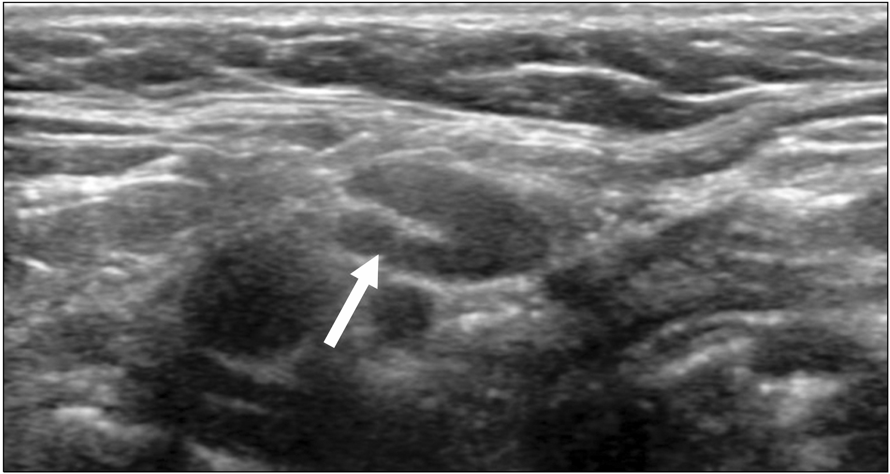
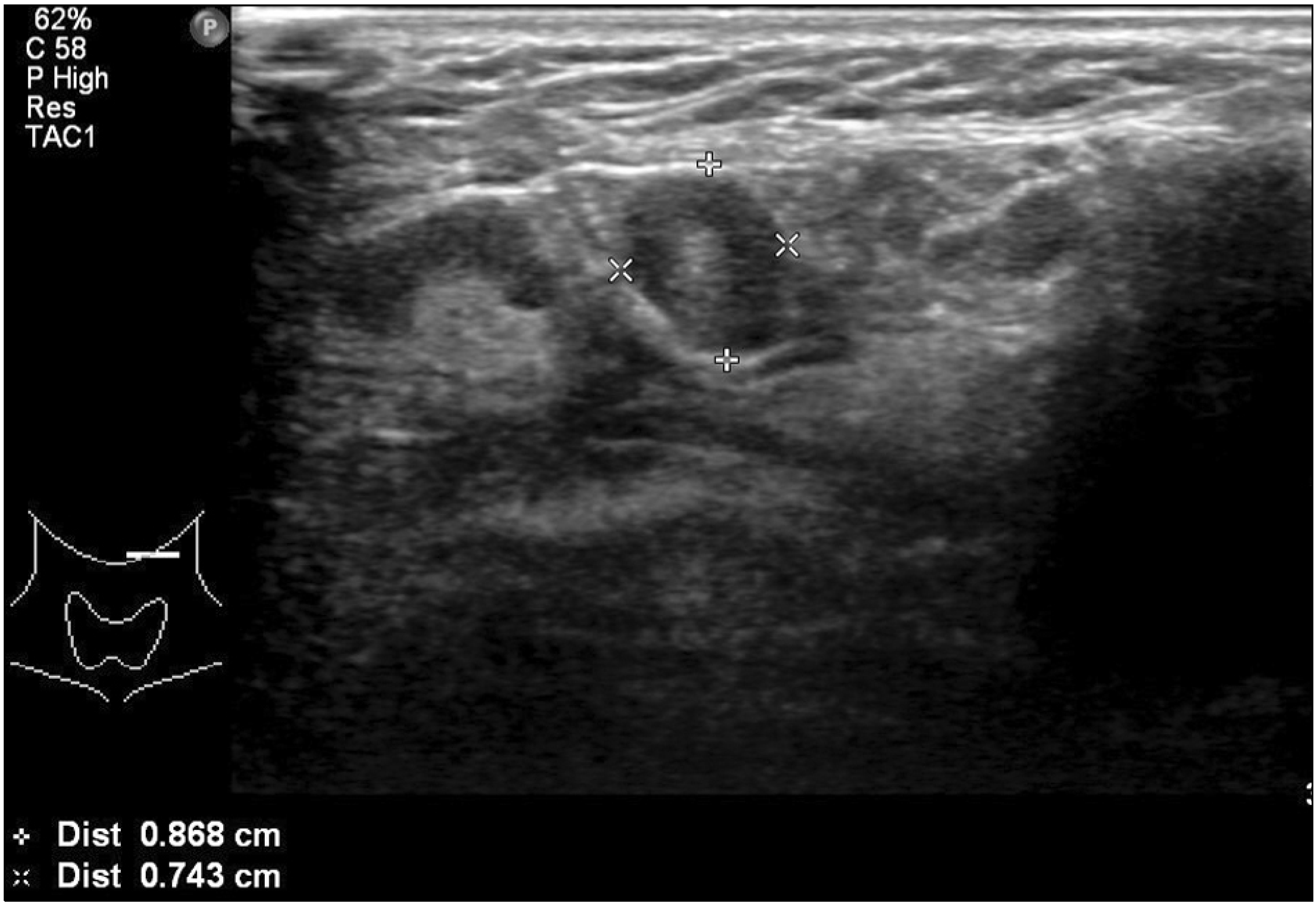
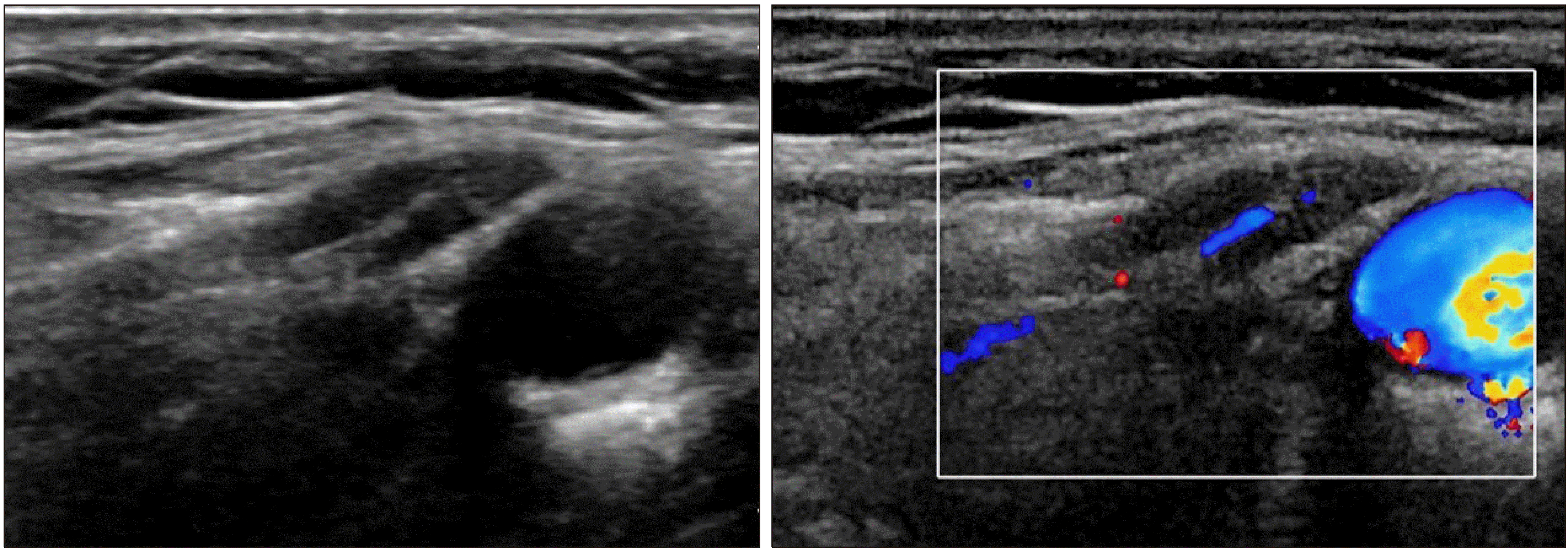
정상 림프절의 전형적인 초음파 소견은 대개 편평한 난원형의 모양(Fig. 1)이나 턱밑샘(submandibular gland), 귀밑샘(paro-tid gland) 림프절에서는 특별한 기저 질환 없이 원형의 모양을 보일 수 있다(Fig. 2). 그리고, 주위 연부 조직보다 균일하게 낮은 에코를 보이는 피질을 관찰할 수 있으며, 중심부에는 주위 연부 조직과 연결된 높은 에코의 림프절문(hilum)을 관찰할 수 있다. 색도플러 초음파검사에서는 림프절문이 위치하는 중심부에서 방사형의 혈류 분포를 관찰할 수 있는데 림프절문의 혈류 유무 판정은 전이 림프절과 반응 림프절의 구분에 도움을 줄 수 있다(Fig. 3).

3. 반응성 림프절염(Reactive lymphadenitis)

경부 림프절 비대의 가장 흔한 형태 중 하나이며 대부분 바이러스 또는 세균에 의한 상기도 감염 또는 두경부의 염증, 감염 이후에 이차적으로 발생하나 원인 병원체를 확인할 수 있는 경우는 매우 제한적이다. 어린이나 청소년에서 흔하며 양측성으로 오는 경우도 흔하다.
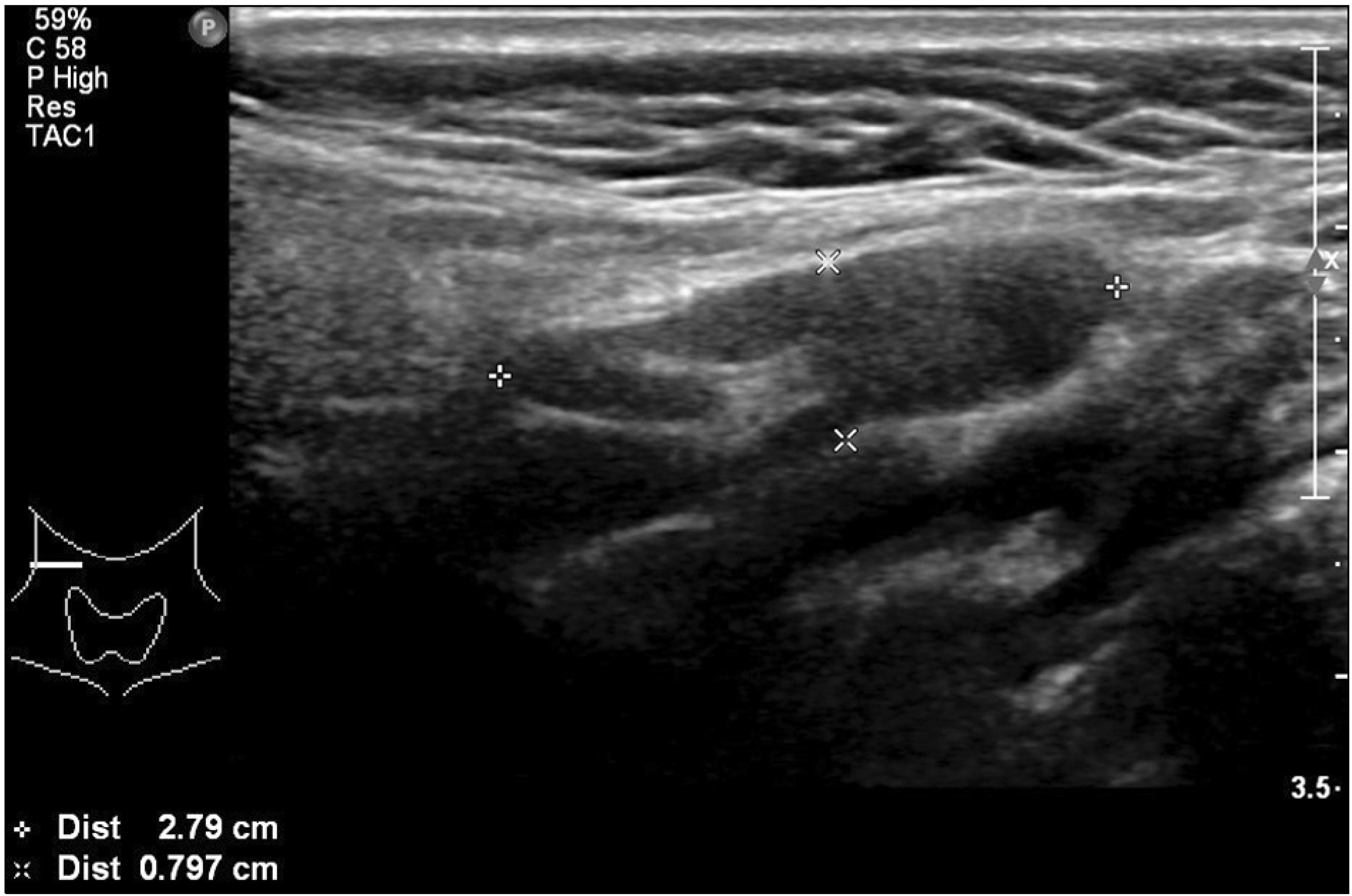
반응성 림프절염의 전반적인 초음파 소견은 앞에서 언급한 정상 림프절의 초음파 소견과 같다. 난원형이며 균일한 낮은 에코의 피질을 가지고 높은 에코의 림프절문이 대개 잘 보인다. 반응성 림프절염의 크기는 매우 다양하고 특히 어린이의 경우 2 cm 이상 커지는 경우도 흔하기 때문에 단일 장축(long-axis) 지름의 타 질환과의 감별진단을 위한 진단적 가치는 떨어진다. 따라서, 타 질환과의 감별을 위해 주로 사용하는 림프절 크기 인자는 단축(short-axis)의 지름과 장축 지름/단축 지름 비율(long-axis/short-axis ratio, L/S ratio, Solibiati index)을 많이 사용한다.(5) 대개 반응성 림프절염의 크기는 단축 지름이 8 mm를 초과하지 않고 모양은 L/S 비율이 2 이상인 난원형을 보인다(Fig. 4). 림프절문은 대략 75% 정도에서 관찰되며 관찰되지 않는 경우도 드물지 않게 있다. 이 경우 색도플러 초음파를 이용하여 중심부 혈류를 관찰하면 진단에 도움이 된다(Fig. 5).

4. 기쿠치병(Kikuchi disease, histiocytic necrotizing lymphadenitis)

기쿠치병은 조직구괴사성림프절염으로 1972년에 일본에서 처음 언급된 질환이며 현재까지도 정확한 발병 기전은 알려져 있지 않다.(6) 호발 연령은 주로 40세 미만의 젊은 연령층이며 우리나라 등 아시아에 흔하고 여자에서 빈도가 약간 높지만 소아에서는 남자의 빈도가 약간 높은 것으로 알려져 있다. 임상 양상은 매우 다양하여 전신 증상이 없는 경우부터 고열과 전신통, 심한 경우 드물게 혈구탐식성조직구증식증(hemophagocytic lymphohistiocytosis)까지도 동반하며 가장 흔한 임상 증상은 경부 림프절 비대이다. 대부분 평균 1-4개월 내에 저절로 소실되지만 6개월까지도 임상 증상이 지속될 수 있다.(7)
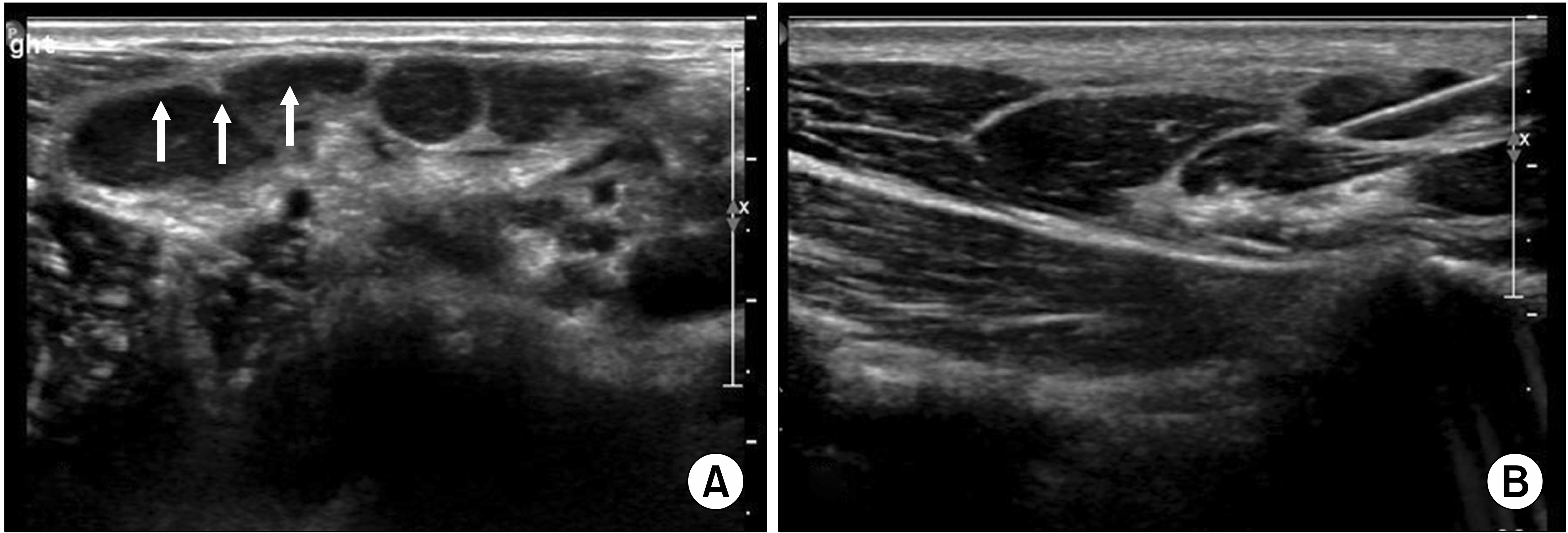
기쿠치병에 동반되는 경부 림프절염의 진단을 위한 특이적 초음파 소견은 없으며 병의 초기에는 앞에서 언급한 반응성 림프절염의 소견을 보인다. 주로 편측의 경부에 다발성으로 균일하게 커져 있으며 동일한 에코를 보이는 림프절들을 관찰할 수 있고 level II-V까지 넓게 분포한다. 이들 커진 림프절은 서로 염증 반응에 의해 엉켜 붙어(matted) 있는 경향이며 림프절 내에 농양이나 석회화는 동반하지 않는다.(8) 또한 환자의 약 65%에서 주위 연부 조직으로의 염증성 침윤으로 인해 커져 있는 림프절 주위로 높은 에코의 테두리를 관찰할 수 있다(Fig. 6).
병리학적 진단은 주로 세침흡입세포검사를 시행하는데 진단율이 대략 50% 내외이며 세침흡입세포검사에서 진단이 애매한 경우 중심부바늘생검이나 절개생검술을 시행할 수 있다.(9)

5. 결핵성 림프절염(Tuberculous lymphadenitis)

결핵성 림프절염은 개발도상국에서 흔한 질병이나 선진국인 우리나라의 경우 풍토병으로 유행하고 있는 질환이므로 환자가 편측 경부에 동통 또는 압통을 동반하지 않는 림프절의 비대로 내원 시 반드시 감별을 해야 하는 질환이다.
결핵성 림프절염의 초음파 소견은 매우 다양하다. 초기에는 앞에서 언급한 반응성 림프절 모양을 가질 수 있고 진행 정도에 따라 불균질한 에코 양상, 불분명한 림프절의 경계, 악성 또는 전이성 림프절 소견을 보일 수도 있으며, 석회화 동반, 림프절 내 괴사로 인한 낭성 변화, 농양 형성, 림프절문의 소실, 림프절 간의 군집 형성, 림프절 주위 연부 조직의 높은 에코 변화 등의 소견을 보일 수 있다.
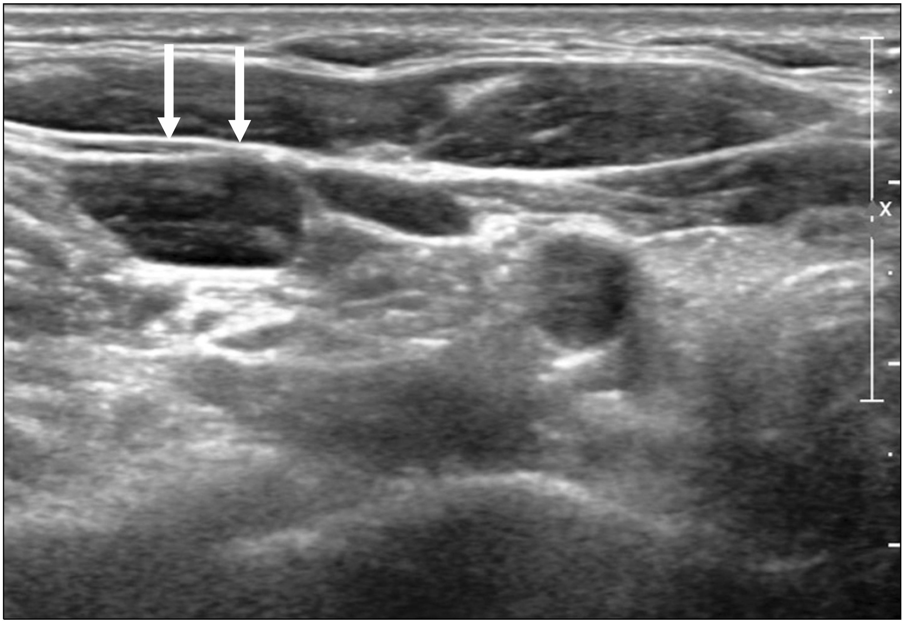
초기 결핵성 림프절염의 소견은 림프절의 비대와 원형 변화이며 약 79%의 결핵성 림프절 환자에서 림프절의 모양이 원형으로 보인다. 림프절문의 소실은 76%-86%에서 보고되며 림프절 내 낭성괴사로 인한 후방음향증강(posterior acoustic enhancement)을 동반하는 경우가 많다(Fig. 7).(10)
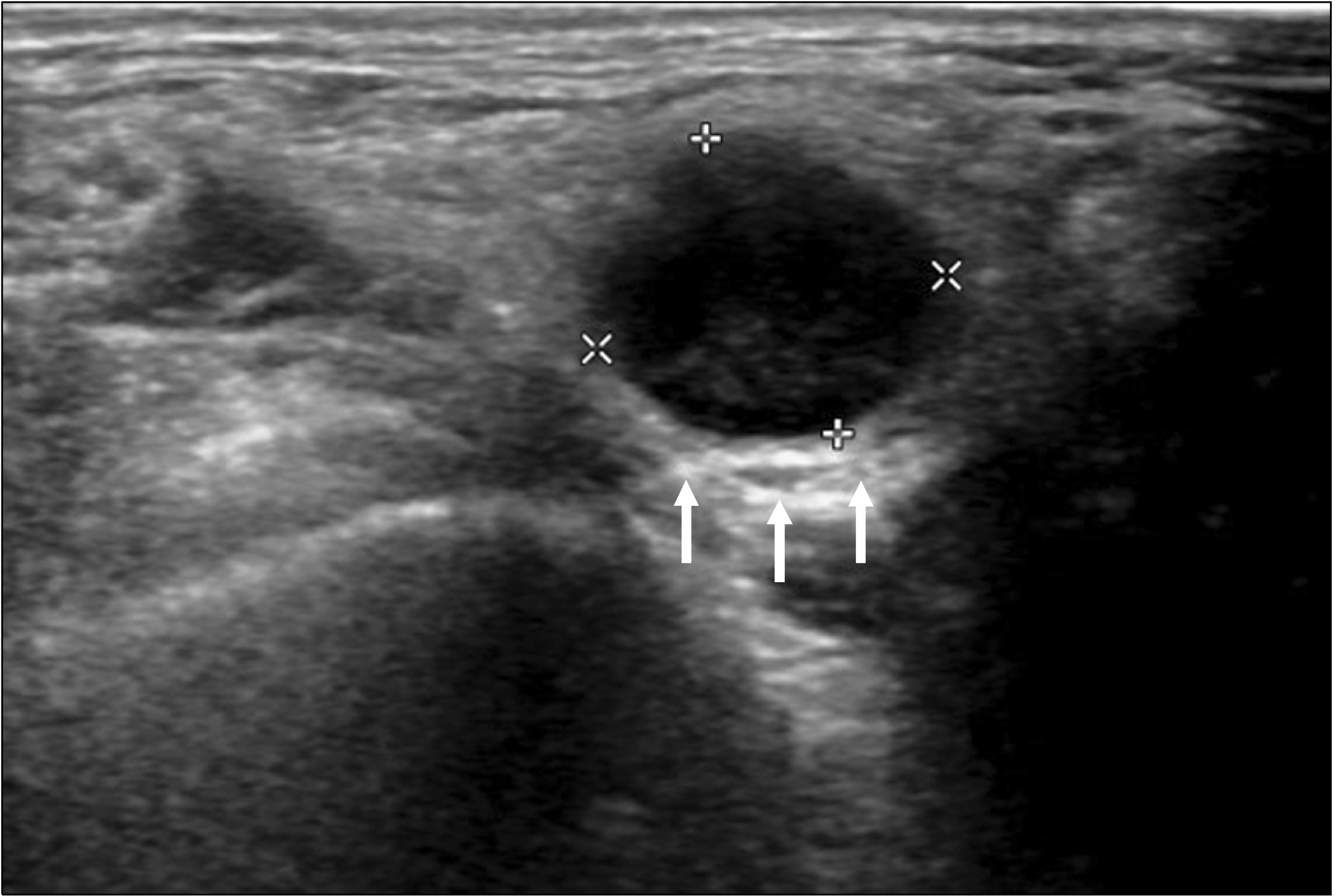
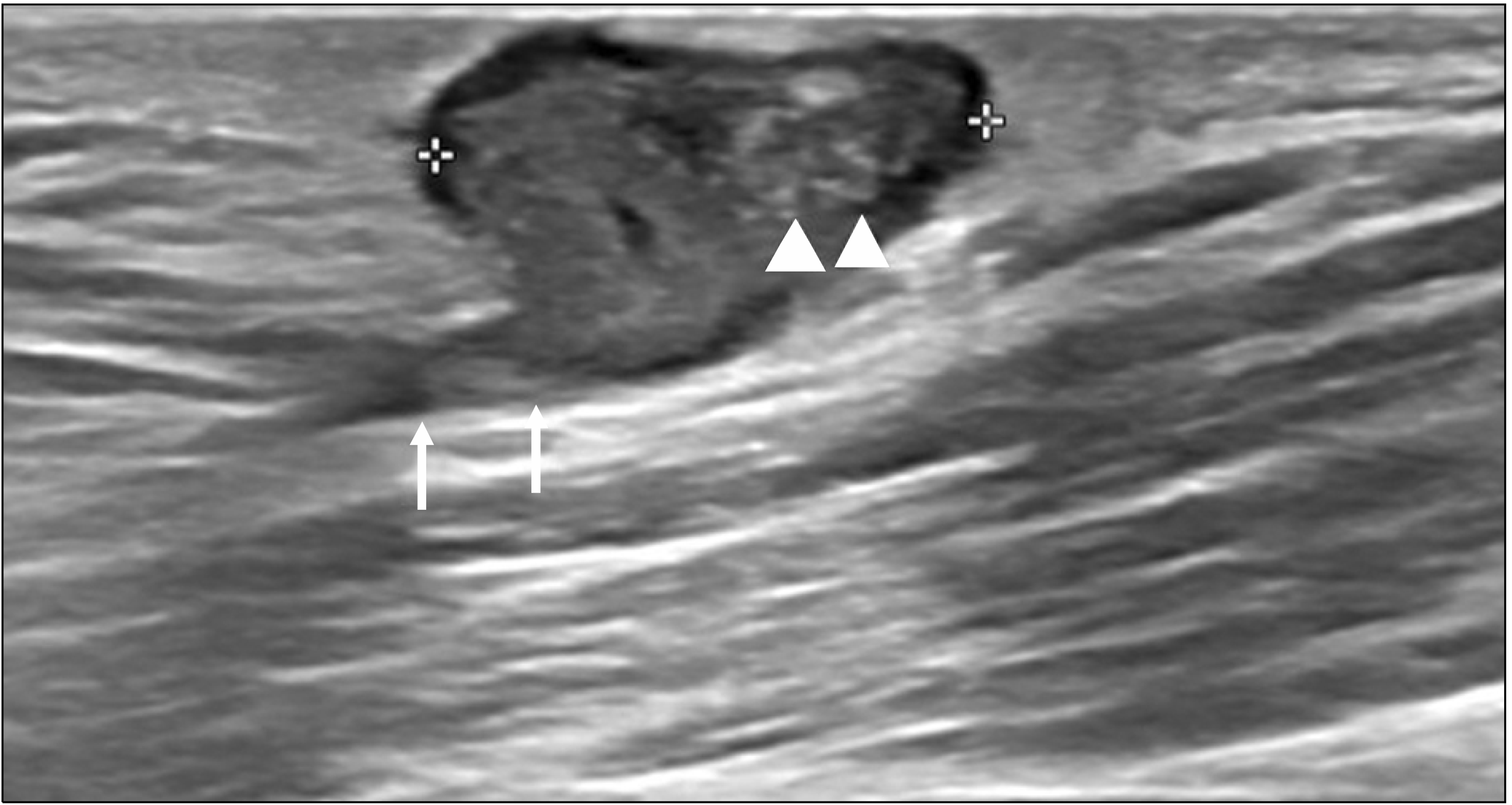
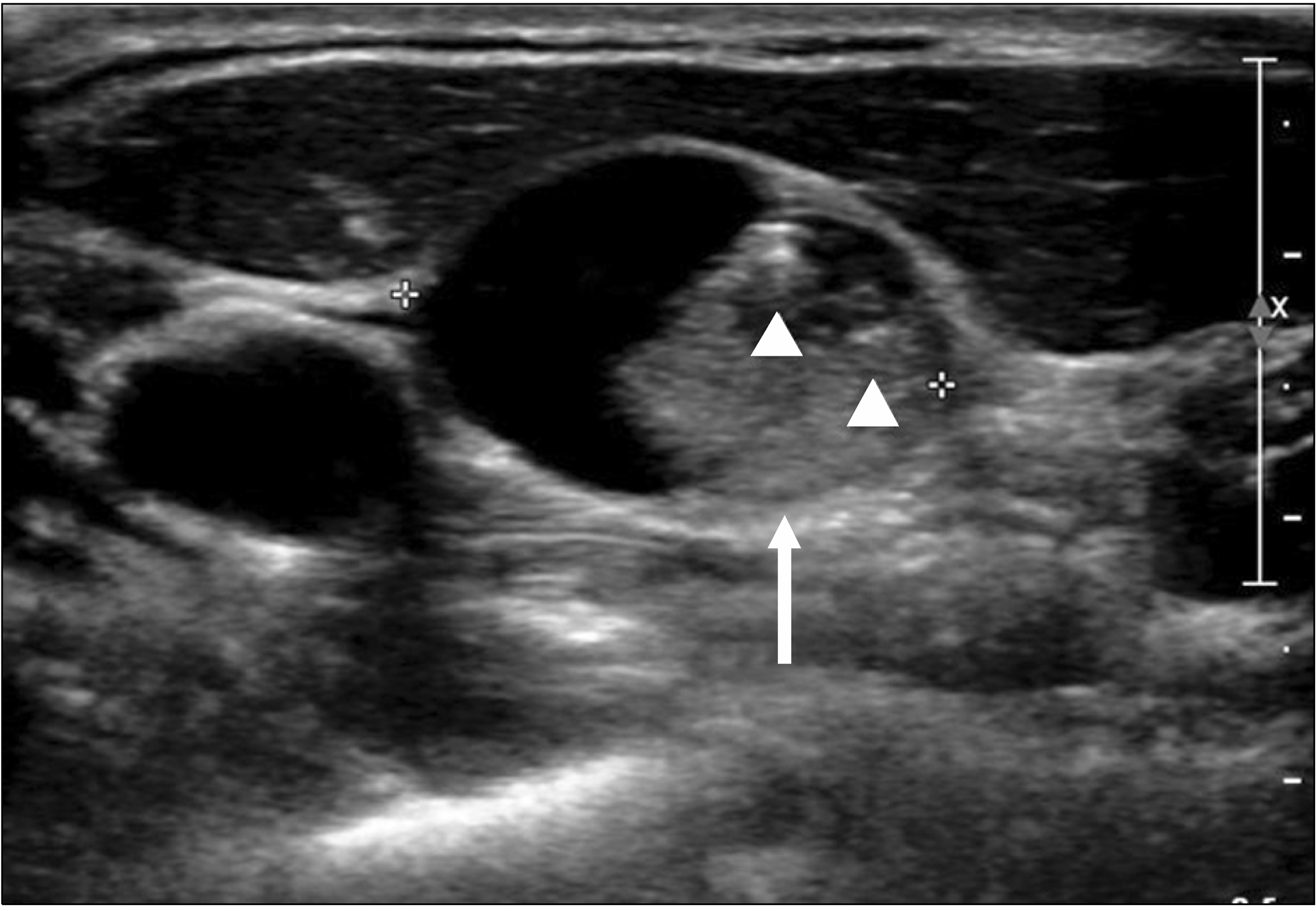
병이 진행됨에 따라 림프절 내에 냉농양(cold abscess)을 형성하는데 이때의 초음파 소견은 경계가 불분명한 낮은 에코의 덩어리로 보이며 내부는 불균질한 에코 양상 또는 석회화 변성에 의한 높은 에코의 점(hyperechogenic spot)들을 관찰할 수 있으며 괴사로 인한 액화 변성으로 부분적인 무에코 소견을 보이는 경우도 있다. 그리고 농양을 형성한 림프절 주위 연부 조직의 부종으로 인해 높은 에코의 테두리가 보인다(Fig. 8). 심한 경우 주위 연부 조직으로의 공동(sinus)을 형성하고 이로 인해 주위 연부 조직에 농양을 형성하기도 하며 피부와 가까이 위치한 경우 누공을 형성하기도 한다(Fig. 9).
진단은 초음파유도하 흡인생검세포검사를 통한 현미경적 세포 검사를 시행할 수 있으나 민감도가 35%로 낮으며 흡인을 통한 결핵균 배양의 민감도는 86%로 높으나 배양에 시간이 많이 걸린다는 단점이 있다.(11) 수술적 절개생검술은 결핵성 림프절염의 진단에 최적 표준 진단법이나 수술 부위의 상처가 잘 낫지 않고 누공을 형성하는 경우가 많은 단점이 있다. 따라서, 최근에는 초음파유도하 중심부바늘생검술을 이용한 검사가 수술적 생검술을 대체하고 있으며 진단의 정확도도 비슷하게 보고되고 있다.(12)

6. 갑상선암의 전이림프절

분화갑상선암의 경부 림프절 전이는 20%-50%로 보고되며 미세전이까지 포함한 경우 90%까지 보고되기도 한다.(13) 따라서, 갑상선암으로 수술이 예정되어 있는 환자의 경우 반드시 수술 전 평가에 경부 림프절의 전이 여부를 포함시켜야 한다. 갑상선암에 의한 측부 경부 림프절 전이는 흔히 level II보다는 level III, IV에 많지만 병변이 갑상선의 상극에 있는 경우 level II나 III로 건너뜀 전이(skip metastasis)가 있을 수 있다.(14)
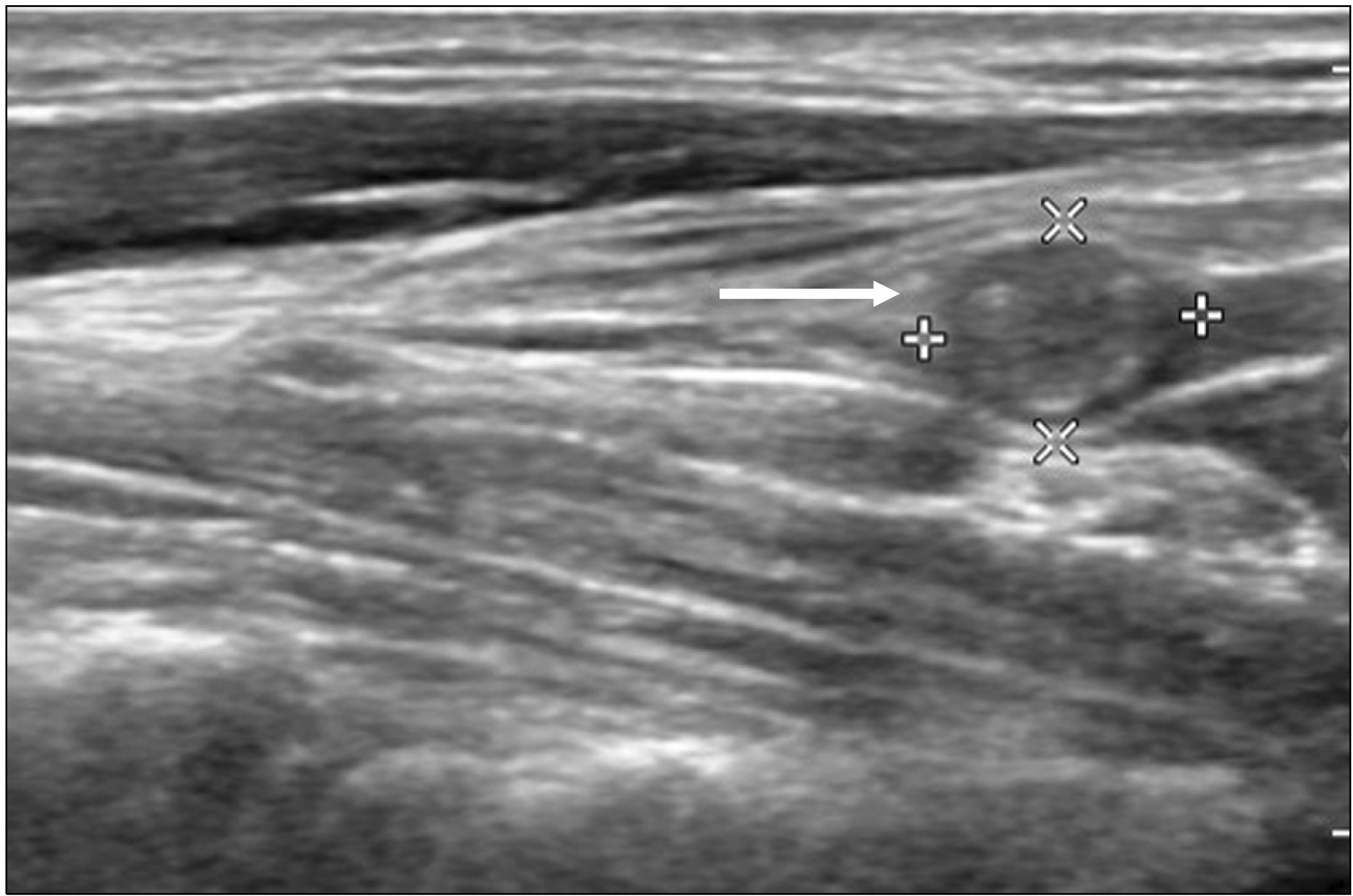
갑상선암 전이림프절의 초음파 소견들은 아래와 같다(Figs. 10-12).(15-19)
- 림프절문의 소실
- 림프절 피질의 높은 에코성 변화
- 림프절의 낭성 변화
- 석회화 동반
- 림프절 내의 변연부 또는 미만성 혈류 증가
위의 초음파 소견들을 바탕으로 2021년 대한갑상선영상의학회에서는 suspicious, indeterminate, 그리고 probably benign으로 림프절을 분류하였다(Table 1).(20) 이 권고안에 의하면 림프절의 낭성 변화, 석회화 동반, 피질에 높은 에코성 변화, 변연부 또는 미만성 혈류 증가 등의 소견 중 하나라도 보이면 전이가 의심되는 결절로 간주한다. 그리고 위의 의심 소견이 보이지 않으면서 고에코의 림프절문이나 림프절문으로의 중심 혈류가 잘 유지되어 있는 경우 양성 림프절로 판단하고 위의 의심 소견들이 없으면서 고에코의 림프절문이나 림프절문으로의 중심 혈류도 보이지 않는 경우 중간 정도의 의심 림프절로 분류하였다. 그리고 갑상선암으로 진단된 환자의 수술 전 검사에서 suspicious 림프절 소견이 보이는 경우 단축의 지름이 3-5 mm 초과, indeterminate 림프절 소견을 보이는 경우 단축의 지름이 5 mm 초과할 때 림프절에 대한 세침흡입검사를 권고한다.(20)
진단은 주로 초음파유도하 세침흡인세포검사로 진단이 되며, 분화갑상선암의 경우 림프절 미세침흡인 후 세척액(wash-out)의 갑상선글로불린(thyroglobulin, Tg) 값을 측정하면(FNA wash-out Tg) 미세침흡인생검 단독보다 예민도가 높고 특이도는 거의 100%를 보이기 때문에 세침흡인세포검사와 함께 FNA wash-out Tg 값을 측정하는 것이 가장 정확한 검사법이라 할 수 있다. FNA wash-out Tg의 검사는 검체를 획득한 바늘에서 FNA를 위해 슬라이드에 검체를 뿌려 세포 병리에 의뢰하고, 나머지 검체를 1 ml의 생리식염수로 희석하여 측정한다.(21) 특히, Tg 측정법은 낭성 변화를 동반한 전이성 림프절의 진단에 도움이 되는데 낭성 변화가 심할 경우 세포 검사를 위한 충분한 세포를 얻기 힘들기 때문이다(Fig. 13). 그러나 갑상선 직하방에 위치한 level VI의 림프절에서는 주위 정상 갑상선 때문에 위양성 결과가 종종 보고되고 있으며 진단의 판정 기준이 되는 절대적 수치는 아직 확립되지 않았으므로 주의를 요하지만 혈정 Tg 수치와 비교하여 의미 있게 높은 경우 양성으로 진단한다.(22)

결 론

경부 림프절의 비대는 여러 가지 원인에 의해 전 연령대에 걸쳐서 흔하게 발생하는 질환이다. 그 원인으로 반응성 림프절 비대, 염증성 림프절 비대, 전이에 의한 림프절 비대 등이 있으며 경부 림프절 비대의 정확한 진단을 위해 환자의 과거력, 혈액학적 검사, 신체 검진, 여러 영상의학적 검사가 필요하며 이 중 초음파 검사는 일차적으로 환자를 평가하는데 간편하고 환자에 유해함 없이 반복적인 검사가 가능하며 실시간으로 세포, 조직 검사까지 가능하므로 경부 림프절 진단에 널리 이용되고 있다.

REFERENCES

1. Stack BC Jr, Ferris RL, Goldenberg D, Haymart M, Shaha A, Sheth S, et al. 2012; American Thyroid Association consensus review and statement regarding the anatomy, terminology, and rationale for lateral neck dissection in differentiated thyroid cancer. Thyroid. 22:501–8. DOI: 10.1089/thy.2011.0312. PMID: 22435914.
crossref
2. Cheung YC, Wan YL, Lui KW, Lee KF. 2000; Sonographically guided core-needle biopsy in the diagnosis of superficial lymphade-nopathy. J Clin Ultrasound. 28:283–9. DOI: 10.1002/1097-0096(200007/08)28:6<283::AID-JCU3>3.0.CO;2-T. PMID: 10867666.
crossref
3. Robbins KT, Medina JE, Wolfe GT, Levine PA, Sessions RB, Pruet CW. 1991; Standardizing neck dissection terminology. Official report of the Academy's Committee for Head and Neck Surgery and Oncology. Arch Otolaryngol Head Neck Surg. 117:601–5. DOI: 10.1001/archotol.1991.01870180037007. PMID: 2036180.
crossref
4. American Institute of Ultrasound in Medicine. 2003; AIUM Practice Guideline for the performance of thyroid and parathyroid ultrasound examination. J Ultrasound Med. 22:1126–30. DOI: 10.7863/jum.2003.22.10.1126. PMID: 14606572.
5. Solbiati L, Osti V, Cova L, Tonolini M. 2001; Ultrasound of thyroid, parathyroid glands and neck lymph nodes. Eur Radiol. 11:2411–24. DOI: 10.1007/s00330-001-1163-7. PMID: 11734934.
crossref
6. Kikuchi M. 1972; Lymphadenitis showing focal reticulin cell hyperplasia with nuclear debris and phagocytes: a clinicopathological study. Nihon Ketsueki Gakkai Zasshi. 35:379–80.
7. Bosch X, Guilabert A, Miquel R, Campo E. 2004; Enigmatic Kikuchi- Fujimoto disease: a comprehensive review. Am J Clin Pathol. 122:141–52. DOI: 10.1309/YF081L4TKYWVYVPQ. PMID: 15272543.
8. Yoo JL, Suh SI, Lee YH, Seo HS, Kim KM, Shin BK, et al. 2011; Gray scale and power Doppler study of biopsy-proven Kikuchi disease. J Ultrasound Med. 30:957–63. DOI: 10.7863/jum.2011.30.7.957. PMID: 21705728.
crossref
9. Tong TR, Chan OW, Lee KC. 2001; Diagnosing Kikuchi disease on fine needle aspiration biopsy: a retrospective study of 44 cases diagnosed by cytology and 8 by histopathology. Acta Cytol. 45:953–7. DOI: 10.1159/000328370. PMID: 11726124.
10. Chou CH, Yang TL, Wang CP. 2014; Ultrasonographic features of tuberculous cervical lymphadenitis. J Med Ultrasound. 22:158–63. DOI: 10.1016/j.jmu.2014.06.007.
crossref
11. Knox J, Lane G, Wong JS, Trevan PG, Karunajeewa H. 2012; Diagnosis of tuberculous lymphadenitis using fine needle aspiration biopsy. Intern Med J. 42:1029–36. DOI: 10.1111/j.1445-5994.2012.02748.x. PMID: 22372860.
crossref
12. Ryu YJ, Cha W, Jeong WJ, Choi SI, Ahn SH. 2015; Diagnostic role of core needle biopsy in cervical lymphadenopathy. Head Neck. 37:229–33. DOI: 10.1002/hed.23580. PMID: 24375555.
crossref
13. Noguchi S, Noguchi A, Murakami N. 1970; Papillary carcinoma of the thyroid. I. Developing pattern of metastasis. Cancer. 26:1053–60. DOI: 10.1002/1097-0142(197011)26:5<1053::AID-CNCR2820260513>3.0.CO;2-X. PMID: 5476786.
crossref
14. Park JH, Lee YS, Kim BW, Chang HS, Park CS. 2012; Skip lateral neck node metastases in papillary thyroid carcinoma. World J Surg. 36:743–7. DOI: 10.1007/s00268-012-1476-5. PMID: 22354485.
crossref
15. Rosário PW, de Faria S, Bicalho L, Alves MF, Borges MA, Purisch S, et al. 2005; Ultrasonographic differentiation between metastatic and benign lymph nodes in patients with papillary thyroid carcinoma. J Ultrasound Med. 24:1385–9. DOI: 10.7863/jum.2005.24.10.1385. PMID: 16179622.
crossref
16. Leboulleux S, Girard E, Rose M, Travagli JP, Sabbah N, Caillou B, et al. 2007; Ultrasound criteria of malignancy for cervical lymph nodes in patients followed up for differentiated thyroid cancer. J Clin Endocrinol Metab. 92:3590–4. DOI: 10.1210/jc.2007-0444. PMID: 17609301.
crossref
17. Park JS, Son KR, Na DG, Kim E, Kim S. 2009; Performance of preoperative sonographic staging of papillary thyroid carcinoma based on the sixth edition of the AJCC/UICC TNM classification system. AJR Am J Roentgenol. 192:66–72. Erratum. DOI: 10.2214/AJR.07.3731. PMID: 19098181.
crossref
18. Sohn YM, Kwak JY, Kim EK, Moon HJ, Kim SJ, Kim MJ. 2010; Diagnostic approach for evaluation of lymph node metastasis from thyroid cancer using ultrasound and fine-needle aspiration biopsy. AJR Am J Roentgenol. 194:38–43. DOI: 10.2214/AJR.09.3128. PMID: 20028903.
19. Yoo RE, Kim JH, Bae JM, Hwang I, Kang KM, Yun TJ, et al. 2020; Ultrasonographic indeterminate lymph nodes in preoperative thyroid cancer patients: malignancy risk and ultrasonographic findings predictive of malignancy. Korean J Radiol. 21:598–604. DOI: 10.3348/kjr.2019.0755. PMID: 32323505. PMCID: PMC7183832.
20. Ha EJ, Chung SR, Na DG, Ahn HS, Chung J, Lee JY, et al. 2021; 2021 Korean Thyroid Imaging Reporting and Data System and imaging-based management of thyroid nodules: Korean Society of Thyroid Radiology consensus statement and recommendations. Korean J Radiol. 22:2094–123. DOI: 10.3348/kjr.2021.0713. PMID: 34719893. PMCID: PMC8628155.
21. Kim MJ, Kim EK, Kim BM, Kwak JY, Lee EJ, Park CS, et al. 2009; Thyroglobulin measurement in fine-needle aspirate washouts: the criteria for neck node dissection for patients with thyroid cancer. Clin Endocrinol (Oxf). 70:145–51. DOI: 10.1111/j.1365-2265.2008.03297.x. PMID: 18466347.
22. Giovanella L, Bongiovanni M, Trimboli P. 2013; Diagnostic value of thyroglobulin assay in cervical lymph node fine-needle aspirations for metastatic differentiated thyroid cancer. Curr Opin Oncol. 25:6–13. DOI: 10.1097/CCO.0b013e32835a9ab1. PMID: 23090642.

Fig. 1
Ultrasonographic findings of normal cervical lymph node: Uniform hypoechoic cortex with oval shape. In the center, hyperechoic hilum (arrow) with connections to the surrounding soft tissue is visible.
jsu-10-1-1-f1.tif
Fig. 2
Ultrasonographic findings of the submandibular lymph node: Lymph node show that it has a round shape with a short axis diameter of 7.4 mm and a long axis diameter of 8.7 mm. The presence of a hyperechoic hilum connected to the surrounding soft tissue is also visible.
jsu-10-1-1-f2.tif
Fig. 3
Color Doppler ultrasonogra-phic finding of normal lymph node: The finding of a normal level 3 lymph node on the right side with visible blood flow along the central hilum on color Doppler ultrasonography.
jsu-10-1-1-f3.tif
Fig. 4
Reactive lymphadenitis: The lymph node has a long-axis diameter of approximately 28 mm, but the short-axis diameter does not exceed 8 mm. The long-axis/short-axis ratio (L/S ratio) is over 2, and an echogenic hilum connected to the surrounding soft tissue is well observed.
jsu-10-1-1-f4.tif
Fig. 5
Central blood flow in reactive lymphadenitis: An 18-year-old female patient visited with enlarged left cervical lymph nodes for several days. Although the hilum could not be clearly observed on ultrasound, central blood flow was observed on color Doppler. Reactive lymphadenitis was subse-quently diagnosed by fine needle aspiration cytology.
jsu-10-1-1-f5.tif
Fig. 6
Kikuchi lymphadenitis: An 18-year-old male patient presented with enlarged right cervical lymph nodes for 1 month. The ultrasound showed uniformly enlarged and matted lymph nodes, with a hyperechoic halo observed around the enlarged lymph node due to changes in the surrounding soft tissue caused by the inflammatory response (A, arrows). A core needle biopsy was performed, and Kikuchi lymphadenitis was subsequently diagnosed (B).
jsu-10-1-1-f6.tif
Fig. 7
Early cervical tuberculous lymphadenitis: The lymph node appears to have a round shape with an L/S ratio close to 1 and the hilum is not visible. Posterior acoustic enhancement is shown due to cystic necrosis within the lymph node (arrows).
jsu-10-1-1-f7.tif
Fig. 8
A 63-year-old male patient has been diagnosed with left cervical tuberculosis lymphadenitis, and the ultrasound findings show several echo-genic spots within a hypoechoic mass. Additionally, a hyperechoic halo is visible due to edema of the surrounding soft tissues (A). At level V, partial anechoic findings due to necrosis are shown (B, arrow).
jsu-10-1-1-f8.tif
Fig. 9
Left level II cervical lymph node, tuberculous lymph node that formed a sinus with the surrounding soft tissue (arrows) and formed a fistula to the skin and also accompanied by heterogeneous calcifications (arrow heads).
jsu-10-1-1-f9.tif
Fig. 10
Metastatic lymph nodes due to papillary thyroid carcinoma: The patient has been diagnosed with left thyroid papillary carcinoma. On ultrasound, a metastatic lymph node (arrow) with round shape and echogenic changes is observed and microcalcifications within the left level IV lymph node is also noted.
jsu-10-1-1-f10.tif
Fig. 11
Metastatic lymph nodes due to papillary thyroid carcinoma: The L/S ratio was more than 2, which is within the normal range, but the hilum was not visible and the lymph node showed heterogeneous high echogenic change (A, arrow), with diffuse blood flow throughout the lymph node (B). On fine needle aspiration cytology, this lymph node has been diagnosed by metastatic papillary thyroid carcinoma.
jsu-10-1-1-f11.tif
Fig. 12
Metastatic lymph nodes due to papillary thyroid carcinoma: Cystic changes was seen in the lymph node, and solid nodules (arrow) with microcalcifications (arrow heads) were visible within the cystic changed lymph node. On fine needle aspiration cytology, this lymph node was diagnosed by metastatic papillary thyroid carcinoma.
jsu-10-1-1-f12.tif
Fig. 13
Diagnosis of metastatic lymph nodes with cystic changes: The patient was diagnosed with right thyroid papillary carcinoma. On neck ultrasonography, lymph node with cystic change was observed in the right lateral neck (arrows). Fine needle aspiration was performed, but an accurate diagnosis could not be made due to low cellularity. However, the washout-Tg value was measured as >500 ng/ml, and it was diagnosed as metastatic lymph node of papillary thyroid carci-noma after surgery.
jsu-10-1-1-f13.tif
Table 1
US-Based Risk Stratification for Cervical Lymph Node Metastasis in Patients with Possible or Proven Thyroid Carcinoma (20)
Category USG Malignancy risk (%)
Suspicious Any of four suspicious features 73-88
- Cystic change 97-100
- Echogenic foci (calcifications) 86-100
- Cortical hyperechogenicity (focal/diffuse) 79-96
- Abnormal vascularity (peripheral/diffuse) 77-84
Indeterminate Loss of echogenic hilum and hilar vascularity 20
Probably benign Echogenic hilum or hilar vascularity <3
TOOLS
Similar articles