This article has been
cited by other articles in ScienceCentral.
Abstract
This study examined the effects of a vertical incision on postoperative edema after third molar extraction. The study design was that of a comparative split-mouth approach. Evaluation was performed via magnetic resonance imaging (MRI). Two patients with homogeneous bilateral impacted mandibular third molars were enrolled. These patients underwent facial MRI within 24 hours after simultaneous extraction surgery. Modified triangular flap and enveloped flap incisions were made. Postoperative edema was evaluated by MRI and was assessed according to anatomical space. The two pairs of homogeneous extractions demonstrated that vertical incisions were associated qualitatively and quantitatively with extensive postoperative edema. The edema associated with these incisions spread toward the buccal space, beyond the buccinator muscle. In conclusion, a vertical incision with mandibular third molar extraction was related to edema in the buccal space and the fascial space, which contributed to clinical facial swelling.
Keywords: Flap design, Incision, Mandibular third molar, Postoperative outcome
I. Introduction
Extraction of the mandibular third molar is one of the most common dental surgeries. Postoperative swelling is a common complication after surgery that affects the physical, psychological, and aesthetic quality of life for a few days. The adverse effects on quality of life were reported to be three-fold higher with postoperative complications
1. Even if facial swelling is not prominent after surgery, postoperative edema may occur from exudates or transudation resulting from surgery. Because postoperative edema and pain are significantly less severe with smaller incisions
2, postoperative edema related to surgical incisions is an area of interest. Some studies reported more severe postoperative swelling with a vertical incision
3-5. However, others showed that surgical flaps do not appear to have a significant role in edema
6-8.
When evaluating postoperative swelling after third molar extraction, several limitations need to be overcome, including the surgical control strategy. Strategic considerations concern the incision, odontomy, ostectomy, suture pattern and technique, and postoperative care along with surgical difficulty and the quantifying method for evaluating swelling
2,7. Several different techniques have been used to measure postoperative swelling including visual measurement
5,7, patient questionnaire
8, direct measurement by a ruler
9, and magnetic resonance imaging (MRI)
10. Among these, MRI is the most objective and reproducible assessment tool, but the high cost may limit its use. In 2021, Jeong et al.
10 suggested qualitative analysis by MRI by classifying postoperative edema based on its anatomic division: one anatomic component (buccinator muscle) and four fascial spaces (supra-periosteum space, buccal space, parapharyngeal space, and lingual space). These researchers reported that most postoperative edema spread though the buccinator muscle and supra-periosteum space
10 and suggested that an incision that avoided these anatomic structures could reduce the level of postoperative edema.
A few studies have reported significant adverse effects of third molar surgery in a comparison of incision techniques
2,6. Kleinheinz et al.
11 made proposals concerning incision design according to cadaver results based on the vascular area that traverses parallel to the alveolar ridge in the vestibulum. Depending on the degree of inflammation and micro-leakage, vertical incisions may create discontinuity of these vascular structures and affect postoperative edema
12. Buccal envelope flaps without a vertical incision and modified triangular flaps with a vertical incision have been used as traditional approaches for impacted third molars.
Few studies have evaluated postoperative swelling by MRI, the most accurate diagnostic tool. Several issues deserve consideration when comparing surgical approaches, including surgical difficulty. Kim et al.
13 proposed a more practical classification system that considered the validity of the existing classification. In 2020, Ku et al.
14 validated the modified difficulty index for an impacted third molar based on the molar’s three-dimensional relationships in space and with the ramus, as well as its depth, associated pathologic conditions, and surgical time with a unified extraction method. The present study examined the effects of a vertical incision on comparative split-mouth MRI evaluation of postoperative edema after third molar extraction with homogeneous difficulty based on the three-dimensional modified difficulty index.
II. Cases Report
The Institutional Review Board of the Armed Forces Capital Hospital approved this study (AFCH-20-IRB-012). This study was conducted according to the principles of the Declaration of Helsinki for research on humans. Written and verbal informed consent was obtained from each patient.
Two patients with homogeneous bilateral impacted mandibular third molars assigned the same grade based on the modified difficulty index by computed tomography
14 were enrolled in this study. These patients had undergone facial MRI within 24 hours after extraction surgery under general anesthesia.
1. Surgical technique
One expert surgeon performed all surgical procedures under general anesthesia. After infiltrating the surgical field with 1.8 mL lidocaine (2% lidocaine HCl; Huons), simple extraction of both maxillary third molars was performed without an incision. For mandibular surgical extraction, an incision was made and included an operculectomy. The operculum is tissue overlying the impacted tooth composed of highly differentiated epithelial cells. Through operculectomy, the cemento-enamel junction of the impacted tooth was accessible, allowing surgical extraction without a distal extent incision. Based on the de-epithelialization concept, this improved the healing potential of the open wound
15.
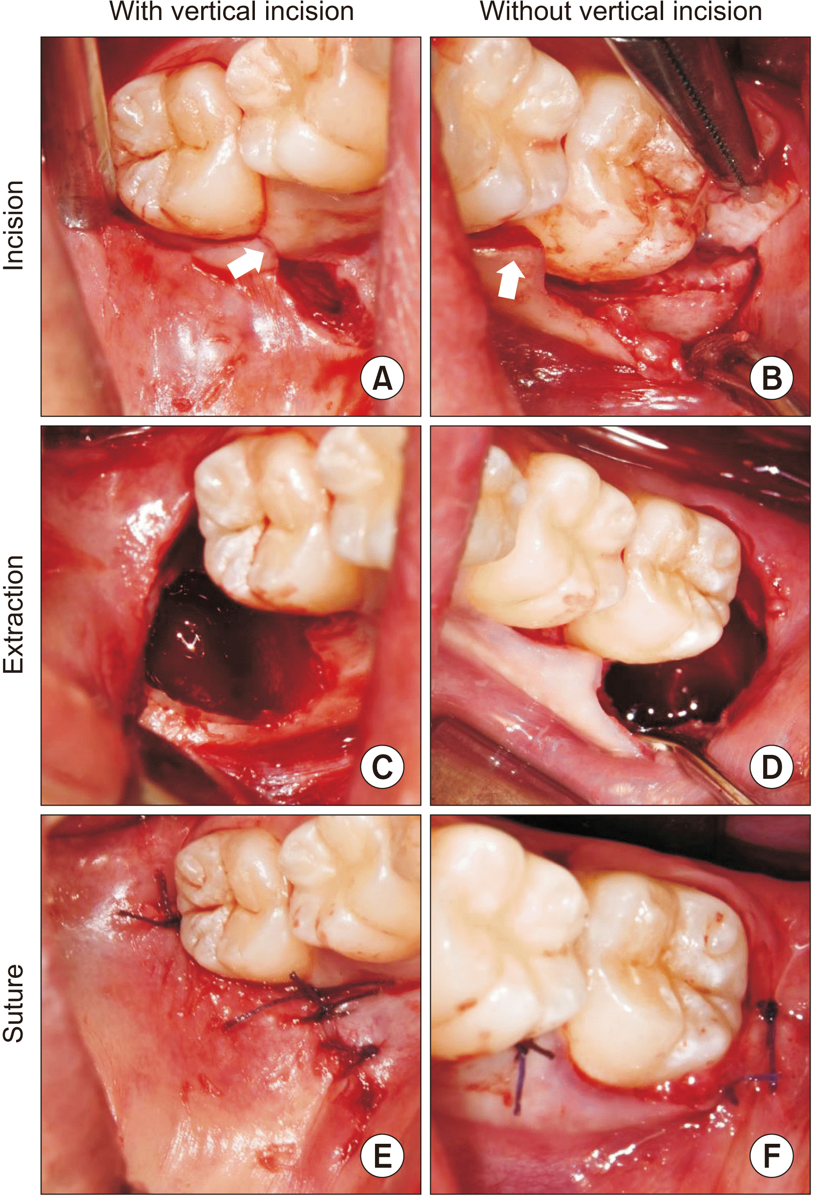
The mesial extent incision from the operculectomy was a randomly constructed modified triangular flap and included a vertical incision on the mesial line angle of the second molar.(
Fig. 1. A) Alternatively, an envelope flap on the mesial papilla of the second molar excluded the need for vertical incision.(
Fig. 1. B) After exposing the mandibular third molar via ostectomy, an odontomy was made using a 2.0 mm round burr with a RemB straight micro-drill (Stryker); the tooth was extracted with curettage of granulation tissue.(
Fig. 1. C, 1. D) The collagen plug (AteloPlug; Hyundai Bioland) was covered to prevent bleeding and food packing
15. The medial extent incision and the distal portion of the second molar were sutured.(
Fig. 1. E, 1. F) The surgical time was calculated from the start of the incision to the last suture using digital photographs
14.
Postoperatively, an ice pack was applied on both sides. The patients were prescribed antibiotics (625 mg, amoxicillin, three times a day; Ilsung Pharmaceutical) and a non-steroidal anti-inflammatory drugs (500 mg, dexibuprofen, three times a day; Samil Pharmaceutical) for seven days and a daily mouth rinse with chlorhexidine solution.
2. Postoperative edema evaluation
Two patients underwent MRI (Discovery MR750; GE HealthCare) with an eight-channel neurovascular phased-array coil (GE Medical System) for use after molar extraction.
The standardized imaging protocol comprised axial, coronal, and sagittal T1-weighted-echo sequence with fat suppression (repetition time [TR], 3.5 ms; echo time [TE], 1.6 ms; matrix, 288×160; section thickness, 5 mm; field-of-view [FOV], 40 mm×28 mm); axial, coronal and sagittal T2-weighted nonfat-saturated fast spin-echo sequence (TR, 2800 ms; TE, 90 ms; matrix, 320×224; gap, 1 mm; slice thickness, 5 mm; FOV, 34 mm×34 mm); and T1-weighted fast spoiled gradient recalled echo (fast SPGR) and T2-weighted 3D fast imaging employing steady-state acquisition (3D FIESTA) sequences.
Postoperative edema was determined according to the spaces around the mandibular third molar, the buccinator muscle, buccal space, parapharyngeal space, and lingual space (sublingual and submandibular space) by a high signal on T2-weighted MRI and a low signal on T1-weighted MRI.
3. Patient No. 1: Right vertical incision and left enveloped incision
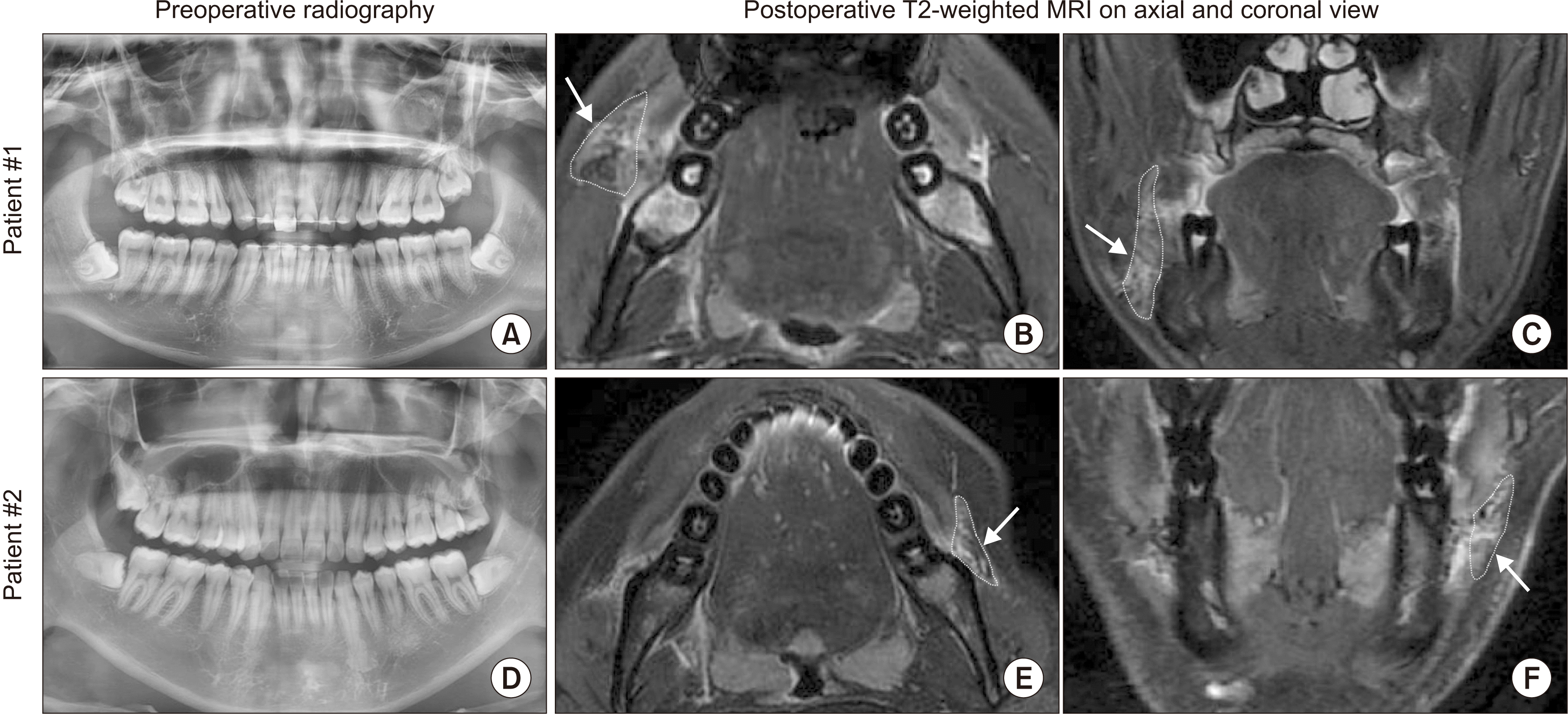
A 20-year-old male underwent surgery for homogeneous impacted mandibular third molars.(
Fig. 2. A) His right third molar was extracted with a vertical incision over 31 minutes, and the left without a vertical incision over 29 minutes.(
Fig. 1) MRI was performed seven hours after the extractions and showed extensive postoperative edema on the right side.(
Fig. 2. B, 2. C) Edema on the left side spread to the buccinator muscle only, but edema on the right-side spread to the buccinator muscle and buccal space. The patient reported generalized mild swelling with visual analog scale (VAS) score of 1.
4. Patient No. 2: Left vertical incision and right enveloped incision
A 21-year-old male underwent surgery for homogeneous impacted mandibular third molars.(
Fig. 2. D) His left third molar was extracted with a vertical incision over 12 minutes, and the other was extracted without a vertical incision over 14 minutes. MRI was performed five hours after the extractions and showed extensive postoperative edema on the left side.(
Fig. 2. E, 2. F) The edema on the right-side spread to the buccinator muscle only, but the edema on the left side spread to the buccinator muscle and buccal space. Clinically, the patient complained of swelling and pain on the left side for four days, with a VAS score of 4 on the left and 1 on the right.
III. Discussion
Regarding postoperative swelling, considerable attention has been given to radiography, dental morphology and depth of impaction, patient age, and experience of the surgeon, but few studies have analyzed incision design
2. In this split-mouth study, the effects of a vertical incision were noted while controlling other variables. Two pairs of homogeneous extractions showed that a vertical incision was qualitatively and quantitatively related to extensive postoperative edema and additional buccal space edema.
The results of some randomized controlled trials (RCTs) conducted on postoperative swelling after a vertical incision (triangular flap) have been controversial. In 2007, Kirt et al.
3 reported greater extraoral swelling after a vertical incision. In 2012, Baqain et al.
4 found significantly greater facial swelling and reduction of mouth opening after a vertical incision. In 2021, Xie et al.
5 observed milder swelling in the absence of a vertical incision. In accordance with the RCTs for evaluating postoperative swelling, the two patients in this study showed more severe postoperative edema on the vertical incision side. This edema spread to the buccal space, which can cause facial swelling
10.
In contrast to the second patient who complained of facial swelling and pain on the left side with a vertical incision, the first patient did not complain of facial swelling and pain, even though MRI showed more severe postoperative edema on the right, vertical incision side. Thus, clinical postoperative edema, which is expressed as facial swelling or patient discomfort, may not reflect the actual amount of edema around the surgical site. Studies have suggested that there is no relationship between postoperative swelling and flap design, although that could be due to underestimated postoperative edema measured from facial swelling according to method
7-9.
In the present study, a vertical incision caused more severe postoperative edema, supporting an anatomic rationale. The discontinuity of the parallel vascular system could impair circulation and cause leakage of edema toward adjacent fascial spaces, such as buccal, parapharyngeal, and lingual spaces
10-12. Among these spaces, the buccal space might be associated with postoperative facial swelling because of its location on the buccal side of the mandible. Although facial swelling and discomfort vary by patient depending on the extent of postoperative edema and anatomic characteristics, a flap design without a vertical incision is recommended to prevent postoperative spread of edema to the buccal space.
This study qualitatively showed that a vertical incision was associated with edema spreading to the buccal space. In addition to the incision design, the extraction time was longer and the extent of postoperative edema was greater in the second patient. A prolonged operation time can increase postoperative complications
16. In general, a triangular flap with a vertical incision could provide a comprehensive surgical field that could reduce the operation time. Further study is needed to determine if flap design can prevent edema spread to the fascial space even if extensive swelling occurred due to aggressive surgical damage or prolonged operation time.
In conclusion, MRI can qualitatively evaluate postoperative edema by anatomical space. Within the limitations of this study, the flap design without a vertical incision helped prevent the spread of edema toward the buccal space after mandibular third molar extraction.


 PDF
PDF Citation
 Print
Print


 XML Download