Journal List > J Korean Assoc Oral Maxillofac Surg > v.49(2) > 1516082645

Lee, Lee, and Kim: Oral spindle cell/sclerosing rhabdomyosarcoma on mandible with anaplastic lymphoma kinase expression mimicking inflammatory myofibroblastic tumor

Abstract

Oral spindle cell/sclerosing rhabdomyosarcoma (SCRMS) with anaplastic lymphoma kinase (ALK) expression is extremely rare, and its diagnosis is very challenging in the absence of clinical or pathological indicators. This case presented with gingival swelling and alveolar bone resorption and was suspected clinically to be periodontitis. A biopsy was performed and, due to immunoreactivity with ALK, the patient was misdiagnosed with inflammatory myofibroblastic tumor. However, based on the combined histological and immunohistochemical features, a revised diagnosis of SCRMS with ALK expression was finally concluded. We believe that this report makes a significant contribution to the precise diagnosis of this rare disease for proper treatment.

I. Introduction

Rhabdomyosarcoma (RMS) is a rare malignancy and commonly occurs in the head and neck region but is uncommon in the oral cavity1. Spindle cell/sclerosing RMS (SCRMS) is a subtype of RMS, constituting 3%-10% of RMS cases, affecting both children and adults and characterized by cellular fascicles of spindle cells and/or sclerosing morphology2. Of the 184 cases of adult RMS in one study, only 8.7% were diagnosed with SCRMS3. Unlike SCRMS in childhood, SCRMS in adults exhibits poor outcomes4. Oral SCRMS with anaplastic lymphoma kinase (ALK) expression is extremely rare, and its diagnosis is very challenging in the absence of clinical or pathological indicators. We report a case of SCRMS with ALK expression in the mandibular gingiva of a 48-year-old male.

II. Case Report

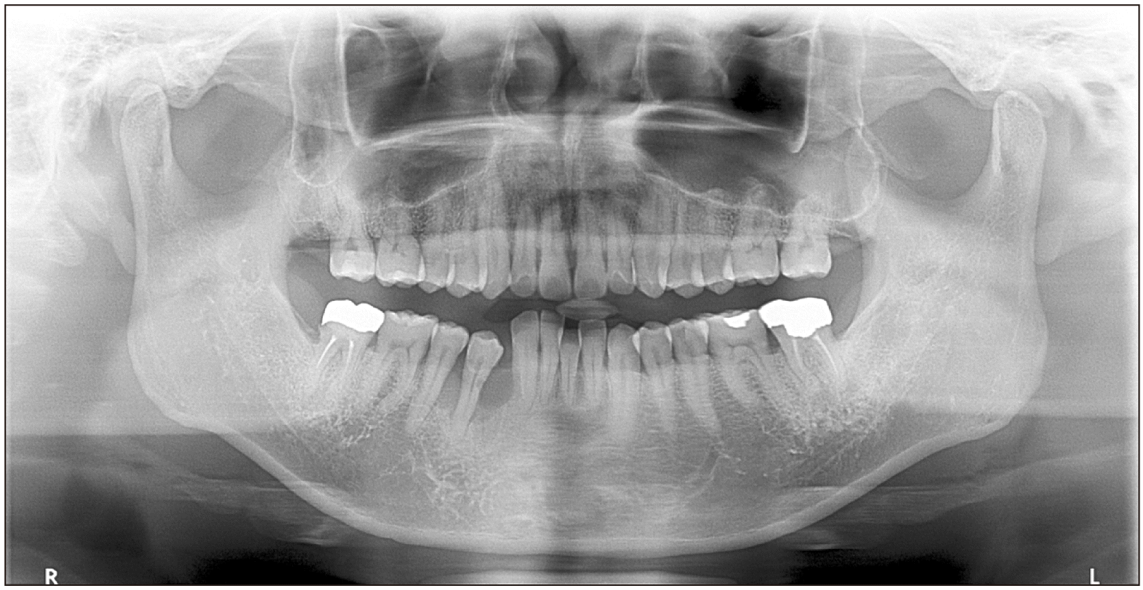
A 48-year-old male without any underlying diseases was referred to our department of periodontology due to an abnormality in the right buccal gingiva after a scaling procedure at a local clinic. Since no improvement in symptoms occurred after periodontal treatment, the patient was referred to the department of oral and maxillofacial surgery. A panoramic radiograph revealed alveolar bone resorption of #43 to #45.(Fig. 1) A biopsy was performed, and diagnosis of spindle cell tumors, including inflammatory myofibroblastic tumors (IMT), was based on immunoreactivity for ALK. Magnetic resonance imaging (MRI) and CECT (contrast-enhanced computed tomography) were performed but revealed no abnormal findings. Due to the swelling, redness, and pain in the affected area without improvement in symptoms, marginal mandibulectomy was performed on the patient under general anesthesia.
The pathologic findings were the same in all biopsied and resected specimens. All microscopic slides were reviewed with additional immunohistochemical staining and a thorough literature review. Histological examination revealed fascicular proliferation of spindled tumor cells at the subepithelial portion of the gingiva.(Fig. 2. A) Tumor cells contained eosinophilic cytoplasm and possessed blunted, ovoid, or fusiform nuclei with inconspicuous nucleoli and occasional mitotic figures.(Fig. 2. B) Sclerosing or focal myxoid stroma was noted. The overlying gingival mucosa exhibited erosion accompanied by mixed inflammatory infiltrate with abundant plasma cells. The inflammatory infiltrate was prominent below the erosive mucosa (Fig. 2. C) but was inconspicuous in the deep portion of the tumor. The marginal mandibulectomy specimen revealed that the tumor cells had invaded the mandibular bone trabeculae. On immunohistochemistry, the tumor cells were strongly and diffusely positive for vimentin, desmin (Fig. 2. D), and myoD1 (Fig. 2. E) and were strongly and multifocally positive for smooth muscle actin (SMA), caldesmon, ALK (Fig. 2. F), and cytokeratin. Tumor cells were negative for epithelial membrane antigen (EMA), myoglobin, S100 protein, and p53. The Ki67-labeling index was approximately 20%. Based on the combined histological and immunohistochemical findings, a revised diagnosis of SCRMS with ALK expression was concluded. No recurrent lesions were observed for 8 months after surgery. Postoperative PET/CT (positron emission tomography/computed tomography) and MRI revealed no residual tumors and no distant or lymph node metastases.

III. Discussion

ALK positivity is more common in IMT, but several reports have expanded on the variety of non-IMT soft tissue that expresses ALK, specifically a subset of RMS5-9. According to Pillay et al.10, 28% of RMS cases were reported to express ALK. The main histological differential diagnosis in the present case included IMT, leiomyosarcoma, and spindle cell squamous cell carcinoma. Inflammatory cell infiltrate and ALK positivity can lead to an inappropriate diagnosis of IMT. However, inflammation was mostly associated with mucosal erosion. A lack of inflammation in the center of the tumor is unusual in IMT. Moreover, myoD1 positivity is very specific for RMS and can confirm its diagnosis. A tumor with proliferation of fascicular spindle cells immunoreactive for desmin and SMA mimics leiomyosarcoma. However, myoD1 positivity is helpful in distinguishing RMS from leiomyosarcoma. Squamous cell carcinoma may possess spindle cell features, and cytokeratin positivity suggests squamous cell carcinoma. However, the overlying squamous epithelium revealed no evidence of squamous cell carcinoma in situ, and negativity for EMA as well as positivity for desmin and myoD1 can distinguish RMS from squamous cell carcinoma.
Although multiple biopsies were performed in the present case, the pathologic diagnoses were inaccurate due to disease rarity and lack of indicators of ALK-positive SCRMS. However, its precise diagnosis is essential for proper treatment. The current routine therapeutic modalities for patients with SCRMS consist of surgery, chemotherapy, radiation therapy, or a combination2,11. However, the optimal treatment scheme and subsequent prognosis for this rare malignancy remain to be determined, presenting a dilemma for clinicians involved in its treatment. Knowledge of this rare disease entity and an appropriate immunohistochemical panel would be helpful in correct diagnosis. Further studies will be needed to demonstrate whether ALK expression in SCRMS has any significance in ALK inhibitor treatment.

Acknowledgements

The authors would like to thank Dr. Jerad M. Gardner, a dermatopathologist & section head of bone/soft tissue pathology at Geisinger Medical Center, Danville, Pennsylvania, USA, for his valuable comments on this case.

Notes

Authors’ Contributions

J.Y.L. participated in data collection and wrote the manuscript. W.L. participated in data collection and revised the manuscript. M.Y.K. participated in the study design, revision and provided funding acquisition. All authors read and approved the final manuscript.

Consent for Publishing Photographs

Written informed consent was obtained from the patient for publication of this article and accompanying images.

Conflict of Interest

No potential conflict of interest relevant to this article was reported.

Funding

This work was supported by a National Research Foundation of Korea (NRF) grant funded by the Korean government (MSIT) (2021R1C1C1008843).

References

1. Miloglu O, Altas SS, Buyukkurt MC, Erdemci B, Altun O. 2011; Rhabdomyosarcoma of the oral cavity: a case report. Eur J Dent. 5:340–3. DOI: 10.1055/s-0039-1698902. PMID: 21769278. PMCID: PMC3137450.
crossref
2. World Health Organization (WHO) Classification of Tumours Editorial Board. 2020. WHO Classification of tumours: soft tissue and bone tumours. 5th ed. IARC Press.
3. Nascimento AF, Fletcher CD. 2005; Spindle cell rhabdomyosarcoma in adults. Am J Surg Pathol. 29:1106–13. DOI: 10.1097/01.pas.0000158396.57566.5d. PMID: 16006807.
crossref
4. Mentzel T, Kuhnen C. 2006; Spindle cell rhabdomyosarcoma in adults: clinicopathological and immunohistochemical analysis of seven new cases. Virchows Arch. 449:554–60. https://doi.org/10.1007/s00428-006-0284-4. DOI: 10.1007/s00428-006-0284-4. PMID: 17013628.
crossref
5. Corao DA, Biegel JA, Coffin CM, Barr FG, Wainwright LM, Ernst LM, et al. 2009; ALK expression in rhabdomyosarcomas: correlation with histologic subtype and fusion status. Pediatr Dev Pathol. 12:275–83. https://doi.org/10.2350/08-03-0434.1. DOI: 10.2350/08-03-0434.1. PMID: 18788887.
crossref
6. Le Loarer F, Cleven AHG, Bouvier C, Castex MP, Romagosa C, Moreau A, et al. 2020; A subset of epithelioid and spindle cell rhabdomyosarcomas is associated with TFCP2 fusions and common ALK upregulation. Mod Pathol. 33:404–19. https://doi.org/10.1038/s41379-019-0323-8. DOI: 10.1038/s41379-019-0323-8. PMID: 31383960.
crossref
7. Li XQ, Hisaoka M, Shi DR, Zhu XZ, Hashimoto H. 2004; Expression of anaplastic lymphoma kinase in soft tissue tumors: an immunohistochemical and molecular study of 249 cases. Hum Pathol. 35:711–21. https://doi.org/10.1016/j.humpath.2003.12.004. DOI: 10.1016/j.humpath.2003.12.004. PMID: 15188137.
crossref
8. van Gaal JC, Flucke UE, Roeffen MH, de Bont ES, Sleijfer S, Mavinkurve-Groothuis AM, et al. 2012; Anaplastic lymphoma kinase aberrations in rhabdomyosarcoma: clinical and prognostic implications. J Clin Oncol. 30:308–15. https://doi.org/10.1200/jco.2011.37.8588. DOI: 10.1200/JCO.2011.37.8588. PMID: 22184391.
crossref
9. Yoshida A, Shibata T, Wakai S, Ushiku T, Tsuta K, Fukayama M, et al. 2013; Anaplastic lymphoma kinase status in rhabdomyosarcomas. Mod Pathol. 26:772–81. https://doi.org/10.1038/modpathol.2012.222. DOI: 10.1038/modpathol.2012.222. PMID: 23307059.
crossref
10. Pillay K, Govender D, Chetty R. 2002; ALK protein expression in rhabdomyosarcomas. Histopathology. 41:461–7. https://doi.org/10.1046/j.1365-2559.2002.01534.x. DOI: 10.1046/j.1365-2559.2002.01534.x. PMID: 12405914.
crossref
11. Hartmann S, Lessner G, Mentzel T, Kübler AC, Müller-Richter UD. 2014; An adult spindle cell rhabdomyosarcoma in the head and neck region with long-term survival: a case report. J Med Case Rep. 8:208. https://doi.org/10.1186/1752-1947-8-208. DOI: 10.1186/1752-1947-8-208. PMID: 24946859. PMCID: PMC4077640.
crossref

Fig. 1
Preoperative panoramic radiograph.
jkaoms-49-2-96-f1.tif
Fig. 2
A. Photomicrograph reveals proliferation of fascicular spindled tumor cells in the subepithelial portion (H&E staining, ×100). B. Higher magnification of tumor cells showing blunted, elongated nuclei and abundant eosinophilic cytoplasm with inconspicuous inflammation (H&E staining, ×200). C. Abundant inflammatory infiltrate associated with mucosal erosion (H&E staining, ×200). D. Tumor cells diffusely positive for desmin (immunohistochemistry staining, ×200). E. Tumor cells diffusely positive for myoD1 (immunohistochemistry staining, ×200). F. Tumor cells multifocally positive for anaplastic lymphoma kinase (immunohistochemistry staining, ×200).
jkaoms-49-2-96-f2.tif
TOOLS
Similar articles