

 PDF
PDF Citation
 Print
Print


I. Introduction
An ectopic tooth is that which is displaced to a location a considerable distance from its normal anatomic site. Ectopic tooth impaction is a relatively uncommon phenomenon1 in which the eruption is out of the dental apparatus or no part of the tooth is within the eruption zone2. Supernumerary, deciduous, and permanent teeth have been reported in diverse heterotopic positions, including the nasal cavity, maxillary sinus, orbit, palate, mandibular condyle and coronoid process3-6.
Ectopic teeth are rare; however, the molar is the most commonly reported ectopic tooth in the antrum4,7 and the mandible8-10. Veerabhadrappa et al.10 noted that more than two-thirds of patients with ectopic mandibular molars were >40 years of age, and more were female, at a sex ratio of 2.5:1. Ectopic tooth eruption, though not completely understood, may result from abnormal tissue interaction during tooth development9.
An ectopic tooth may be associated with recurrent infections characterized by pain, swelling, and purulent discharge or may be discovered incidentally during other investigations4,5,11. Ectopic molars may be associated with dentigerous cysts, bone expansion, and pain8.
In this study, we report a case series of ectopic teeth in rare locations and highlight the patients’ associated pathology and surgical management.
II. Patients and Methods
We reviewed all the cases of ectopic teeth observed and managed at the Department of Oral and Maxillofacial Surgery of University of Maiduguri Teaching Hospital from January 2011 to December 2020. Approval was obtained from the ethical review committee of University of Maiduguri Teaching Hospital (No. OHRP-IRB00013572 UMTH/REC/22/1090), and the written informed consent was obtained from all patients where necessary. The information retrieved includes biodata, location of the ectopic tooth, symptoms, signs, type of tooth and associated pathology, surgical approach and complications.
III. Results
We recorded 10 cases of ectopic teeth during this period that were outside of the tooth-bearing region of the jaws. One ectopic tooth was discovered incidentally during a radiological investigation for gunshot wound to the face and was removed prophylactically during maxillofacial surgery.
Mean patient age was 23.3 years and ranged from 11-45 years. Most (80.0%) of the patients were males; only two of the patients were females. Six patients (60.0%) had ectopic teeth on the right side. There were no cases of bilateral ectopic teeth.(Table 1)
Canine teeth accounted for 60.0% of the ectopic tooth impactions in the maxilla; the third molar and premolar each accounted for 40.0% of ectopic tooth impactions in the mandible.
In this series, the maxillary antrum was the only location of ectopic impaction in the upper jaw, accounting for 50.0% of all ectopic sites. The lower border of the mandible was the commonest location in the lower jaw (Fig. 1) and accounted for 40.0% of total impactions.
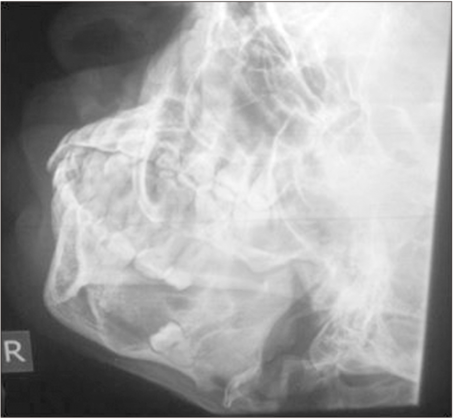
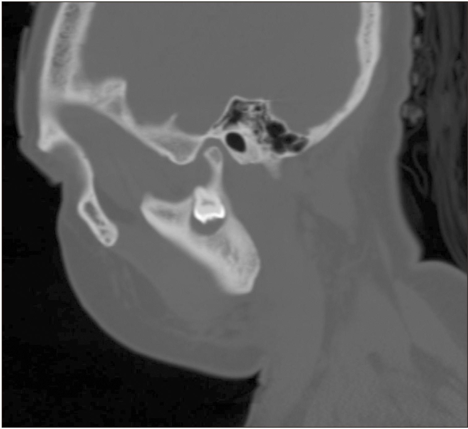
All cases except the ectopic tooth discovered accidentally were symptomatic. Nine patients (90.0%) presented with swelling, five (50.0%) of which were associated with pain. Discharge was purulent in only two cases, and other discharges were suggestive of ruptured cyst content.(Table 1) Fistula and trismus were recorded only in patient No. 9 with ectopic impaction in the sigmoid notch.(Fig. 2)
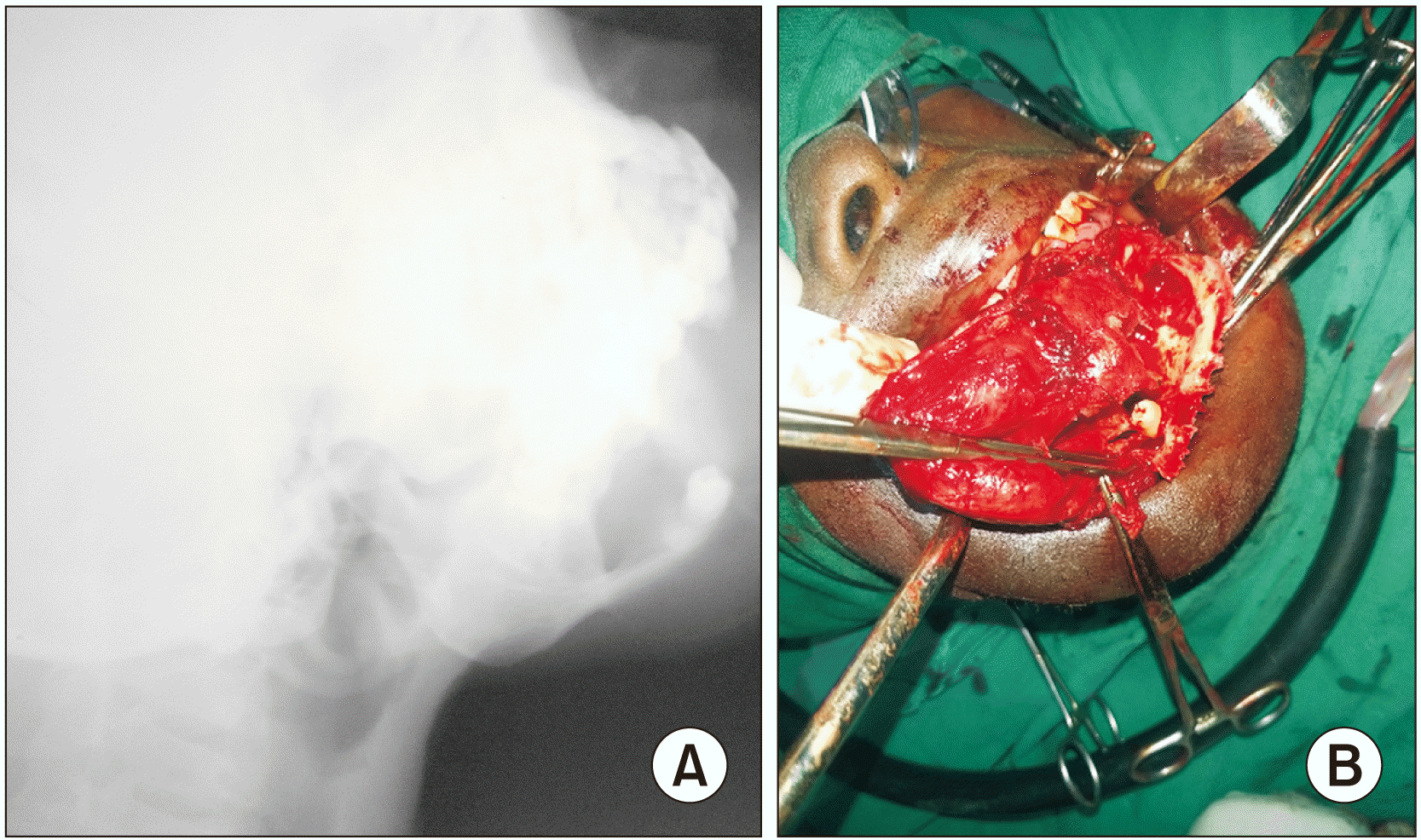
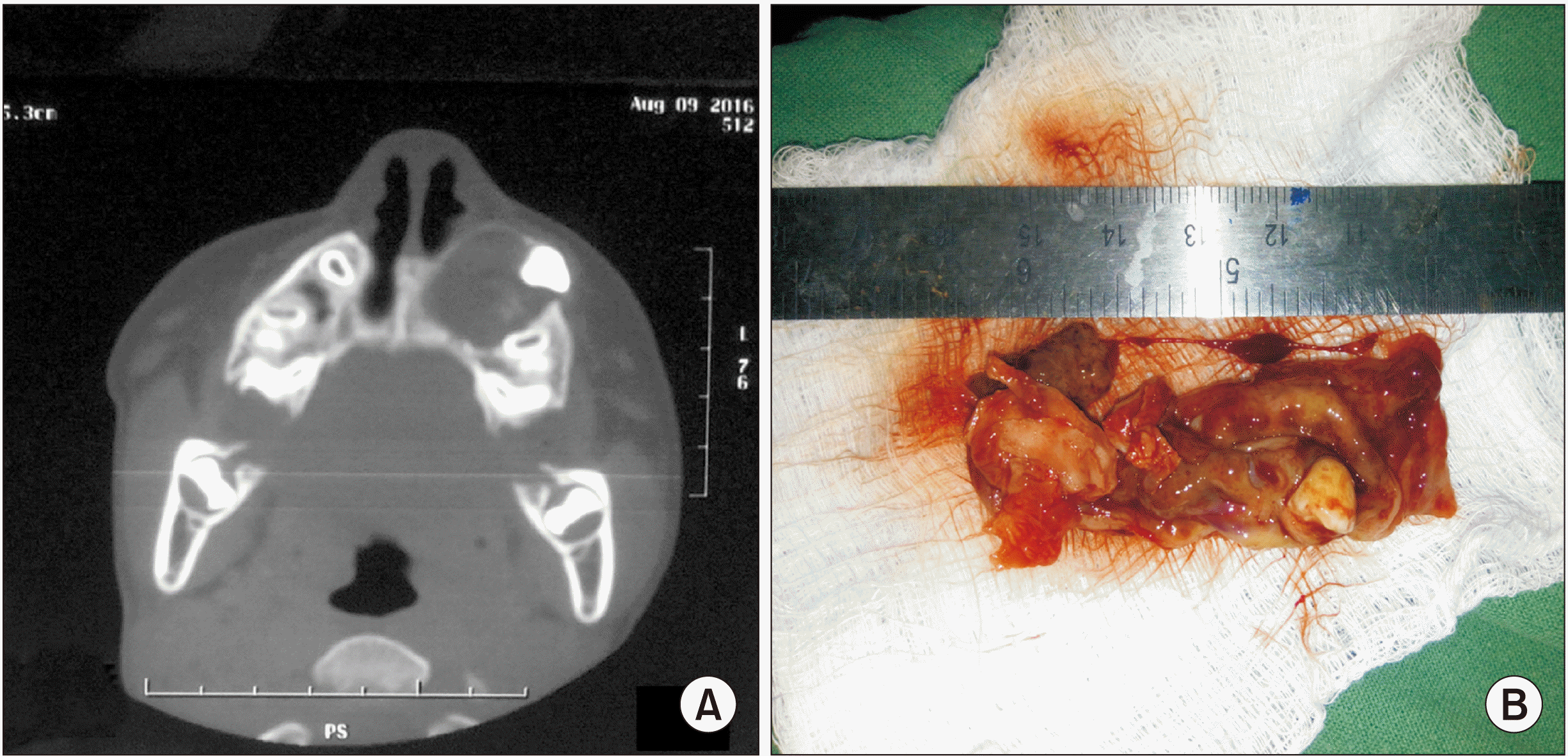
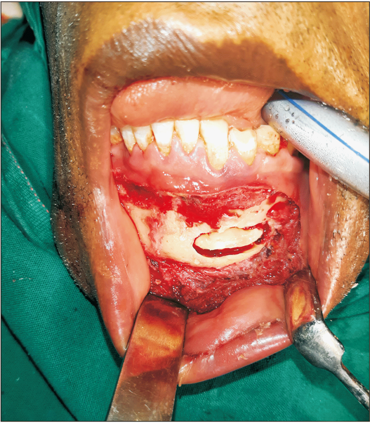
Seven of the treated patients had enucleation/extraction of ectopic teeth carried out via the intraoral approach. We resected an ameloblastoma in patient No. 4 and reconstructed the defect using a titanium plate and an iliac crest bone graft. The crown of the premolar in the lower border of the mandible was visible on the radiograph (Fig. 3. A) and intraoperatively.(Fig. 3. B) All patients with dentigerous cysts in the antrum were treated surgically via the Caldwell-Luc approach. The computed tomography scan taken in patient No. 1 showed an enlarged cystic swelling surrounding a radiopaque, canine tooth-shaped structure (Fig. 4. A) confirmed postoperatively as an ectopic canine tooth with an associated cyst sac.(Fig. 4. B)
Histopathology confirmed dentigerous cysts in 7 patients, with one ameloblastoma and ameloblastic fibroma. The ectopic canine (patient No. 3) found incidentally had no associated pathology.(Fig. 5)
IV. Discussion
Ectopic teeth have been reported in various locations, including the antrum, floor of the orbit, lower border of the mandible, nasal cavity, ramus, coronoid, condyle and sigmoid notch3-6,9,10. The ectopic tooth is a rare entity; only a few have been reported10.
The prevalence of ectopic teeth may be under-reported because some are asymptomatic and only diagnosed incidentally4,12. The under-reporting could also be the result of low reporting of ectopic teeth associated with odontogenic tumors. Veerabhadrappa et al.10 and Fındık and Baykul9 reported a high prevalence of ectopic mandibular teeth in females in their forties. This is contrary to this study’s finding in which most patients were males with a mean age of 30 years.
Dentigerous cysts are relatively common and represent 30% of all jaw cysts13. All ectopic teeth in the maxilla were in the antrum and were associated with a dentigerous cyst; these teeth comprised 71% of the entire cyst. However, 70%-83% of dentigerous cysts have been reported to occur in the mandible12,13. Dentigerous cysts and odontogenic tumors were the two main pathologies associated with ectopic teeth in the mandible. Interestingly, two of the teeth were premolars; and the teeth were not displaced to the lower border of the mandible despite there being a tooth attached to the lower border with incomplete root formation.(Fig. 3. A) The other patient had a retained deciduous molar.
The ectopic teeth in the antrum were all symptomatic in this series and were associated with the dentigerous cysts. The mean age at presentation was 16.4 years. Maxillary dentigerous cysts are usually common in adolescents and those in their twenties and thirties6,7,12; however, one reported case was in a patient as young as four years old14. Zhang et al.’s study of 2,029 dentigerous cysts of the jaws13 demonstrated that dentigerous cysts are rare in the first decade of life and are most common in the second and third decades. The frequency declined with increased age. Our patient with an asymptomatic ectopic tooth in the mandible was diagnosed incidentally at age 45 years. This age demographic differs from the finding that symptomatic ectopic teeth are most commonly diagnosed earlier, in the third decade. Wu et al.2, however, reported a higher mean age of 36.3 years for ectopic teeth in the mandible, even though most cases were symptomatic.
Contrary to other reports1,2,10, all of our mandibular ectopic teeth except one were located at the lower border of the mandible. The high frequency of reported ectopic molars in the condylar and subcondylar regions1,2,10 could be due to the focus of the studies; that focus was predominantly on ectopic third molars. Ectopic teeth at the lower border, perhaps asymptomatic, may have gained little attention in those reports.
The intraoral surgical approach was preferred except for cases requiring access to the sigmoid/subcondylar region. Most surgeons advocate the use of the intraoral approach whenever possible1,2,9,10 particularly due to its superior cosmetic outcome when access and wide surgical field is not compromised. We used the retromandibular approach to remove the ectopic tooth in the subcondylar area after a failed intraoral approach. Cosmesis was important to this patient who required counselling. The extra-oral approach is the most frequently used for ectopic teeth in the mandibular condyle, ramus, angle, and lower border1,2,10. This approach provides better surgical field exposure and direct tooth access, reducing the risk of iatrogenic injuries and excess bone removal10. The risk of complications is not eliminated because damage to the marginal mandibular nerve and other facial nerve branches can occur in extra-oral approaches.
Consequently, some surgeons have used endoscopically assisted extraction of an ectopic mandibular third molar15 and advocate its use for an ectopic tooth in the antrum. The endoscopic approach is conservative and illuminates and magnifies the surgical field. The technique, however, requires expensive endoscopic equipment and advanced training.
V. Conclusion
Ectopic teeth are rare and not always associated with pathology. A dentigerous cyst is the most common pathology of unerupted or impacted teeth, which may present with swelling and other vague symptoms. A high index of suspicion and radiological investigation are necessary for diagnosis. The type of associated pathology and location should inform the decision regarding a surgical approach and possible complications. A more extensive multi-center study is warranted to determine the prevalence of ectopic teeth other than the third molar.
 XML Download