Journal List > Ann Lab Med > v.43(5) > 1516082625

Chuah, Lim, Tan, Pratumvinit, Loh, Vasikaran, Markus, and the APFCB Harmonization of Reference Intervals Working Group: Functional Reference Limits: Describing Physiological Relationships and Determination of Physiological Limits for Enhanced Interpretation of Laboratory Results

Abstract

Functional reference limits describe key changes in the physiological relationship between a pair of physiologically related components. Statistically, this can be represented by a significant change in the curvature of a mathematical function or curve (e.g., an observed plateau). The point at which the statistical relationship changes significantly is the point of curvature inflection and can be mathematically modeled from the relationship between the interrelated biomarkers. Conceptually, they reside between reference intervals, which describe the statistical boundaries of a single biomarker within the reference population, and clinical decision limits that are often linked to the risk of morbidity or mortality and set as thresholds. Functional reference limits provide important physiological and pathophysiological insights that can aid laboratory result interpretation. Laboratory professionals are in a unique position to harness data from laboratory information systems to derive clinically relevant values. Increasing research on and reporting of functional reference limits in the literature will enhance their contribution to laboratory medicine and widen the evidence base used in clinical decision limits, which are currently almost exclusively contributed to by clinical trials. Their inclusion in laboratory reports will enhance the intellectual value of laboratory professionals in clinical care beyond the statistical boundaries of a healthy reference population and pave the way to them being considered in shaping clinical decision limits. This review provides an overview of the concepts related to functional reference limits, clinical examples of their use, and the impetus to include them in laboratory reports.

INTRODUCTION

Reference intervals describe the percentile values of a biomarker measured in a reference population [1, 2]. Ideally, the reference population should be selected from a representative population free of any pathological conditions that are known to affect the biomarker, with samples handled under standardized pre-analytical conditions. Frequently encountered lower and upper reference limits are the 2.5th and 97.5th percentile values, respectively, representing the central 95% of the distribution. Using this strict statistical definition, 5.0% of “apparently healthy” subjects will have biomarker values outside the derived reference interval, equally split between subjects falling outside the upper or lower reference limit. Reference intervals provide population-level statistical limits for a biomarker and are the most commonly used tools for laboratory result interpretation [3].
Within a biological homeostatic system, numerous physiological processes exist that regulate and maintain the stability of the internal environment of the system, particularly in response to changing external conditions [4]. Homeostasis is achieved through self-regulating positive and negative feedback mechanisms, where an increase or reduction in a metabolite concentration, for example, is accompanied by a physiological response to decrease or increase the concentration back to a biological set point. Components of the homeostatic system may be measured in the laboratory and, when profiled, provide a more complete assessment of the physiological state of the subject.
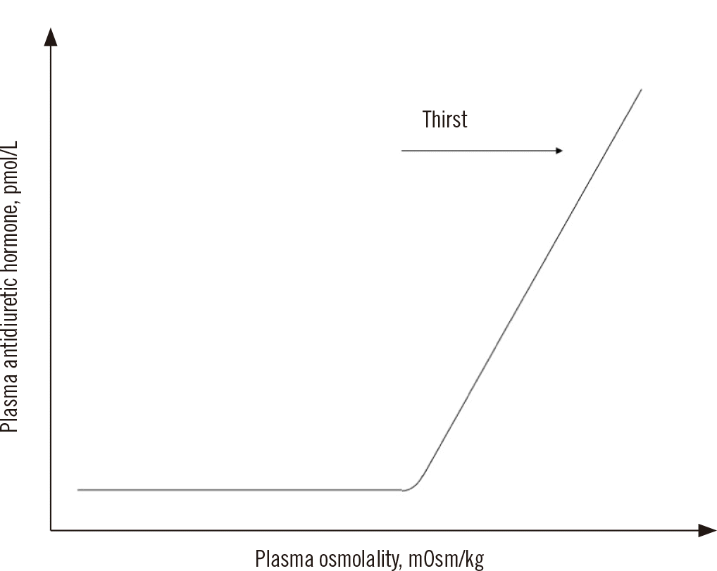
Consider the example of a simplified homeostatic system for water balance, including plasma osmolality and antidiuretic hormone measurements [5]. When plasma osmolality is above a certain threshold, as in the case of dehydration, it stimulates the thirst center in the hypothalamus and the posterior pituitary to release antidiuretic hormone (Fig. 1). The antidiuretic hormone acts on the collecting ducts of the nephrons to increase water resorption, which lowers plasma osmolality. This in turn lowers the stimulus for further antidiuretic hormone secretion and eventually restores plasma osmolality to the homeostatic set point.
An understanding of physiological and homeostatic processes is fundamental for proper laboratory result interpretation and determining the physiological status of the patient [4, 6]. Homeostatic mechanisms are designed to tolerate or compensate for physiological disturbances to a certain degree; however, excessive disruptions may eventually lead to the breakdown of these compensatory mechanisms and the subsequent development of a pathological state.
Reference intervals do not provide information on the relationships between two or more physiologically related biomarkers [7]. This may limit the ability of clinical professionals to gain important insights into the physiological status of the patient. While reference intervals define the statistical boundaries of a reference population, their clinical performance depends on the selection of the reference population (e.g., whether including subclinical/pathological subjects) and the relative distance between non-diseased and diseased population distributions [1, 2, 8]. Furthermore, many clinical conditions exist as a spectrum that ranges from healthy over subclinical disease to overt pathology, and arbitrary partitioning of this continuum may lead to misdiagnosis [6]. This limitation of reference intervals is highlighted in conditions for which a large proportion of the general population harbors a subclinical presentation that may be difficult to identify and exclude.
For example, serum vitamin D insufficiency and deficiency are common in the general population, even in tropical regions [9]. If reference intervals would be established based on the general population, the lower reference limit would likely include a significant proportion of subjects with insufficient vitamin D concentrations and some who are deficient but asymptomatic. This would lead to an inappropriately low lower reference limit, which would fail to identify subjects with vitamin D insufficiency and hinder clinicians from making an appropriate clinical interpretation for optimal treatment. Similar conundrums exist for population-derived serum ferritin reference intervals in women [7].
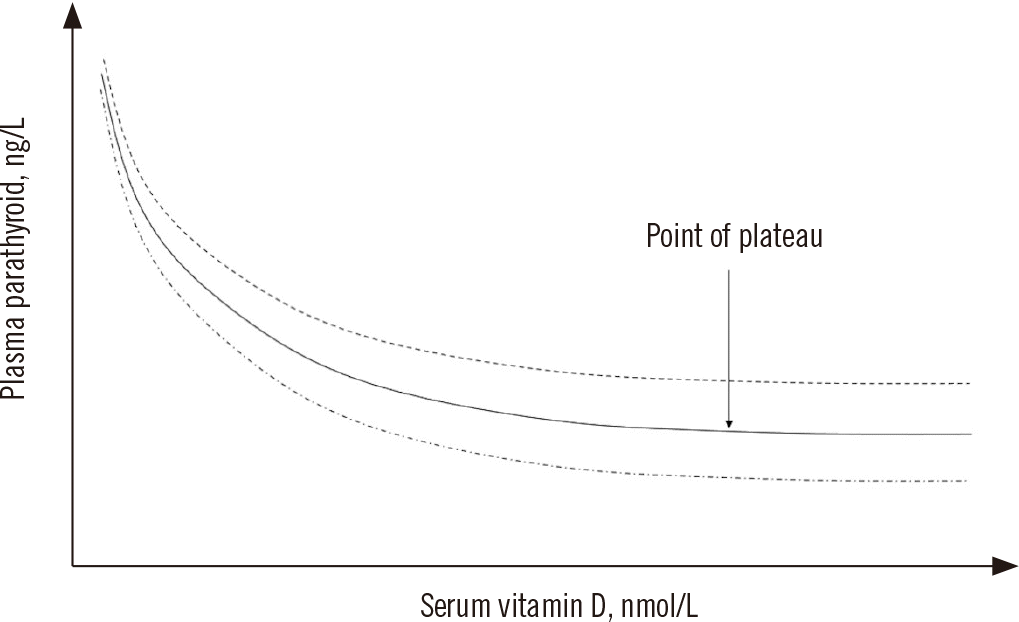
Instead, the interpretation of serum vitamin D concentrations relies on medical decision limits that define deficiency, insufficiency, and sufficiency [10]. These medical decision limits are geared toward identifying subjects for replacement therapy to reduce the risk of rickets, osteomalacia, or osteoporosis [10, 11]. These thresholds are, in part, informed by examination of the physiological relationships among the serum vitamin D concentration, plasma parathyroid hormone concentration, and intestinal calcium and phosphorus absorption [10]. Parathyroid hormone promotes calcium efflux from the bone, decreases calcium loss from the kidneys, and enhances intestinal calcium absorption. Serum vitamin D and plasma parathyroid hormone concentrations have an inverse relationship that plateaus (Fig. 2) [12]. The point at which the plateau begins represents the functional limit of the serum vitamin D concentration at which the parathyroid hormone concentration is maximally suppressed and has been suggested as the basis for defining the optimal vitamin D status [12].

DEFINITIONS OF FUNCTIONAL REFERENCE LIMITS

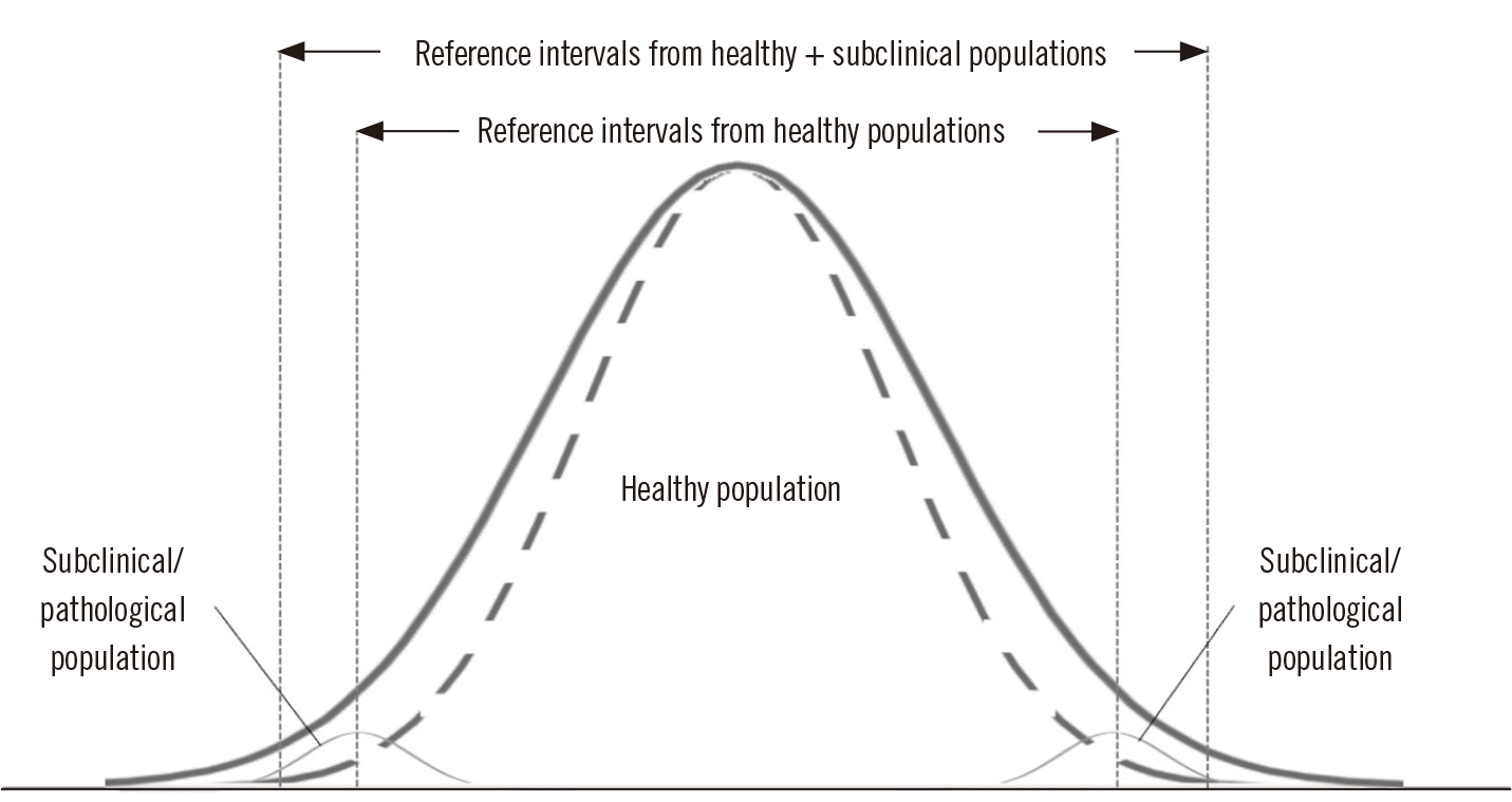
It can be difficult to identify and exclude subjects with subclinical disease or pathological states in reference interval studies because many of these subjects may be asymptomatic. Moreover, the distribution of the subclinical diseased population may overlap with that of the healthy reference population and contribute to the appearance of one overall normal distribution (Fig. 3) [7]. If such subjects are inadvertently included in the reference population, this may broaden the overall population distribution and significantly influence the derived percentile values that lie at the extreme ends of the population distribution, such as the conventional 2.5th and 97.5th percentiles. The application of such artificially broadened reference intervals may result in inappropriate interpretation at the lower, upper, or both limits, depending on the influence of the pathological states on the biomarker. Hence, in the establishment of reference intervals, the inclusion of subclinical and pathological populations is clearly undesirable and contributes “noise” to the reference data and therefore should be avoided to derive clinically appropriate values.
In contrast, functional reference limits seek to describe reference values that span the continuum of healthy, subclinical, and pathological populations, with the inclusion of decompensated populations being considered integral to the definition of functional reference limits. In homeostatic and physiological systems, functional relationships may exist between different components, where changes in one component may positively or negatively affect the others [4]. The relationships among the components are maintained under normal conditions but may significantly change under physiological or pathological stress. Such changes in the interactions among physiological components can be modeled using statistical approaches [7]. A functional reference limit is defined as the numerical threshold at which the relationship between physiologically related components, such as laboratory biomarkers, changes significantly [7]. Here, the term “functional” describes both the biological and statistical relationships between two related physiological components. Biologically, the change in a relationship may represent a significant functional change in a physiological state (e.g., limits of homeostasis or decompensated state) or pathology. The biological relationship is generally specific for a particular physiological system or clinical condition.
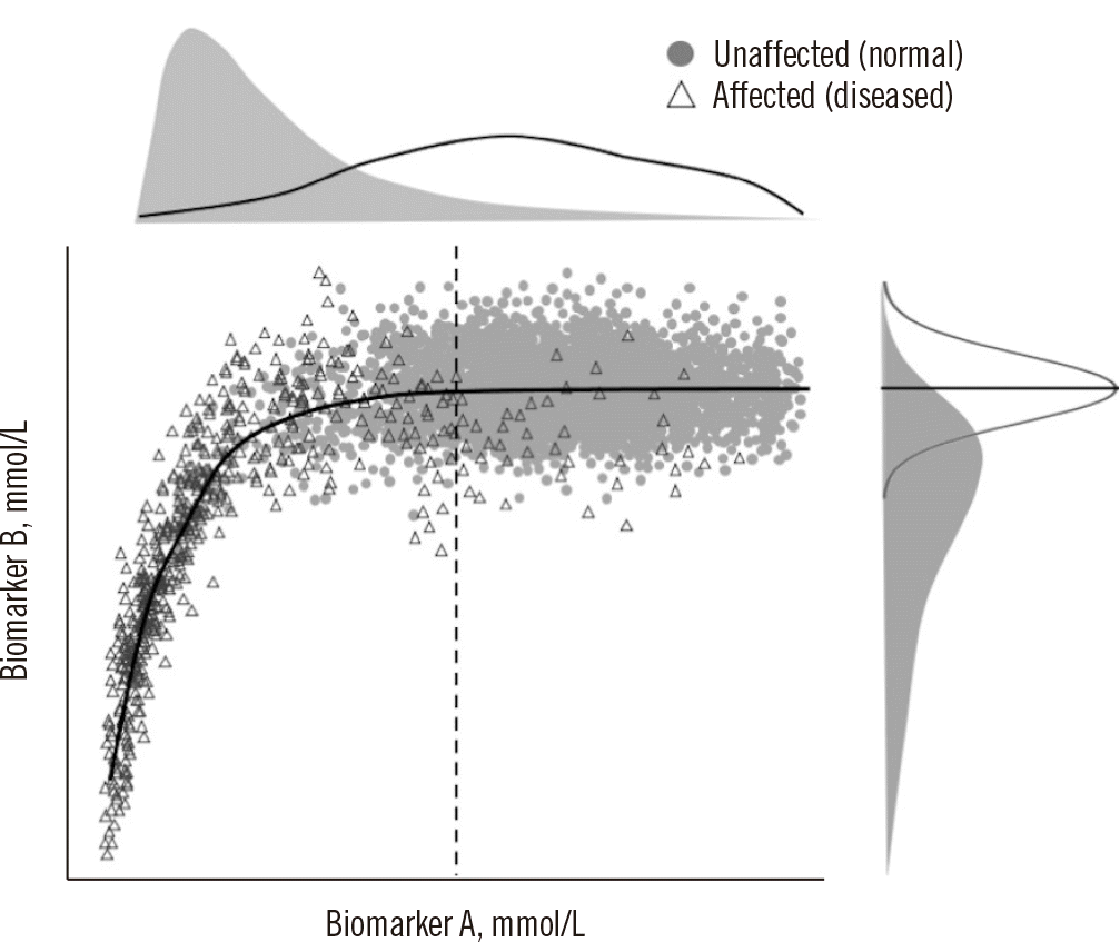
Statistically, this may be represented by a significant change in the curvature of a mathematical function or curve (e.g., an observed plateau) (Fig. 4). The point at which the statistical relationship changes significantly is the point of curvature inflection and can be mathematically modeled from the relationship between the interrelated biomarkers. Unlike reference intervals, functional reference limits may exist for only one side of the measurand concentration distribution, i.e., only one functional reference limit may exist [7].

DIFFERENCES BETWEEN FUNCTIONAL REFERENCE LIMITS AND CLINICAL DECISION LIMITS

At first glance, functional reference limits and clinical decision limits may appear to be similar and can be confused with one another. However, there are important distinctions between them. Clinical decision limits are generally derived from large-scale clinical outcome studies, clinical guidelines (that may be formed from expert opinions), and consensus value statements [2]. They may be defined by diagnostic performance in terms of clinical sensitivity, clinical specificity, and costs of clinical misclassification, which may be optimized differently in different clinical settings and clinical conditions. The observed diagnostic performance is heavily influenced by the distance between biomarker concentrations observed in healthy and diseased populations. Clinical decision limits described in this manner have clearly defined diagnostic performance characteristics and are generally related to the identification of subjects with a specific clinical condition. Examples of clinical decision limits with defined diagnostic performance characteristics include the serum cortisol threshold for diagnosing cortisol excess and the cardiac troponin cutoff for myocardial infarction [13, 14].
Additionally, clinical decision limits can be determined from population-based epidemiological studies. Clinical outcomes are usually derived from large prospective or retrospective cohort studies that examine specific outcomes of interest, such as the risk of complications or effects of applied interventions. Examples of such clinical decision limits include serum lipid concentrations and diabetes diagnostic criteria [15, 16]. Finally, critical values, which are laboratory results representing physiological states at such variance from normal that require prompt action to avert life-threatening adverse events, are also considered clinical decision limits [2]. Critical values may be associated with an increased risk of death or critical states requiring intensive care [17, 18]. An acceptable level of risk for clinical decision limits is generally arbitrarily selected for a clinical scenario or setting [2].
In contrast, functional reference limits are defined by statistical modeling and identification of the point of significant change in the modeled physiological relationship. Hence, they may only indicate the point where homeostasis is disturbed and not the presence of a disease. Functional reference limits also do not predict disease risk or treatment targets to reduce the risk profile of a clinical condition. Data related to functional reference limits may be considered during the formulation of clinical decision limits, as shown in the above example of serum vitamin D clinical decision limits; however, the converse is not true. The differences among reference intervals, clinical decision limits [2], and functional reference limits are summarized in Table 1.

STATISTICAL APPROACHES IN FUNCTIONAL REFERENCE LIMITS

The derivation of functional reference limits generally involves a two-step process [7]. The first is a general overall assessment of the shape (i.e., relationship) between the two interrelated biomarkers through exploratory data analysis. Following the identification of a relationship with an inflection point, further regression modeling may be applied to optimally describe the mathematical relationship.
In the past, the correlation between two interrelated biomarkers has been examined using scatter plots. The “functional reference limit” (although not recognized as such at the time) can then be determined visually [19]. The scatter plot remains a cornerstone in evaluating the relationship between a pair of biomarkers, although more accurate estimations of the relationship can be made using regression models. More recently, advancements in computational power have allowed for a more sophisticated assessment of the relationship between two biomarkers. Some statistical approaches that have been used include quantile regression, a restricted cubic spline, and smoothed curves [20]. In general, the median is used to describe the central tendency of a distribution, as it is more resistant than the mean to outliers in a dataset that may contain more extreme representations of physiological and pathological states. Depending on the statistical model applied, it may be appropriate or desirable to identify and remove extreme values or outliers prior to applying the regression functions.
Quantile regression is an extension of ordinary least squares regression that estimates the conditional quantiles (e.g., the median) of the response variable. This regression framework has the advantage of being more robust to outliers in the dependent variable and can be used when the assumptions of ordinary linear regression are not fulfilled. This regression approach has been used to determine ferritin functional reference limits in children [21]. In the cubic spline approach, the median values of the data distribution across the concentration range are determined at specific intervals or partitions [20]. Subsequently, these median values are fitted using piecewise polynomial regression (in a cubic spline, third-order polynomials are used). This is a relatively simple approximation of a complex relationship, with curve fitting demonstrating good accuracy. However, this approach can produce curves with a “knotted” appearance. A smooth curve is a curve with a continuously turning tangent (i.e., nonlinear component) without singular points with the aim to reduce the influence of extreme values and outliers. Fractional polynomial regression can be used to model relationships and produce a smooth continuous curve fit across a data range [22]. It should be noted that the application of different regression models to fit a curve may produce different impressions of the trends between the biomarker pair [7]. It is important that the investigator and reader familiarize themselves with the physiology of biomarkers and how they interact, also within the spectrum of different clinical conditions, to allow an informed interpretation of the relationship.
Once the optimal statistical model is established, the inflection point can be determined. This may be a single point or region where a significant biological or statistical change occurs and represents the functional reference limit. It can be determined by assessing the statistical significance of the correlation between groups, a defined biological change (e.g., a decrease in the correlated dependent biomarker), the application of clinical criteria (e.g., setting an a priori clinical threshold/concentration for one of the biomarkers), or simple visual inspection of the point of inflection [7]. In yet another approach, piecewise linear regression models are used to split the data into different segments to fit different linear regressions and determine the breakpoint (also termed “knot value”), which is then considered the functional reference limit [23].
At present, there is no guidance on regression models and statistical definitions of the point of inflection to adopt when deriving a functional reference limit [7]. Hence, a methodology may be adopted considering personal preference, familiarity with the technique, data density, data distribution, and functional and statistical relationships between the biomarkers. It is helpful to explore the different statistical models and consider the physiology and pathophysiology of the clinical condition to arrive at an optimal interpretation. In all cases, confidence intervals of the estimated functional reference limits from each of the different regression approaches should be determined to facilitate comparison. Likewise, no guidance is available for the preferred approach to calculating confidence intervals; however, bootstrap approaches have been used in some studies.
Like for conventional reference intervals, it may be advantageous to establish functional reference limits for specific populations. While there is no guidance on the approach to partition data for this purpose, it may be reasonable to consider performing a stratified analysis based on known reference interval partitioning when the physiology of the subpopulation is expected to significantly differ from that of the general population. A comparison of the estimated parameters and confidence intervals with and without partitioning may help inform whether the data and populations can be merged.

INDIRECT APPROACHES USING HISTORICAL LABORATORY VALUES

In previous studies, biomarker relationships were observed using smaller study designs that examined more extreme conditions in healthy subjects or by assessing the pathophysiology of diseased subjects [7, 19]. More recently, efforts have been made to statistically describe the relationship between two physiologically related biomarkers, including the homeostatic response and decompensation point, using big data analysis [20, 23]. This was made possible by the confluence of improvements in laboratory automation, information systems, and statistical computation. Modern-day laboratory automation allows generating high-throughput laboratory data at affordable prices. The accessibility of laboratory services has lowered the threshold for testing and encouraged clinicians to perform tests on subjects with a low clinical index of suspicion [24]. In other words, many laboratory tests are performed on subjects who may be otherwise healthy. Consequently, a full spectrum of laboratory data that reflect the physiological–pathophysiological continuum of a biomarker can be curated.
Moreover, an increasing repertoire of laboratory biomarkers is becoming available, and they may examine different components of a physiological or homeostatic system. These related biomarkers are often bundled in convenient test panels for specific clinical conditions or physiological systems. Examples of such test panels include thyroid function tests, generally composed of thyroid-stimulating hormone and free thyroxine, and anemia panels, which may include serum iron, ferritin, transferrin, total iron-binding capacity, complete blood count, and blood film examination. Clinical test protocols, algorithms, and guidelines have further improved the systematic investigation and identification of related biomarkers. Modern laboratories also serve a wide spectrum of patient populations spanning from growth into adulthood and the progression of pregnancy to delivery at term in both healthy and pathological states.
Laboratory results are stored in laboratory information systems that can be queried to retrieve results. Such laboratory information systems may contain relevant clinical information that accompanied the request or from integrated electronic health records [24]. Clinical and laboratory information allows for better stratification of subjects eligible for analysis. Finally, significant efficiency improvements in computational power and statistical software have made it possible to analyze vast amounts of data retrieved from information systems.
The use of retrospective laboratory data to derive clinically relevant biological values is termed the “indirect approach,” “data-mining approach,” or “big data approach” [24]. Valuable laboratory values derived using this approach include reference intervals [8, 24-26], biological variation data [2731], and critical value thresholds [17, 18, 32]. The indirect approach is particularly well suited to derive laboratory values for measurands (e.g., biological variation data for therapeutic drugs [31]) and specific populations (e.g., pediatric population [17, 26, 29, 30, 33, 34]) that are difficult to prospectively examine and recruit owing to ethical, logistical, and resource challenges.
The requirement for related biomarkers to be measured simultaneously across the spectrum of health and pathology implies that the data-mining approach is particularly suited for the investigation of functional reference limits. These have been increasingly exploited to improve our understanding of physiological and pathophysiological processes. They also provide crucial data to inform the formulation of clinical decision limits [4]. Laboratory professionals are well positioned to unlock the value of existing laboratory data.

CLINICAL EXAMPLES OF INDIRECT APPROACHES TO DERIVE FUNCTIONAL REFERENCE LIMITS

The indirect derivation of functional reference limits is particularly suited for biomarkers that have clear physiological relationships and are investigated in a systematic manner, including nutritional markers and biomarkers of homeostasis, hormone systems, and end-organ dysfunctions. In the following sections, we present and discuss examples of the application of the indirect (data-mining) approach to derive functional reference limits. It is worth reiterating that these functional reference limits are specific to the physiological system, clinical condition, or their combination being investigated.

SERUM FERRITIN AND Hb PARAMETERS IN IRON-DEFICIENCY ANEMIA

Ferritin is an intracellular protein that stores iron and serum ferritin is an indirect biomarker of body iron stores. A low serum ferritin concentration is generally associated with an increased risk of iron-deficiency anemia. The gold standard for defining iron deficiency is histological evidence of depleted iron stores (by histological staining) in a bone marrow aspirate. This approach was used to define the earliest “functional reference limit” for serum iron and the total iron-binding capacity for iron-deficiency anemia [35]. However, the highly invasive nature of bone marrow aspirates and subjective interpretation of depleted iron stores/staining limit the use of this approach.
Recently, investigators have established relationships between serum ferritin concentrations and Hb concentrations and other erythrocyte parameters in large databases to indirectly derive functional reference limits for serum ferritin and iron-deficiency anemia. Åsberg, et al. [36] analyzed data from 12,270 subjects using quantile regression and reported an abrupt downward shift (i.e., point of inflection) in the Hb concentration when the serum ferritin concentration fell <20 μg/L and 30 μg/L for women and men, respectively. Abdullah, et al. [37] applied restricted cubic spline regression to data from 1,257 children aged between 12 and 36 months and found that functional iron deficiency began when serum ferritin was <18 μg/L. Markus, et al. [21] examined laboratory results from 54,896 children and proposed functional reference limits of 26 μg/L for children between 4 months and 13 years and of 39 μg/L for children aged 13-18 years using quantile regression models. Foy, et al. [22] reported a 5% reduction in erythrocyte indices at serum ferritin concentrations of 10-25 μg/L based on data from 58,451 hospitalized adult patients, whereas Sezgin, et al. [20] found a hematological plateau (where the erythrocyte value difference between subsequent correlated ferritin values becomes <1) at a serum ferritin concentration of 10 μg/L. Choy, et al. [38] found that analytical bias in ferritin measurement procedures between the Abbott and Siemens platforms did not significantly influence the functional reference limits. While more studies are required to confirm this result, the analytical bias in ferritin measurement found in this study significantly affects the application of clinical decision limits [38].
Despite the disparate study populations, population age partitions, and statistical definitions, the functional reference intervals established in these studies showed less variability than conventional lower reference limits, which ranged from 1.6 μg/L to 66.1 μg/L, as reported by Sezgin, et al. [7]. This highlights the difficulty in identifying and excluding subjects in the reference population who may harbor factors that influence the biomarker of interest in conventional reference interval studies (e.g., in the case of serum ferritin, inflammatory processes may elevate its concentration). The indirect approach to deriving serum ferritin functional reference limits based on Hb concentrations and erythrocyte parameters has allowed the observation of progression to physiological decompensation of the different erythrocyte components [20, 21].

SERUM VITAMIN D, CALCIUM, PARATHYROID HORMONE, AND ALKALINE PHOSPHATASE CONCENTRATIONS IN BONE DISEASE

The diagnoses of vitamin D insufficiency (75 nmol/L) and deficiency (30 nmol/L) are based on clinical decision limits. Shah, et al. [23] extracted laboratory results of 11,855 non-pregnant adult subjects without chronic kidney disease or other hormonal disorders from a laboratory information system. They used piecewise linear regression to examine the data to identify breakpoints (“knot values”) between serum vitamin D and serum calcium, parathyroid hormone, phosphate, and alkaline phosphatase concentrations. Breakpoints were identified at a serum vitamin D concentration of 30 nmol/L, which correlated with serum calcium, parathyroid hormone, and alkaline phosphatase concentrations, although no plateau was observed beyond 75 nmol/L serum vitamin D. The authors challenged the labeling of subjects with vitamin D concentrations of 30-75 nmol/L as being in an insufficient state, given the lack of a breakpoint (or limit) at these concentrations. While secondary hyperparathyroidism was present in 34% of subjects with serum vitamin D ≤30 nmol/L, hypocalcemia was present in only 6% of the study subjects, with the majority having no biochemical abnormalities [23].

REPORTING OF FUNCTIONAL REFERENCE LIMITS

Laboratory reports are generally required to be supplied with an appropriate interpretative tool to assist the end user in correctly interpreting laboratory results. This may be in the form of reference intervals, clinical decision limits, or interpretative comments. Notwithstanding this requirement and its general fulfillment by laboratories, nearly 8% of primary care clinicians are uncertain of the interpretation of laboratory reports provided [39, 40]. This may lead to an increased risk of erroneous clinical decision making and delayed clinical care. Therefore, it is unsurprising that nearly 40% of malpractice claims are associated with inappropriate laboratory result interpretation [41, 42].
Despite the obvious usefulness of functional reference limits in aiding laboratory result interpretation, they are not routinely supplied in laboratory reports. This may be because of a general lack of familiarity with this laboratory interpretative tool. The provision of functional reference limits in graphical form and appropriate interpretative text will help laboratory professionals appreciate the physiology and pathophysiology of clinical conditions, which are crucial for the evaluation of laboratory results [6].
The challenge in providing functional reference limits is to position the patient’s laboratory results relative to the functional curve. Additionally, interpretative comments should be supplied according to the specific clinical condition mentioned or question raised in the laboratory request, as functional reference limits are specific to a clinical condition. The reporting of functional reference limits in laboratory reports is an area in which laboratory professionals can create added clinical value by filling a clinical gap. Clinical decision limits are generally translated into clinical practice guidelines by the relevant specialty using clinical trial data, with limited laboratory professional input [2]. As highlighted by the serum vitamin D scenario, the publication of functional reference limits can provide clinical evidence based on physiological/pathophysiological evaluations instead of arbitrary risk thresholds [2]. Such data may be considered to be orthogonal supporting evidence for risk-based outcomes that can be used to determine clinical decision limits.

LIMITATIONS OF FUNCTIONAL REFERENCE LIMITS

Some biomarkers may have no functional correlations with other biomarkers that are routinely measured in clinical laboratories. For such biomarkers, functional reference limits cannot be determined (e.g., for some tumor markers) [7]. The lack of a well-described approach for deriving functional reference limits increases the probability of generating different values for the same clinical condition [7]. This includes the minimal data size (particularly if partitioning is required), the strength of correlation required to define the presence of a relationship, the degree of change in curvature, the statistical model used, and the statistical definition of the point of inflection, and sensitivity analysis may play a role in revealing the robustness of the model output. More research is required to allow better-informed processes and practices to produce better-harmonized functional reference limits.

CONCLUSIONS

Functional reference limits describe key changes in the physiological relationship between a pair of physiologically related components. Conceptually, they reside between reference intervals, which describe the statistical boundaries of a single biomarker within the reference population, and clinical decision limits that are often linked to the risk of morbidity or mortality and are often set as thresholds. Functional reference limits provide important physiological and pathophysiological insights that can aid laboratory result interpretation.
Laboratory professionals are in a unique position to harness data from laboratory information systems to derive clinically relevant values. Increasing research on and reporting of functional reference limits in the literature will enhance their contribution to laboratory medicine and widen the evidence base used in clinical decision limits, which are currently almost exclusively contributed to by clinical trials. Their inclusion in laboratory reports will enhance the intellectual value of laboratory professionals in clinical care beyond the statistical boundaries of a healthy reference population and pave the way to them being considered in shaping clinical decision limits.

ACKNOWLEDGEMENTS

None.

Notes

AUTHOR CONTRIBUTIONS

Loh TP, Vasikaran S, and Markus C conceived the review. Chuah TY, Loh TP, Lim CY, Tan RZ, and Markus C researched the literature and collected, analyzed, and presented the data. Pratumvinit B critically reviewed the manuscript and provided input. All authors contributed to manuscript writing, editing, and reviewing and approved the final manuscript.

CONFLICTS OF INTEREST

None declared.

REFERENCES

1. Miller WG, Horowitz GL, Ceriotti F, Fleming JK, Greenberg N, Katayev A, et al. 2016; Reference intervals: strengths, weaknesses, and challenges. Clin Chem. 62:916–23. DOI: 10.1373/clinchem.2016.256511. PMID: 27230874.
crossref
2. Ozarda Y, Sikaris K, Streichert T, Macri J. IFCC Committee on Reference intervals and Decision Limits (C-RIDL). 2018; Distinguishing reference intervals and clinical decision limits - a review by the IFCC Committee on Reference Intervals and Decision Limits. Crit Rev Clin Lab Sci. 55:420–31. DOI: 10.1080/10408363.2018.1482256. PMID: 30047297.
crossref
3. Jones G, Barker A. 2008; Reference intervals. Clin Biochem Rev. 29(Suppl 1):S93–7.
4. Billman GE. 2020; Homeostasis: the underappreciated and far too often ignored central organizing principle of physiology. Front Physiol. 11:200. DOI: 10.3389/fphys.2020.00200. PMID: 32210840. PMCID: PMC7076167.
crossref
5. Rondon-Berrios H, Berl T. 2019; Physiology and pathophysiology of water homeostasis. Front Horm Res. 52:8–23. DOI: 10.1159/000493233. PMID: 32097945.
crossref
6. Sikaris KA. 2014; Physiology and its importance for reference intervals. Clin Biochem Rev. 35:3–14.
7. Sezgin G, Loh TP, Markus C. 2021; Functional reference limits: a case study of serum ferritin. J Lab Med. 45:69–77. DOI: 10.1515/labmed-2020-0127.
crossref
8. Tan RZ, Markus C, Vasikaran S, Loh TP. APFCB Harmonization of Reference Intervals Working Group. 2022; Comparison of 8 methods for univariate statistical exclusion of pathological subpopulations for indirect reference intervals and biological variation studies. Clin Biochem. 103:16–24. DOI: 10.1016/j.clinbiochem.2022.02.006. PMID: 35181292.
crossref
9. Hawkins R. 2013; Total 25-OH vitamin D concentrations in Chinese, Malays and Indians. Ann Lab Med. 33:156–8. DOI: 10.3343/alm.2013.33.2.156. PMID: 23479565. PMCID: PMC3589645.
crossref
10. Holick MF, Binkley NC, Bischoff-Ferrari HA, Gordon CM, Hanley DA, Heaney RP, et al. 2011; Evaluation, treatment, and prevention of vitamin D deficiency: an Endocrine Society clinical practice guideline. J Clin Endocrinol Metab. 96:1911–30. DOI: 10.1210/jc.2011-0385. PMID: 21646368.
crossref
11. Morris HA. 2014; Vitamin D activities for health outcomes. Ann Lab Med. 34:181–6. DOI: 10.3343/alm.2014.34.3.181. PMID: 24790904. PMCID: PMC3999315.
crossref
12. Ginde AA, Wolfe P, Camargo CA Jr, Schwartz RS. 2012; Defining vitamin D status by secondary hyperparathyroidism in the U.S. population. J Endocrinol Invest. 35:42–8.
13. Kirk LF Jr, Hash RB, Katner HP, Jones T. Cushing's disease: clinical manifestations and diagnostic evaluation. Am Fam Physician. 2000; 62:1119–27. 1133–4.
14. Carlton E, Greenslade J, Cullen L, Body R, Than M, Pickering JW, et al. 2016; Evaluation of high-sensitivity cardiac troponin I levels in patients with suspected acute coronary syndrome. JAMA Cardiol. 1:405–12. DOI: 10.1001/jamacardio.2016.1309. PMID: 27438316.
crossref
15. Grundy SM, Stone NJ, Bailey AL, Beam C, Birtcher KK, Blumenthal RS, et al. 2019; 2018 AHA/ACC/AACVPR/AAPA/ABC/ACPM/ADA/AGS/APhA/ASPC/NLA/PCNA guideline on the management of blood cholesterol: a report of the American College of Cardiology/American Heart Association Task Force on Clinical Practice guidelines. Circulation. 139:e1082–143. DOI: 10.1161/CIR.0000000000000624.
16. American Diabetes Association Professional Practice Committee. 2022; 2. Classification and diagnosis of diabetes: standards of medical care in Diabetes-2022. Diabetes Care. 45:S17–S38. DOI: 10.2337/dc22-S002. PMID: 34964875.
17. Tan EH, Yang Z, Li Y, Metz MP, Loh TP. 2019; Outcome-based critical result thresholds in the adult patient population. Am J Clin Pathol. 152:177–84. DOI: 10.1093/ajcp/aqz026. PMID: 31067292.
crossref
18. Du H, Markus C, Metz M, Feng M, Loh TP. 2018; Derivation of outcome-based pediatric critical values. Am J Clin Pathol. 149:324–31. DOI: 10.1093/ajcp/aqx165. PMID: 29474506.
crossref
19. Agha F, Akhter P, Khan RA. 1987; Serum ferritin levels in apparently healthy subjects. J Pak Med Assoc. 37:63–6.
20. Sezgin G, Monagle P, Loh TP, Ignjatovic V, Hoq M, Pearce C, et al. 2020; Clinical thresholds for diagnosing iron deficiency: comparison of functional assessment of serum ferritin to population based centiles. Sci Rep. 10:18233. DOI: 10.1038/s41598-020-75435-5. PMID: 33106588. PMCID: PMC7589482.
crossref
21. Markus C, Saxon B, Metz M. 2019; Ferritin as a functional biomarker of iron status in children and young adults. Br J Haematol. 184:640–2. DOI: 10.1111/bjh.15127. PMID: 29468653.
crossref
22. Foy BH, Li A, McClung JP, Ranganath R, Higgins JM. 2020; Data-driven physiologic thresholds for iron deficiency associated with hematologic decline. Am J Hematol. 95:302–9. DOI: 10.1002/ajh.25706. PMID: 31849101.
crossref
23. Shah S, Chiang C, Sikaris K, Lu Z, Bui M, Zebaze R, et al. 2017; Serum 25-hydroxyvitamin D insufficiency in search of a bone disease. J Clin Endocrinol Metab. 102:2321–8. DOI: 10.1210/jc.2016-3189. PMID: 28379394.
crossref
24. Jones GRD, Haeckel R, Loh TP, Sikaris K, Streichert T, Katayev A, et al. 2018; Indirect methods for reference interval determination - review and recommendations. Clin Chem Lab Med. 57:20–9. DOI: 10.1515/cclm-2018-0073. PMID: 29672266.
crossref
25. Kang T, Yoo J, Jekarl DW, Chae H, Kim M, Park YJ, et al. 2023; Indirect method for estimation of reference intervals of inflammatory markers. Ann Lab Med. 43:55–63. DOI: 10.3343/alm.2023.43.1.55. PMID: 36045057. PMCID: PMC9467833.
crossref
26. Sung JY, Seo JD, Ko DH, Park MJ, Hwang SM, Oh S, et al. 2021; Establishment of pediatric reference intervals for routine laboratory tests in Korean population: a retrospective multicenter analysis. Ann Lab Med. 41:155–70. DOI: 10.3343/alm.2021.41.2.155. PMID: 33063677. PMCID: PMC7591287.
crossref
27. Tan RZ, Markus C, Vasikaran S, Loh TP. APFCB Harmonization of Reference Intervals Working Group. APFCB Harmonization of Reference Intervals Working Group. Comparison of two (data mining) indirect approaches for between-subject biological variation determination. Clin Biochem. 2022; 105-106:57–63. DOI: 10.1016/j.clinbiochem.2022.04.015. PMID: 35489473.
crossref
28. Tan RZ, Markus C, Vasikaran S, Loh TP. APFCB Harmonization of Reference Intervals Working Group. 2022; Comparison of four indirect (data mining) approaches to derive within-subject biological variation. Clin Chem Lab Med. 60:636–44. DOI: 10.1515/cclm-2021-0442. PMID: 35107229.
crossref
29. Loh TP, Ranieri E, Metz MP. 2014; Derivation of pediatric within-individual biological variation by indirect sampling method: an LMS approach. Am J Clin Pathol. 142:657–63. DOI: 10.1309/AJCPHZLQAEYH94HI. PMID: 25319981.
30. Loh TP, Metz MP. 2015; Indirect estimation of pediatric between-individual biological variation data for 22 common serum biochemistries. Am J Clin Pathol. 143:683–93. DOI: 10.1309/AJCPB7Q3AHYLJTPK. PMID: 25873502.
crossref
31. Chai JH, Flatman R, Teis B, Sethi SK, Badrick T, Loh TP. 2019; Indirect derivation of biological variation data and analytical performance specifications for therapeutic drug monitoring activities. Pathology. 51:281–5. DOI: 10.1016/j.pathol.2018.12.418. PMID: 30803737.
crossref
32. Yang Z, Tan EH, Li Y, Lim B, Metz MP, Loh TP. 2019; Relative criticalness of common laboratory tests for critical value reporting. J Clin Pathol. 72:325–8. DOI: 10.1136/jclinpath-2018-205549. PMID: 30593460.
crossref
33. Loh TP, Metz MP. 2015; Trends and physiology of common serum biochemistries in children aged 0-18 years. Pathology. 47:452–61. DOI: 10.1097/PAT.0000000000000274. PMID: 26126034.
crossref
34. Loh TP, Antoniou G, Baghurst P, Metz MP. 2014; Development of paediatric biochemistry centile charts as a complement to laboratory reference intervals. Pathology. 46:336–43. DOI: 10.1097/PAT.0000000000000118. PMID: 24798150.
crossref
35. Krause JR, Stolc V. 1980; Serum ferritin and bone marrow biopsy iron stores. II. Correlation with low serum iron and Fe/TIBC ratio less than 15%. Am J Clin Pathol. 74:461–4. DOI: 10.1093/ajcp/74.4.461. PMID: 7424828.
crossref
36. Åsberg A, Mikkelsen G, Thorstensen K, Åsberg AE. 2013; Lower hemoglobin with lower ferritin: it is not just a question of anemia. Scand J Clin Lab Invest. 73:622–6. DOI: 10.3109/00365513.2013.835863. PMID: 24047331.
crossref
37. Abdullah K, Birken CS, Maguire JL, Fehlings D, Hanley AJ, Thorpe KE, et al. 2017; Re-evaluation of serum ferritin cut-off values for the diagnosis of iron deficiency in children aged 12-36 months. J Pediatr. 188:287–90. DOI: 10.1016/j.jpeds.2017.03.028. PMID: 28431746.
crossref
38. Choy KW, Sezgin G, Wijeratne N, Calleja J, Liwayan R, Rathnayake G, et al. 2022; Assessment of analytical bias in ferritin assays and impact on functional reference limits. Pathology. 54:302–7. DOI: 10.1016/j.pathol.2021.06.123. PMID: 34538480.
crossref
39. Mrazek C, Lippi G, Keppel MH, Felder TK, Oberkofler H, Haschke-Becher E, et al. 2020; Errors within the total laboratory testing process, from test selection to medical decision-making - a review of causes, consequences, surveillance and solutions. Biochem Med (Zagreb). 30:020502. DOI: 10.11613/BM.2020.020502. PMID: 32550813. PMCID: PMC7271754.
crossref
40. Hickner J, Thompson PJ, Wilkinson T, Epner P, Sheehan M, Pollock AM, et al. 2014; Primary care physicians' challenges in ordering clinical laboratory tests and interpreting results. J Am Board Fam Med. 27:268–74. DOI: 10.3122/jabfm.2014.02.130104. PMID: 24610189.
crossref
41. Gandhi TK, Kachalia A, Thomas EJ, Puopolo AL, Yoon C, Brennan TA, et al. 2006; Missed and delayed diagnoses in the ambulatory setting: a study of closed malpractice claims. Ann Intern Med. 145:488–96. DOI: 10.7326/0003-4819-145-7-200610030-00006. PMID: 17015866.
crossref
42. Kachalia A, Gandhi TK, Puopolo AL, Yoon C, Thomas EJ, Griffey R, et al. 2007; Missed and delayed diagnoses in the emergency department: a study of closed malpractice claims from 4 liability insurers. Ann Emerg Med. 49:196–205. DOI: 10.1016/j.annemergmed.2006.06.035. PMID: 16997424.
crossref

Fig. 1
Relationship between plasma osmolality and plasma antidiuretic hormone concentration. Not drawn to scale.
alm-43-5-408-f1.tif
Fig. 2
Relationship between serum vitamin D and plasma parathyroid hormone concentrations represented by a regression line (solid line) and its 95% confidence interval (dashed lines). Not drawn to scale.
alm-43-5-408-f2.tif
Fig. 3
Inclusion of subclinical/pathological populations may inappropriately broaden lower and upper reference limits. The dashed lines represent the distribution of the healthy population while the solid line represents the broadened distribution when subclinical/pathological populations are included.
alm-43-5-408-f3.tif
Fig. 4
Relationship between two interrelated biomarkers in different overlapping populations, normal subjects (gray circles), and pathological subjects (open triangles). The concentrations of biomarkers A and B both increase until a point where they start to plateau (change in curvature, dashed vertical line), which is considered the functional reference limit.
alm-43-5-408-f4.tif
Table 1
Differences among reference intervals, clinical decision limits, and functional reference limits, adapted from [2]
Type of interpretative tool Reference intervals Clinical decision limits Functional reference limits
Number of values Statistical limits (two values) Clinical threshold (typically one value) Biological and/or statistical limit (typically one value)
Derivation A biological characteristic of the unaffected population A decision regarding a clinical condition An inflection point of a functional relationship between two interrelated biomarkers
Population General population Clinical population General and clinical populations
Based on 95% central interval of the reference distribution Clinical outcome studies, guidelines and consensus values, ROC curves, predictive values Numerical threshold at which the relationship between physiologically related components, such as laboratory biomarkers, changes significantly
Defined by Laboratory experts Clinicians and laboratory experts Laboratory experts
Most prominent experts Laboratory experts Clinical experts Laboratory experts
Consensus standard Well defined To be developed To be developed
TOOLS
Similar articles