Abstract
The ultimate purpose of eye movement is to maintain clear vision by ensuring that images of observed objects are focused on the fovea in the retina. Accurate evaluation of ocular movements, including nystagmus and saccadic intrusions, provides very useful information for determining the overall function and abnormality of the complex oculomotor system, from the peripheral vestibular system to the cerebrum. Eye movement tests are therefore essential for the accurate diagnosis of patients who complain of dizziness and imbalance. They help to predict lesion locations from the peripheral vestibular system to the central cerebral cortex and play an important role in differentiation from other diseases. The methodology of recording and interpreting ocular movements using video-oculography are described in this review article.
When there is an abnormality in the oculomotor system, the eyeball does not stay in the desired position and the image of the object deviates from the fovea centralis, so eye movement occurs to correct this.1 These eye movements include saccades, smooth pursuit, vestibulo-ocular reflex (VOR), and optokinetic nystagmus, in which both eyes move in the same direction (version), and vergences such as convergence and divergence, in which the two eyes move in opposite directions. They are also classified into fast and slow movements according to their velocity.2 The function of moving the gaze to a new object is accomplished through the rapid saccade eye movement, and the function of keeping the image fixed in the fovea centralis to stabilize without slippage is the slow smooth-pursuit eye movement.3,4
Wells generated afterimages of eye movement and recorded the slow and fast components of nystagmus in 1792.5 Schott and Meyers then introduced the concept of electronystagmography (ENG) for the first time in the 1920s, and after the principle of ENG based on the potential difference between the cornea and retina was identified by Mowrer in 1936, it began to be actively used in eye-movement recordings.6 ENG has the advantages of being easy to apply and relatively accurate, but it is affected by the surrounding environment such as the pathological condition of the retina, skin condition, and laboratory lighting, and the disadvantages of inaccurate vertical eye movement measurements and the inability to measure torsional eye movements.7 The magnetic search coil developed by Robinson in 1963 could measure torsional eye movements, but its clinical use was restricted by the high cost, inconvenience from requiring anesthesia in the eye, and the risk of corneal damage.8 As a method to overcome the shortcomings of these tests, video-oculography (VOG) is currently widely used in clinical practice, which records eye movements using an infrared camera and analyzes image data using a computer. Eye-movement recordings have made it possible to objectively and quantitatively evaluate eye movement abnormalities found in various neurological disorders.6
This review article describes the methodology and interpretation of the eye-movement recordings consisting of spontaneous and induced nystagmus, and positional nystagmus.
Eye-movement recording is used to evaluate ocular movements, and are expressed by synthesizing the information from the vestibular, visual, and the proprioceptive systems.9 This is important for differentiating between central and peripheral causes in patients with dizziness and balance disorders.10 Peripheral nystagmus often presents as horizontal torsional nystagmus directed to the opposite side of the lesion.11 According to Alexander’s law, nystagmus worsens when looking toward the opposite side of the lesion (healthy side), and improves when looking toward the lesion side, and can be suppressed through fixation.12 Central nystagmus is generally not inhibited by or may be stronger from fixation, and the direction of nystagmus may change depending on the gaze direction.13 Pure-vertical or torsional nystagmus suggests that the lesion is located centrally.13 In case of abnormal findings in saccades, smooth pursuits, and optokinetic nystagmus, the presence of central causative disease must be determined. Since abnormal saccades are commonly observed in cerebellar lesions and degenerative brain disorders, differential diagnosis can be aided by recording the ocular movements.14 Diverse anatomical structures involved in smooth-pursuit eye movements, and because eye movements are performed through complex interconnections, abnormalities in smooth pursuit may be caused by lesions at various sites such as the cerebrum, cerebellum, and brainstem; degenerative brain diseases and cerebellar disorders in particular can cause abnormalities.15
Eye-movement recordings in laboratories are measured as objective values to enable accurate judgment and recording, such as the degree of and changes in abnormal eye movement.16 Nystagmus and abnormal eye movements are affirmative indicators that reflect abnormalities in the nervous system in patients with dizziness and balance disorders. Recording of ocular movements also plays a decisive role in determining whether there an organic disease is present, and can help to determine the lesion site and differentiate between causative diseases.17 A laboratory oculomotor examination is necessary when visual observations are unclear, and clinical changes must be recorded and precise interpretation judged with consideration of the neurological examinations is required.16
Patient examinations should include determining the duration of the nystagmus, oscillation of objects in the visual field (oscillopsia), normality of vision, and the presence of other neurological symptoms. Oscillopsia is commonly observed in acquired nystagmus and is rare in congenital nystagmus. If the patient has oscillopsia, it should be determined if it is aggravated when looking at nearby or distant objects, and the worsening pattern according to the gaze direction should also be identified. The presence of spontaneous eye movements in childhood, strabismus, and history of eye surgery are important in diagnosing congenital nystagmus. The use of drugs such as anticonvulsants is particularly important information to obtain from the medical history.
The vestibular and oculomotor systems are closely connected, and one of their most common roles is to fix an object in the view of the fovea centralis of the retina to obtain clear vision.1 When there are abnormal neural circuits, the eyeball does not stay exactly in the desired position and may exhibit abnormal ocular movements that slowly depart the focus from the gaze point and then quickly correct it, which is called nystagmus.1 Nystagmus can be tested relatively easily in hospital ward and outpatient settings. Physicians can evaluate the direction and degree of nystagmus (spontaneous nystagmus with fixation) using Frenzel goggles (spontaneous nystagmus without fixation) while the patients sits and looks straight at a target.
The classification of nystagmus is very complicated because it considers the lesion site, pattern, cause, and triggering factors. Nystagmus is clinically classified into spontaneous nystagmus and induced nystagmus according to the pattern of occurrence, and each manifestation of nystagmus can be categorized into central and peripheral nystagmus according to the lesion site, which is helpful in determining the examination method, treatment, and prognosis of patients.
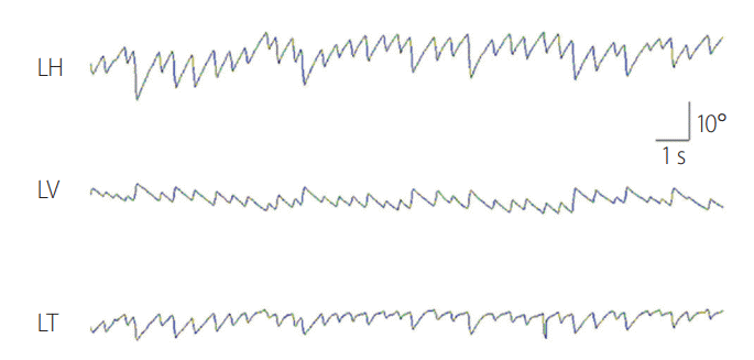
Spontaneous nystagmus refers to nystagmus presenting without a specific trigger. Induced nystagmus can be caused by horizontally or vertically changing the gaze direction (gaze-evoked nystagmus [GEN]) or shaking the head parallel to the plane of the horizontal semicircular canal (head-shaking nystagmus [HSN]). Induced nystagmus also refers to nystagmus caused by other types of stimulation, such as vibration (vibration-induced nystagmus), hyperventilation (hyperventilation-induced nystagmus), or postural change (positional nystagmus).18-21 Both spontaneous and induced nystagmus can be objectively examined using VOG. The VOG system recognizes the pupil and records the position of the eye in three directions (horizontal, vertical, and torsional) to confirm the direction, velocity, and waveform of nystagmus.6
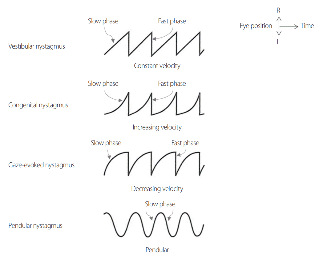
The linear waveform of nystagmus is mostly observed in peripheral vestibular disorders. An exponential decrease in the velocity of the slow phase indicates abnormality of the neural integrator. However, in the case of congenital nystagmus, the velocity of the slow phase increases exponentially. Pendular nystagmus also has a waveform comprising only slow components, and oscillates without the fast ones (Fig. 2).24
Spontaneous nystagmus is inspected by the patient wearing VOG goggles looking directly at a target 1 m away while sitting on a chair. This procedure should be performed in a dark room without other light sources. Calibration is essential to ensure accurate recordings. An examiner asks the patient to keep looking at the target for a certain duration and then records the spontaneous nystagmus. Changes in eye movements are then assessed after covering the entire field of view of the patient (spontaneous nystagmus without fixation) for the same duration.25
Spontaneous nystagmus is observed in peripheral vestibular system disorders as horizontal-torsional nystagmus directed to the side opposite to the lesion. The degree of nystagmus becomes worse without fixation and when looking toward the side opposite to the lesion.11,12 Central forms of spontaneous nystagmus often appear irrespective of fixation, and vertical and torsional nystagmus predominates in some cases.13 However, since spontaneous nystagmus that presents in a centrally located lesion frequently looks like it forms the peripheral vestibular disorder, the additional characteristics of nystagmus should be assessed. The primary cause should then be differentiated through a detailed neurological examination focused on the oculomotor system.26
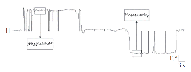
GEN is nystagmus induced when the eyeball looks at the target point out of the primary position. It reflects the function of the neural integrator, which keeps the gaze position in place out of the primary position. The eyes do not maintain a fixated eccentric position and begin to drift back to a primary position when the neural integrator malfunctions, which GEN then compensates for. The slow phase of GEN appears in an exponentially decreasing form (Fig. 3).18
In order to inspect horizontal GEN, the target is shifted from a primary position to 20-30° to the right and left. The gaze of the patient follows the target for about 20 seconds. For vertical GEN, the target is shifted from a primary position to 10-20° above and below in the same way.27
GEN is a type of nystagmus that occurs when the eyeball is out of the primary position and looking to one side. Tonic contraction of the extraocular muscles that can counteract the elasticity of the tissue around the eyeball to return the eyeball to its original position is required to maintain the eyeball deviating to one side.27 Continuous nervous system excitation is required for this, and the structure responsible for this mechanism is called the neural integrator.27 This integrator maintains contraction of the extraocular muscles by converting a command (pulse) for the velocity of the eye movement into information (step) for the position of the eyeball to induce the VOR, saccades, and smooth pursuit.28 The nucleus prepositus hypoglossi, medial vestibular nucleus, and the cerebellar flocculus play roles as neural integrators in horizontal eye movements, and the interstitial nucleus of Cajal is involved in vertical and rotational ones.27 When the function of the neural integrator is degraded, the eyes do not remain at the desired position and move toward the center, causing the fast phase of nystagmus to compensate for this abnormal eye movement.29 The time constant of eye movement is typically 20-70 seconds in normal conditions, but this decreases to less than 1 second if the neural integrator is abnormal.30 GEN is often suggestive of lesions in the lobe and connections of the cerebellum.31 The waveform of nystagmus has a characteristic decelerating slow phase.32
GEN should be differentiated from end-point nystagmus, which occurs when the eyes have a tendency to look in one direction.27 End-point nystagmus often disappears after a few instances, and it is not a pathological phenomenon because it can even be observed in normal conditions due to fatigue when looking in one direction for more than 30 seconds. 33 In addition, if it is observed only in the horizontal direction with an amplitude of less than 4°, even if nystagmus is continuously present, it cannot be regarded as a pathological phenomenon and may appear differently in the two eyes.33 GEN is suggested when the amplitude of the nystagmus is greater than 4° or it is fixed asymmetrically in both directions.13,34 The most notable difference is the change in the slow-phase velocity in a dark room. The velocity of the slow phase nearly doubles in end-point nystagmus, whereas in GEN there is almost no change.13,35 When clinically evaluating GEN, the eyeball should not deviate more than 30° from the center to avoid confusion with end-point nystagmus.36 Since the maximum range of horizontal eyeball movement in healthy adults is about 45°, the range from the center to the maximum lateral gaze is generally divided into three parts so that the eyeball position does not exceed two-thirds. When the target is too close, nystagmus or saccadic oscillation may occur due to convergence, so it is better to place the target as far away as possible. The most common cause of GEN is drug use (e.g., anticonvulsants, sedatives, or alcohol), which should therefore be checked in patients with GEN. Both horizontal and vertical drug-induced types of GEN are common.
HSN is inspected by covering the view of the patient in a sitting position. Then, with the head bent forward parallel to the horizontal semicircular canal, an examiner shakes the head 20-30 times at a speed of about 2 Hz and observes the nystagmus. Since the patients can involuntarily close their eyes during the examination, they should be instructed to keep them open.37
HSN is rare or very subtle in healthy patients. In unilateral vestibular disorders, HSN can appear toward the affected side according to the asymmetry of excitatory signals that accumulate in the central velocity storage mechanism. HSN is also observed in the presence of central lesions, typically in lateral medullary infarction. It is directed toward the lesion side regardless of the direction of spontaneous nystagmus. In the presence of cerebellar lesions, HSN can appear perpendicular to the direction of the head-shaking (upward or downward, which is referred to as perverted HSN).19,38
Hyperventilation-induced nystagmus is inspected by the patient breathing deeply and quickly to induce adequate hyperventilation (1 Hz for 30-60 seconds) while in a sitting position, and the examiner then observing the nystagmus. Since the eyes could be involuntarily closed during hyperventilation, patients should be instructed to keep them open.39
Hyperventilation-induced nystagmus often occurs in the demyelinating lesion within the vestibular nerves. Hyperventilation reduces the partial pressure of carbon dioxide and H+ in the blood, which results in metabolic alkalization, and the nerve conduction velocity increases as the ionized calcium concentration temporarily decreases. The demyelinated vestibular nerve therefore temporarily increases nerve conduction after hyperventilation, causing excitatory nystagmus toward the lesion side. In particular, because hyperventilation-induced nystagmus occurs in more than 50% of acoustic neuroma cases, it can be used for screening.40 Focal ischemia due to vasospasm, effects on the vestibular compensatory tract, intracranial pressure changes, and epilepsy are also discussed as mechanisms by which hyperventilation induces or enhances nystagmus. When the same symptoms are caused by pressure changes in the external auditory canal, it is called Hennebert’s sign, and the mechanism is known to be similar.39
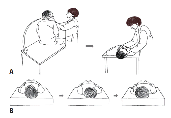
The patient is seated on a bed when inspecting positional nystagmus. The eye movement is first observed for about 30 seconds or more while the head is bowed forward by at least 30° (head-bending nystagmus). The head is then raised and pointed forward to determine if there is a reverse movement. With the head facing forward, the patient is asked to lie down and the eye movements are observed for the same amount of time (lying-down nystagmus). During this, a pillow is used to keep the neck flexed at about 30° from the ground to keep the horizontal semicircular canal parallel to the ground surface. The head is then rotated to the right, front, and left in order by 90° each, and the eye movements are observed for the same amount of time in each position (supine head-roll test). The patient then sits back up, has their head moved to a straight position, and then lies down flat. At this time, the head should be tilted back by 20-30° from the examination table (neck extension), and eye movements should be observed for about 30 seconds (straighthead-hanging test). The patient is then asked to sit down to check for reversing nystagmus. From a seated position, the head is then turned 45° to the right and the patient lies down on their back. At this time, the head should be tilted back by 20-30° from the examination table (neck extension), and eye movements should be checked for about 30 seconds (right-side Dix-Hallpike test). The patient then sit backs and reverse movement is checked for. The same maneuver is then repeated with the head turned 45° to the left (left-side Dix-Hallpike test). If nystagmus is observed at either stage, it should be observed for a sufficient time until it disappears or constant-velocity nystagmus is maintained for more than 30 seconds (Fig. 4).21
Confirming the presence of positional nystagmus is essential for diagnosing benign paroxysmal positional vertigo (BPPV). The Dix-Hallpike and supine head-roll tests are often used to diagnose BPPV in the posterior and horizontal semicircular canals, respectively (Fig. 4).41 In the case of posterior semicircular canal BPPV, torsional-upward nystagmus toward the lesion side is observed in the Dix-Hallpike test, and the nystagmus often disappears within 1 minute.42 When seated again, torsional-downward nystagmus occurs in the opposite direction.42 In the case of horizontal semicircular canal BPPV, geotropic or apogeotropic horizontal nystagmus is observed during the supine head-roll test.43 Geotropic nystagmus beats toward the ground, which is often caused by the otoliths in the canals (canalolithiasis), and the side with the stronger nystagmus is the lesion side. Apogeotropic nystagmus that beats away from the ground is caused by otoliths attached to the macula (cupulolithiasis), and the side with the weaker nystagmus is the lesion side.44 If the strength of the nystagmus is similar in both directions on the supine head-roll test, the direction of nystagmus during head-bending and lying down may be helpful for confirming the lesion side.45
Positional nystagmus and vertigo are mostly caused by peripheral lesions, but may be rarely observed in cases of central lesions if they are located near the fourth ventricle.46 Positional nystagmus caused by central lesions is mostly vertical (upbeat or downbeat) nystagmus, and is often accompanied by other neurological abnormalities.47 Positional nystagmus without vertigo is suggestive of a lesion located centrally.48 Even if positional downbeat nystagmus is observed on the head-hanging test, but the vertigo is mild, the possibility of a lesion in the cerebellum should be considered.48 Common causes of central positional vertigo include multiple sclerosis, cerebellar atrophy, cerebellar tumors, and Chiari malformations.46
Nystagmus is characterized by repetitive movements of the eyes, initiated by slow phases. Although physiologic nystagmus can occur, most cases of nystagmus are associated with underlying pathology. When examining for pathologic nystagmus, a systematic study of changes in fixation, eye position, and head position should be performed. Nystagmus can be triggered by head shaking and hyperventilation. Although nystagmus is typically described based on the direction of the quick phases, it is the slow phases that reveal the underlying disorder. In clinical practice, differentiating between peripheral and central nystagmus is of the utmost importance. By carefully observing nystagmus, clinicians can gain valuable insights into the underlying pathology.
REFERENCES
1. Downey DL, Leigh RJ. Eye movements: pathophysiology, examination and clinical importance. J Neurosci Nurs. 1998; 30:15–22. quiz 23-24.

2. Leigh RJ, Zee DS. The neurology of eye movements. 5th ed. New York: Oxford University Press;2015. p. 10–12.
6. Haslwanter T, Clarke AH. Eye movement measurement: electro-oculography and video-oculography. Handbook of Clinical Neurophysiology. 2010; 9:61–79.
7. Young LR, Sheena D. Survey of eye movement recording methods. Behavior Research Methods & Instrumentation. 1975; 7:397–429.
8. Robinson DA. A method of measuring eye movemnent using a scieral search coil in a magnetic field. TBME. 1963; 10:137–145.
9. Strupp M, Glasauer S, Jahn K, Schneider E, Krafczyk S, Brandt T. Eye movements and balance. Ann N Y Acad Sci. 2003; 1004:352–358.
10. Baloh RW. Differentiating between peripheral and central causes of vertigo. Otolaryngol Head Neck Surg. 1998; 119:55–59.
11. Serra A, Leigh RJ. Diagnostic value of nystagmus: spontaneous and induced ocular oscillations. J Neurol Neurosurg Psychiatry. 2002; 73:615–618.
12. Robinson DA, Zee DS, Hain TC, Holmes A, Rosenberg LF. Alexander’s law: its behavior and origin in the human vestibulo-ocular reflex. Ann Neurol. 1984; 16:714–722.

13. Strupp M, Hüfner K, Sandmann R, Zwergal A, Dieterich M, Jahn K, et al. Central oculomotor disturbances and nystagmus: a window into the brainstem and cerebellum. Dtsch Arztebl Int. 2011; 108:197–204.
14. Tilikete C, Pélisson D. Ocular motor syndromes of the brainstem and cerebellum. Curr Opin Neurol. 2008; 21:22–28.

15. Keller EL, Heinen SJ. Generation of smooth-pursuit eye movements: neuronal mechanisms and pathways. Neurosci Res. 1991; 11:79–107.

16. Bedell HE, Stevenson SB. Eye movement testing in clinical examination. Vision Res. 2013; 90:32–37.

18. Cannon SC, Robinson DA. Loss of the neural integrator of the oculomotor system from brain stem lesions in monkey. J Neurophysiol. 1987; 57:1383–1409.

19. Katsarkas A, Smith H, Galiana H. Head-shaking nystagmus (HSN): the theoretical explanation and the experimental proof. Acta Otolaryngol. 2000; 120:177–181.

20. Ohki M, Murofushi T, Nakahara H, Sugasawa K. Vibration-induced nystagmus in patients with vestibular disorders. Otolaryngol Head Neck Surg. 2003; 129:255–258.

22. Kavanagh KT, Babin RW. Definitions and types of nystagmus and calculations. Ear Hear. 1986; 7:157–166.

23. Barnes GR. A procedure for the analysis of nystagmus and other eye movements. Aviat Space Environ Med. 1982; 53:676–682.

25. Levo H, Aalto H, Petteri Hirvonen T. Nystagmus measured with video-oculography: methodological aspects and normative data. ORL J Otorhinolaryngol Relat Spec. 2004; 66:101–104.

26. Karatas M. Central vertigo and dizziness: epidemiology, differential diagnosis, and common causes. Neurologist. 2008; 14:355–364.
27. Rett D. Gaze-evoked nystagmus: a case report and literature review. Optometry. 2007; 78:460–464.

28. Abel LA, Dell’osso LF, Daroff RB. Analog model for gaze-evoked nystagmus. IEEE Trans Biomed Eng. 1978; 25:71–75.

29. Robinson DA. Eye movement control in primates. The oculomotor system contains specialized subsystems for acquiring and tracking visual targets. Science. 1968; 161:1219–1224.
30. Robinson DA. The use of control systems analysis in the neurophysiology of eye movements. Annu Rev Neurosci. 1981; 4:463–503.

31. Lee H, Kim HA. Anatomical structure responsible for direction changing bilateral gaze-evoked nystagmus in patients with unilateral cerebellar infarction. Medicine (Baltimore). 2020; 99:e19866.

33. Abel LA, Parker L, Daroff RB, Dell’Osso LF. End-point nystagmus. Invest Ophthalmol Vis Sci. 1978; 17:539–544.
35. Eizenman M, Cheng P, Sharpe JA, Frecker RC. End-point nystagmus and ocular drift: an experimental and theoretical study. Vision Res. 1990; 30:863–877.

36. Eizenman M, Sharpe JA. End point, gaze-evoked, and rebound nystagmus. In : Sharpe JA, Barber HO, editors. The vestibulo-ocular reflex and vertigo. New York: Raven Press;1993. p. 257–267.
37. Kamei T. Two types of head-shaking tests in vestibular examination. Acta Otolaryngol Suppl. 1988; 458:108–112.

38. Asawavichiangianda S, Fujimoto M, Mai M, Desroches H, Rutka J. Significance of head-shaking nystagmus in the evaluation of the dizzy patient. Acta Otolaryngol Suppl. 1999; 540:27–33.
39. Minor LB, Haslwanter T, Straumann D, Zee DS. Hyperventilation-induced nystagmus in patients with vestibular schwannoma. Neurology. 1999; 53:2158–2168.

40. Kheradmand A, Zee DS. The bedside examination of the vestibulo-ocular reflex (VOR): an update. Rev Neurol (Paris). 2012; 168:710–719.

41. Parnes LS, Agrawal SK, Atlas J. Diagnosis and management of benign paroxysmal positional vertigo (BPPV). CMAJ. 2003; 169:681–693.
42. Schmal F, Stoll W. Diagnosis and management of benign paroxysmal positional vertigo. Laryngorhinootologie. 2002; 81:368–380.
43. Singh J, Bhardwaj B. Lateral semicircular canal BPPV…are we still ignorant? Indian J Otolaryngol Head Neck Surg. 2020; 72:175–183.

44. Vannucchi P, Giannoni B, Pagnini P. Treatment of horizontal semicircular canal benign paroxysmal positional vertigo. J Vestib Res. 1997; 7:1–6.

45. Kim CH, Kim YG, Shin JE, Yang YS, Im D. Lateralization of horizontal semicircular canal canalolithiasis and cupulopathy using bow and lean test and head-roll test. Eur Arch Otorhinolaryngol. 2016; 273:3003–3009.


 PDF
PDF Citation
 Print
Print


 XML Download