Journal List > Blood Res > v.58(s1) > 1516082580

Lee: Recent advances in diagnosis and therapy in systemic mastocytosis

Abstract

Mastocytosis is a heterogeneous neoplasm characterized by accumulation of neoplastic mast cells in various organs. There are three main types: cutaneous mastocytosis (CM), systemic mastocytosis (SM), and mast cell sarcoma. CM mainly affects children and is confined to the skin, whereas SM affects adults and is characterized by extracutaneous involvement, with or without cutaneous involvement. Most cases of SM have an indolent clinical course; however, some types of SM have aggressive behavior and a poor prognosis. Recent advances in the understanding of the molecular changes in SM have changed the diagnosis and treatment of aggressive and advanced SM subtypes. The International Consensus Classification and World Health Organization refined the diagnostic criteria and classification of SM as a result of accumulation of clinical experience and advances in molecular diagnostics. Somatic mutations in the KIT gene, most frequently KIT D816V, are detected in 90% of patients with SM. Expression of CD30 and any KIT mutation were introduced as minor diagnostic criteria after the introduction of highly sensitive screening methods. SM has a wide spectrum of clinical features, and only a few drugs are effective at treating advanced SM. Currently, the mainstay of SM treatment is limited to the management of chronic symptoms related to release of mast cell mediators. Small-molecule kinase inhibitors targeting the KIT-downstream and KIT-independent pathways were recently approved for treating advanced SM. I describe recent advances in diagnosis of SM, and review the currently available and emerging therapeutic options for SM management.

INTRODUCTION

Mastocytosis is a group of disorders characterized by substantial increases in the number of mast cells (MCs) in the skin and internal organs. The first report of an MC disorder was a case of urticaria pigmentosa (UP) in a 2-year-old patient reported by Nettleship and Tay in 1869 [1]. The first reported case of systemic mastocytosis (SM) was an autopsy of a one-year-old infant who died of diffuse organ infiltration by MCs in 1949 [2].
Mastocytosis is a rare hematologic disorder characterized by the clonal expansion and accumulation of neoplastic MCs in various organs, including the bone marrow (BM), skin, gastrointestinal (GI) tract, liver, and/or spleen [3]. Whereas cutaneous mastocytosis (CM) mainly affects children and is almost always limited to the skin [4, 5], SM is distinguished from CM by its extracutaneous manifestations, with or without skin involvement, which is associated with multi-organ failure and a poor prognosis in adults [6]. Several classification schemes have been developed to provide guidelines on the prognosis and treatment of mastocytosis. In the 2016 revision of the World Health Organization (WHO) classification of myeloid neoplasms, mastocytosis was no longer considered as a subgroup of myeloproliferative neoplasms owing to its unique clinical and pathological features and highly variable disease course [7]. In the 2016 WHO classification system, which was validated in a large retrospective cohort study [6], SM was divided into 5 sub-groups: indolent SM (ISM), smoldering SM (SSM), aggressive SM (ASM), SM with an associated hematologic neoplasm (SM-AHN), and MC leukemia (MCL) [7]. Based on the presence of B and/or C findings, SM is divided into non-advanced SM (non-AdvSM), including ISM, bone marrow mastocytosis (BMM), and SSM; and advanced SM (AdvSM), including ASM, SM-AHN, and MCL [3]. The presence of activating somatic mutations in the KIT gene and aberrant immunophenotypes in clonal MCs are crucial biological markers of SM. The International Consensus Conference (ICC) Group [8] and WHO [9], published new classifications which recognize several distinct subtypes of CM and SM in 2022 (Table 1).

PATHOPHYSIOLOGY

MCs are immune-effector cells that play a key role in immunoglobulin E (IgE)-mediated inflammatory reactions. MCs are involved in multiple cellular processes including host defense in acquired and innate immunity, allergic reactions, wound healing, fibrosis, angiogenesis, and autoimmune diseases. MCs do not circulate in their mature form and are found in subepithelial connective and mucosal tissue, and around blood vessels [10]. Their estimated lifespan is months, in contrast to that of other leukocytes [11, 12]. MCs are derived from hematopoietic progenitor cells in the BM and peripheral blood (PB), which express CD34, CD117 (KIT), and CD13 [13]. MC development from CD34+ progenitors is dependent on the interaction between KIT and its ligand, stem-cell factor [14]. In addition to promoting the MC development, stem-cell factor directly promotes the release of MC-derived mediators and augments MC mediator release in response to IgE and antigen stimulation [15]. MCs can be activated by IgE-dependent or -independent mechanisms, and function as central effector cells in allergic reactions, and play a role in innate immunity, angiogenesis, and the coagulation cascade. MCs release preformed mediators, including histamine, tryptase, and heparin, and newly synthesized mediators such as leukotrienes, prostaglandins, and cytokines [16]. MC activation syndrome (MCAS) is a heterogeneous group of disorders characterized by episodic MC activation symptoms in more than two organ systems, that respond to MC mediator-directed agents [17]. An elevated serum tryptase level is the best marker of MCAS [18].
Mastocytosis is a clonal disorder of MC progenitors driven by a somatic gain-of-function mutation in KIT resulting in pathological accumulation and activation of MCs in tissue. The most common mutation, KIT D816V in exon 17 [19], is present in >90% of cases of adult SM [20]. In contrast, KIT mutations are only found in approximately 30% of cases of childhood CM, and several different codons are affected [21]. The majority of adults diagnosed with ISM, which is compatible with a normal lifespan, have a KIT D816V mutation [22]. In contrast, patients with AdvSM harboring the same mutation have a much poorer prognosis [23]. Moreover, the results of tyrosine kinase inhibitor (TKI) studies targeting the KIT D816V mutation in SM have been disappointing [24, 25]. These findings suggest that additional KIT-independent factors are involved in the patho-physiology. Several additional somatic mutations (TET2, SRSF2, ASXL1, CBL, RUNX1, and RAS) have been identified in patients with AdvSM (Table 2) [26]. The type and number of mutations in patients with multimutated SM correlate with prognosis, drug response, and survival [27]. Such additional changes may be co-expressed not only with the KIT D816V mutation but also in other myeloid lineages, especially in patients with SM-AHN or SM with an associated myeloid neoplasm. The presence of multimutated myeloid non-MC-lineage progenitors of the granulocyte-macrophage colony forming unit (CFU-GM) type suggests an initial clonal expansion at an early stage of hematopoietic development, with a subsequent phenotype modification toward SM owing to later acquisition of KIT D816V. In contrast, ISM and SSM are rarely associated with CFU-GM mutations, which may partially explain their excellent prognosis [28].

CLINICAL FEATURES

The symptoms and signs of SM are diverse, depending on the organs affected and MC-derived mediators involved. Skin lesions are a prominent clinical feature of mastocytosis. UP/Maculopapular CM (MPCM) lesions appear as small yellowish-tan to reddish-brown macules or slightly raised papules that can exhibit the Darier sign (swelling and redness of skin after brisk friction to a lesion) [29]. Identical skin lesions are also observed in SM and the skin lesions are described as mastocytosis in the skin (MIS). In patients with MIS, BM examination is required to differentiate between SM and CM [30]. Some patients with AdvSM lack typical cutaneous lesions. In patients without MIS, the diagnosis of SM is often confirmed after a BM biopsy for signs such as unexplained anaphylaxis, angioedema, organomegaly, skeletal lesions, and/or elevated serum tryptase level [31]. Flushing, itching, or blistering are also reported in patients with SM as MC-mediator related symptoms (MC-MRS).
Other organ biopsies can be performed to make a diagnosis when organ involvement in SM is suspected [30]. MC-MRS may be mild, extensive, or life threatening. Many patients experience recurrent episodes of unexplained anaphylaxis or systemic reactions after insect bites [32, 33]. In patients with severe anaphylaxis, serum tryptase levels increase substantially and MCAS may be detected. Vague, nonspecific constitutional symptoms, such as fatigue, general weakness, flushing, fever, and weight loss, could may be present in patients with mastocytosis [34]. Additionally, patients with SM may experience organ dysfunction associated with MC infiltration and MC-derived mediators. GI symptoms including nausea, vomiting, abdominal pain, and diarrhea are commonly associated with both non-AdvSM and AdvSM. Peptic ulcer disease is thought to reflect enhanced gastric acid secretion owing to increased histamine release [35]. Patients with SM may also develop osteopathy, often in the form of advanced osteopenia or osteoporosis. In advanced SM, features of MC infiltration, such as marked cytopenia, lymphadenopathy, hepatosplenomegaly, ascites, hypalbuminemia, malabsorption, or pathologic fractures, may be present (Table 3) [34].

DIAGNOSIS AND RISK STRATIFICATION

BM aspiration and biopsy should be performed if SM is suspected based on a combination of MC-MRS, adult-onset MIS regardless of serum tryptase level, and elevation of serum tryptase level (>20 ng/mL). The serum tryptase level can transiently increase during anaphylactic events. Basal tryptase levels should be assessed at least 48 hours after resolution of all MC-MRS. Once the diagnosis of SM is made, an additional staging workup should be performed, including assessment of BM morphology, immunohistochemistry, and flow cytometry to document the expression of CD2, CD25, and CD 30 in neoplastic MCs. Complete blood count with differential counts, blood chemistry, coagulation parameters, and IgE levels are also useful for staging. Bone scan/osteodensitometry and computed tomography can be used to evaluate extracutaneous organ involvement. If disease progression is suspected, staging, including BM study, blood tests, molecular analysis, and imaging studies should be repeated to assess organ damage [31].
The ICC [8] and WHO [9] released revised criteria for diagnosing SM in 2022. In addition to the KIT D816V activating mutation, mutations at other locations of KIT gene were added to the minor diagnostic criteria. Aberrant expression of CD30 in addition to CD2/CD25 in PB, BM or other extracutaneous organs, and a serum tryptase level >20 ng/mL in the absent of hereditary alpha-tryptasemia are additional newly incorporated minor diagnostic criteria (Table 4). The burden of disease criteria (B findings), which are used to differentiate SMM from ISM, and C findings, representing SM-induced organ damage, which are used to define ASM, are listed in Table 5. A C finding counts as SM-related organ damage only if it is caused by massive local MC infiltration, which should be documented by biopsy if possible. Cytoreductive therapy should be initiated to prevent further SM-related organ damage [36]. The proposed diagnosis and classification of SM is described in Fig. 1 based on these diagnostic criteria (Table 46). In the absence of a KIT mutation, particularly in patients with eosinophilia, myeloid/lymphoid neoplasms with eosinophilia and tyrosine kinase (TK) gene fusions should be excluded. The KIT D816V variant allele frequency (VAF) is correlated with disease activity and the SM subtype. A KIT V816V VAF ≥10% is associated with multilineage involvement or AHN in ISM, or SM masked by a myeloid neoplasm [37] and suggests a high tumor burden [38]. Therefore, KIT D816V VAF ≥10% in BM or PB leukocytes was introduced as a B finding in the 2022 WHO criteria (Table 5). Additional non-KIT mutations (e.g., SRSF2, ASXL1, RUNX1 and/or DNMT3A) are hallmarks of AdvSM [27] and are independent prognostic factors of progression and overall survival (OS) [39, 40]. Moreover, next-generation sequencing (NGS) can help predict which patients may progress to a more aggressive disease or harbor an undetected AHN [41]. Therefore, use of NGS to detect other KIT and non-KIT mutations, and KIT D816V mutation analysis with VAF quantitation, prior to and during therapy are warranted for patients with SM.
Patients with ISM have a median survival of 198 months, which is similar to that of the general population [6]. In the European Competence Network on Mastocytosis (ECNM) cohort, progression to AdvSM was rare, and there was no significant difference in survival and transformation rate to AdvSM between patients with ISM (1.2%), SSM (1.8%), and BMM (0.1%) [42]. Quality of life is the most important factor to consider when planning treatment for patients with non-AdvSM [43]. The OS of patients with AdvSM ranges from a few months to several years, with a median OS of <4 years [44], and the worst outcomes are observed in patients with MCL (OS, <2–31.6 mo) [6, 45, 46].
Several multiparameter prognostic risk stratification systems have been developed and validated to predict the progression and survival of patients with SM (Table 7). For non-AdvSM, these include the Spanish Network on Mastocytosis (REMA) score for ISM [39] and the International Prognostic Scoring System for Mastocytosis (IPSM) [47]. The IPSM, Mutation-Adjusted Risk Score (MARS) [44], Global Prognostic Score (GPS) [48], and Mayo Alliance Prognostic System (MAPS) [49] are used for risk stratification of AdvSM. Clinical parameters such as age >60 years, anemia, thrombocytopenia, elevated serum tryptase, alkaline phosphatase, and β2-microglobulin levels, and mutational status, are used to evaluate the risk of SM. MARS is the only validated multiparameter WHO-independent prognostic score for advanced SM [44].

GENERAL CONSIDERATIONS IN TREATING SM

Treatment of SM should be personalized according to the symptom burden. Treatment options for AdvSM include observation alone, symptom-directed management, supportive care, and cytoreductive therapy for MC debulking. Patients without AdvSM, or symptoms or signs of disease progression, should be advised about the avoidance of factors that may trigger symptoms (such as extreme temperature, physical exertion, nonsteroidal anti-inflammatory drugs, alcohol, contrast dye, and anesthetics) [50] and given prophylactic histamine receptor (H) blocker therapy [51]. Anaphylaxis can develop after insect bites and stings. In this situation, epinephrine autoinjectors and instructions regarding their use can be provided to patients with a history of anaphylaxis [52]. Symptom-directed management focusing on controlling MC-MRS, MIS, and osteopathy should be considered in all patients with SM. Most patients with recurrent severe MC-MRS respond to a combination of H1 and H2 antagonists. The choice of therapy should be based on the patient’s symptoms (Table 8). In women and those treated with long-term corticosteroids, prevention and treatment of osteopenia and osteoporosis should be considered. Patients with a T score <-2, should be treated with bisphosphonate (BPP). In cases refractory to BPP therapy, cytoreductive therapy with low-dose interferon-α (IFN-α), cladribine, or receptor activator of NF-κB ligand (RANKL) inhibitors are additional therapeutic options [51]. In patients with non-AdvSM with severe anaphylaxis and signs of disease progression, cladribine is usually effective in reducing the MC burden. Some patients experience significant symptoms despite combinations of high-dose antimediator treatment, and have a poor quality of life. In these refractory patients cladribine can also be used [53]. Currently TKIs targeting KIT D816V in patients with non-AdvSM are being assessed within a trial context only [54-56].
The treatment of patients with non-AdvSM should focus on prevention and control of anaphylaxis, MC-MRS, and osteoporosis. However, patients with AdvSM sometimes require MC cytoreductive therapy to improve disease-related organ dysfunction [23]. Recent clinical trials have revealed encouraging outcomes following treatment with small-molecule TKIs that target the activation loop mutants of the KIT receptor, supporting the hypothesis that KITD816V represents the driver mutation for SM. Over the past decade the KIT-targeted TKIs, midpstaurin and avapritinib have shown superior efficacy and OS than older cytoreductive treatments such as IFN-α and cladribine. In patients with pure MCL and ASM (without an AHN component), treatment with midostaurin or avapritinib can result in complete remission (CR) or partial remission (PR). Patients may develop cytopenia due to the SM disease burden and myelosuppression due to TKIs. In these cases, a dose reduction and support with blood products, erythropoiesis-stimulating agents, thrombopoietin agonists, and granulocyte-colony-stimulating factor may be needed to enable continuation of TKI therapy. For eligible patients who achieve a CR, allogeneic hematopoietic stem cell transplantation (allo-HSCT) should be considered to potentiate the effects of treatment. The current treatment algorithm for patients with SM-AHN recommends separate treatment to each component, as if the other is not present. This usually leads to treatment of the AHN component, as most studies suggest that disease progression is related to the AHN component. Sequential treatment of each component may be preferred; however, the optimal treatment sequence remains unknown.

CURRENTLY AVAILABLE AND EMERGING TREATMENT OPTIONS

Cytoreductive therapy

In patients with slowly progressive AdvSM, cytoreductive therapy with IFN-α or cladribine is the first-line treatment, and these agents are effective at reducing the MC burden and alleviating the C findings [57]. A retrospective French study of 20 patients (16 with ASM and 4 with ISM) treated with IFN-α for a minimum of 6 months, resulted in 100% response rate with MC-MRS improvement in 65% [58]. Another retrospective review of 47 patients treated with IFN-α, with or without prednisolone, at the Mayo Clinic showed an overall response rate (ORR) of 60% in the ASM group and 45% in the SM-AHN group, with a median response duration of 12 months (range, 1–67 mo). The absence of MC-MRS before treatment was associated with a poorer response [59]. Despite its effectiveness, the role of IFN-α in SM has been limited due to the high incidence of adverse events, lack of reproducibility, and relapse within a short period of IFN-α discontinuation. Currently, IFN-α may be of benefit in a minority of patients with lytic lesions/osteoporosis as bone mineral density is the only C finding [60]. Cladribine has been used to treat all SM subtypes but is most commonly used in patients with rapidly progressive AdvSM where rapid debulking of the disease is required [23]. In the Mayo Clinic study, the ORR was 55%, with a median response duration of 11 months (range, 3–74 mo). The presence of circulating immature myeloid cells was significantly associated with inferior response to cladribine (0% vs. 75%). The major toxicities include myelosuppression and infection [59]. A French nationwide retrospective study of 68 patients over a decade provided evidence that cladribine is an effective for treating SM, with an ORR of 72% (92% in non-AdvSM, and 50% in AdvSM). After a median follow-up of >10 years, the median response duration was 3.71 years (range, 0.1–8 yr) for non-AdvSM and 2.47 years (range, 0.5–8.6 yr) for AdvSM. The most frequent grade 3/4 toxicities were cytopenia and opportunistic infections [61]. AHN-directed therapy is sometimes required in patients with SM-AHN. Hydroxyurea (HU) is used to control leukocytosis, thrombocytosis, and hepatosplenomegaly. The ORR was 19%, and the median duration of response was 31.5 months (range, 5–50 mo) [59].

Allogeneic hematopoietic stem cell transplantation

All types of AHN should be regarded as secondary to SM. Similarly, in SM-associated acute myeloid leukemia (SM-AML), AML should be treated as in other patients with secondary AML. For most patients, high-dose chemotherapy and allo-HSCT are routinely recommended [57]. In patients with AHN, the presence of co-existing SM is often a poor prognostic sign. Moreover, the presence of KIT D816V in AML is associated with a worse prognosis [62]. A multicenter retrospective cohort study of 57 patients with SM who underwent allo-HSCT (38 SM-AHN, 7 ASM, and 12 MCL) in 2014 found a 70% ORR and a 16% CR [63]. Among the 30% of patients who did not respond, 21% had stable disease, and 9% had primary refractory disease. All 38 patients with SM-AHN had a CR for the AHN component; however, 10 patients (26%) had an AHN relapse, and 5 of these patients died. The OS outcome of allo-HSCT varies by AdvSM subtype. Patients with SM-AHN, ASM, and MCL had 3-year survival rates of 74%, 43%, and 17%, respectively. Patients with MCL and reduced-intensity allo-HSCT had poorer OS than patients with myeloablative allo-HSCT. Treatment-related mortality at 6 months and 1 year was 11% and 20%, respectively, and was highest in patients with MCL. These data suggest that allo-HSCT may improve survival in some patients, particularly those with SM-AHN. Allo-HSCT is needed to optimize the outcome. With the availability of modern TKIs, more patients have achieved pathological CR, and allo-HSCT has become more widely used in eligible patients [64].

Imatinib

Imatinib inhibits ABL1, KIT, and PDGFR [65]. Although imatinib has shown efficacy against wild-type (WT) KIT and certain transmembrane domain mutations (F52C) in vitro, it does not inhibit SM-associated KIT D816V [66]. Most patients with SM harbor an activating KIT D816V mutation, which makes them resistant to imatinib treatment, but some patients with mutations in domains other than the TK domain are sensitive to imatinib [67]. In a clinical trial involving patients with SM, the ORR was 18%, and the median duration of response was 19.6 months (range, 9–69 mo). Of patients treated with imatinib 86% had KIT D816V mutations. The ORR in mutation-positive and mutation-negative patients was 17% and 33%, respectively [59]. Imatinib is effective in patients with well-differentiated SM harboring the F522C transmembrane mutation [68, 69]. Imatinib was approved by the US Food and Drug Administration (FDA) in 2005 for patients with AdvSM without KIT D816V or with an unknown mutational status.

Midostaurin

Midostaurin is a multitarget TKI that targets not only KIT D816V, but also WT KIT, PDGFR α/β, VGFR2, and FLT3. Midostaurin inhibits both IgE-dependent release of histamine and growth of neoplastic MCs bearing various mutant forms of KIT [70]. Midostaurin was approved for patients with AdvSM based on data reported in 2016 from an international multicenter phase 2 clinical trial of 89 patients with ≥1 C findings which demonstrated favorable efficacy and safety [71]. The ORR was 60% (45% major response, 15% partial response). The median response duration, OS, and progression-free survival (PFS) were 24.1 months, 28.7 months, and 14.1 months, respectively. The response rates by AdvSM subtype were 75%, 58%, and 50% for ASM, SM-AHN, and MCL, respectively, regardless of prior therapy, presence of combined AHN, or KIT-D816V status. Midostaurin therapy was associated with improved C findings, including improved hemoglobin levels and platelet counts and weight gain. Significant decreases in tryptase levels (-58%) and BM MC burden (-59%) and spleen size were also observed. Mild GI adverse events were reported in 82% of patients at all grades, with 6–8% reporting grade 3–4 symptoms. The main adverse symptoms were nausea and vomiting, and most patients tolerated midostaurin with antiemetic treatment. Myelosuppression was manageable with dose reduction and growth factor support. The median PFS and OS were 14.1 and 28.7 months, respectively. A clinical trial which evaluated impact of molecular markers on survival showed that a ≥25% KIT D816V RNA-expressed allele burden reduction was significantly independently associated with improved OS [72]. A registry-based analysis also confirmed that survival was high with midostaurin than with cladribine therapy in patients with AdvSM [73]. Owing to these favorable results, midostaurin has become a first-line therapy for patients with AdvSM, and the FDA and European Medicines Agency approved midostaurin for patients with AdvSM in 2017.

Avapritinib

Avapritinib is a highly selective oral type 1 multi-kinase inhibitor of KIT D816V with a 10-fold lower 50% inhibitory concentration than midostaurin, measured using a KIT D816V inhibition assay [74]. It also inhibits PDGFRα with negligible activity against WT KIT and is effective in patients with unresectable or metastatic gastrointestinal stromal tumor (GIST) harboring a PDGFRA D842V mutation in exon 18 [75]. Avapritinib was approved in 2021 by the FDA for adults with AdvSM (recommended platelet count ≥50× 109/L), based on the results of the phase 1 EXPLORER trial [76] and an interim analysis of the phase 2 PATHFINDER trial [77].
The EXPLORER trial consisted of a dose escalation phase evaluating doses of 30–400 mg daily in patients with AdvSM with ≥1 eligible organ-damage finding. The subsequent dose-expansion phase evaluated dosing cohorts of 200 and 300 mg daily, and 200 mg daily was selected as the recommended phase 2 dose. During a median follow-up of 23 months, the median OS was not reached. The estimated 24-month OS in all patients with AdvSM was 76% (100%, 67%, and 92% for the ASM, SM-AHN, and MCL subtypes, respectively). The estimated PFS was 84% and 63% at 12 and 24 months, respectively. The comparative survival in all patients with AdvSM in the global midostaurin trial was 53% (86%, 49%, and 26% for ASM, SM-AHN, and MCL, respectively) [71]. The modified IWG-MRT-ECNM criteria [78] were used for response evaluation. CR, CR with partial hematologic recovery (CRh) (hemoglobin level, 8.1–9.9 g/dL; platelet count, 50–100×109/L; and neutrophil count, 0.5–1.0×10⁹/L), PR, and clinical improvement (CI) were observed in 15%, 21%, 34%, and 6% of response-evaluable patients, respectively. Partial hematologic recovery could be attributable to the myelosuppressive effect of avapritinib or the presence of concomitant AHN. Marked decreases were observed in BM MC burden (≥50% reduction in 92%), serum tryptase (≥50% reduction in 99%), spleen volume (≥35% reduction in 82%), and KIT VAF (≥50% reduction in 80%), and a complete molecular remission (CMR) (a new response marker for AdvSM, measured using digital droplet polymerase chain reaction with a limit of detection of 0.17%) was achieved in 30% of patients. Patients with SRSF2/ASXL/ RUNX1 (S/A/R) mutations or a baseline MARS ≥2 had shorter survival. In a separate analysis of the phase 1 EXPLORER trial exploring impact of mutations on avapritinib efficacy, 20% of patients progressed during treatment with a median follow-up of 23 months, and in most cases, the progression was driven by KIT-D816V negative AHN clones. Progression of the SM component was infrequent, and re-emergence of KIT D816V was rare. Survival was more favorable in patients with lower MARS scores. These data provide a rationale for further study of avapritinib in combination with AHN-directed therapy [79].
Data from the phase 2 PATHFINDER trial demonstrated the efficacy and safety of avapritinib at a starting dose of 200 mg once daily in patients with AdvSM, excluding patients with SM-AML and high-risk myelodysplastic syndromes [77]. In an interim analysis of the PATHFINDER study with 32 response-evaluable patients, the ORR was 75% according to the modified IWG-MRT-ECNM criteria, with a median follow-up of 10.4 months. CRh, PR, and CI were reported in six (19%), 10 (31%) and 8 (25%) patients, respectively. Responses were observed in all AdvSM subtypes, regardless of exposure to prior therapy or the presence of high-risk mutations in the S/A/R panel. Significant reductions in the serum tryptase level, BM MC burden, and KIT-D816V VAF level of at least 50% from baseline were observed in 93%, 88%, and 60% of patients, respectively. The baseline C findings improved for pleural effusion (83%), splenomegaly (79%), and ascites (57%). The median PFS and OS in the safety population (N=62) were not reached by data cutoff. The estimated 12-month PFS and OS rates were 79% and 86%, respectively, with a median follow-up of 7 months (range, 5.6–8.1 mo) and 52 patients (84%) still receiving treatment.
In the EXPLORER study, 9 (13%) patients experience intracranial bleeding (ICB), including intraparenchymal hemorrhage and subdural hematoma, as a substantial adverse event. Seven of the 9 patients had thrombocytopenia (platelet count <50×109/L). Owing to the increased incidence of ICB in patients with thrombocytopenia the EXPLORER trial protocol was amended to exclude enrollment of new patients with severe thrombocytopenia, increase monitoring of the platelet count, and to provide guidelines for dose adjustment and platelet transfusions [76]. Peripheral edema (45–50%) and periorbital edema (48–65%) were the most common adverse events (AEs) and were managed with diuretics and dose reduction. Grade 3 neutropenia, thrombocytopenia, and anemia were observed in 19–24%, 16–41%, and 16–36% of the patients, respectively. Grade 1/2 GI adverse events included diarrhea (23–43%), nausea (18–42%), and vomiting (18–32%). Grade 1/2 cognitive impairment occurred in 20–30% of patients, which presented as mild memory impairment. Prompt protocol amendment resulted in a lower AEs and ICB rate (1.6%) in the PATHFINDER study than in the EXPLORER study. No treatment-related deaths occurred in the PATHFINDER study, whereas six treatment-related deaths (8.7%) occurred in the EXPLORER study [76, 77].
Compared with a historical cohort of patients with AdvSM treated with the best available therapy, patients treated with avapritinib had significantly improved survival, a longer duration of treatment (23.8 vs 5.4 mo; P<0.001), and a 60% greater mean difference in the percent maximum decrease in serum tryptase levels [80]. A pooled analysis of the phase I EXPLORER trial and interim phase 2 PATHFINDER trial revealed an ORR of 72% (CRh, 28%; PR, 28%; CI, 15%). The estimated median OS of all the patients with AdvSM was 46.9 months [81]. The ORR among the 31 previously treated patients was 71%, including a CR/CRh rate of 19%. The OS at 12 and 24 months in these patients was 80% and 65%, respectively, and the median OS was not reached after a median follow-up of 17.7 months [82].

BLU-263

BLU-263 is a next-generation TKI that shows selectivity and potency comparable to those of avapritinib, with limited central nervous system penetration, which may lower the risk of cognitive changes and ICB. In a phase 1 trial, BLU-263 was safe, with linear pharmacokinetics across all tested doses and a half-life allowing once-daily dosing [83]. A randomized, double-blind, placebo-controlled phase 2/3 trial assessing the efficacy and safety of BLU-263 in patients with ISM whose symptoms are not adequately controlled by standard therapies is ongoing (HARBOR, NCT04910685) [54]. Given the concern about the potential risk of ICB in patients with AdvSM receiving avapritinib, BLU-263 needs to be evaluated in patients with AdvSM. A phase 1/2, two-arm trial evaluating the safety and efficacy of BLU-263 in patients with AdvSM and SM-AHN, both as a monotherapy and in combination with AHN-directed therapy, is currently underway [84].

Bezuclastinib

Bezuclastinib is an oral, highly selective type I TKI with potent activity against KIT exons 9, 11, 17, and 18, including D816V. Besides targeting KIT D816V, bezuclastinib was designed to avoid closely related kinases, such as PDGFRα, PDGFRβ, wild-type KIT, VEGFR2 (KDR), and CSF1R (FMS). Additionally, minimal brain penetration and no central nervous system toxicities have been observed with bezuclastinib in preclinical studies [85]. A phase 1/2 study of patients with advanced solid tumors, including GIST, showed that bezuclastinib has clinical activity and a tolerable safety profile. Patients treated with bezuclastinib had reduction in exon 17 mutational burden, which was associated with a reduction in tumor burden [86]. These findings support the investigation of bezuclastinib as a potential therapy for KIT-driven diseases, such as GIST and SM. Bezuclastinib is currently being evaluated in a multicenter, phase 2, open-label clinical trial (Apex, CGT9486, NCT04996875) to evaluate the safety, efficacy, pharmacokinetics, and pharmacodynamics of bezuclastinib in patients with AdvSM. Preliminary safety and efficacy data from the Apex trial suggest that bezuclastinib is well-tolerated and shows encouraging early signs of clinical activity, as demonstrated by meaningful reductions in serum tryptase, MC burden, and KIT D816V VAF [87]. A phase 2/3 clinical trial in patients with ISM/SSM (Summit, NCT05186753) is also in progress [55].

CONCLUSION

Understanding of SM has evolved owing to better classification, identification of good prognostic/indolent disease subsets, and routine availability of molecular testing. The outcomes for patients with AdvSM have significantly improved since the introduction of novel TKIs. Avapritinib can generate molecular remission of KIT D816V, and has more favorable long-term outcomes than historical treatments, including midostaurin. Treating SM-AHN, in which the prognosis is usually driven by AHN, remains a major challenge, and the presence of a complex mutational profile beyond KIT D816V may promote progression and resistance. How to combine KIT inhibition with AHN-targeted therapy should be a major focus of clinical trials for AdvSM. HSCT should be considered as a potentially curative option in patients with AHN. Prospective trials are required to assess the usefulness of allo-HSCT combined with novel TKIs. New diagnostic or predictive biomarkers, therapeutic strategies, and validation of better clinical scoring systems for assessing both prognosis and response are also questions that need to be addressed.

Notes

Authors’ Disclosures of Potential Conflicts of Interest

No potential conflicts of interest relevant to this article were reported.

REFERENCES

1. Nettleship E. 1869; Rare form of urticaria. Br Med J. 2:323–4.
2. Ellis JM. 1949; Urticaria pigmentosa; a report of a case with autopsy. Arch Path (Chic). 48:426–35.
3. Valent P, Akin C, Metcalfe DD. 2017; Mastocytosis: 2016 updated WHO classification and novel emerging treatment concepts. Blood. 129:1420–7. DOI: 10.1182/blood-2016-09-731893. PMID: 28031180. PMCID: PMC5356454.
4. Caplan RM. 1963; The natural course of urticaria pigmentosa. Analysis and follow-up of 112 cases. Arch Dermatol. 87:146–57. DOI: 10.1001/archderm.1963.01590140008002. PMID: 14018418.
5. Uzzaman A, Maric I, Noel P, Kettelhut BV, Metcalfe DD, Carter MC. 2009; Pediatric‐onset mastocytosis: a long term clinical follow‐up and correlation with bone marrow histopathology. Pediatr Blood Cancer. 53:629–34. DOI: 10.1002/pbc.22125. PMID: 19526526. PMCID: PMC2786499.
6. Lim KH, Tefferi A, Lasho TL, et al. 2009; Systemic mastocytosis in 342 consecutive adults: survival studies and prognostic factors. Blood. 113:5727–36. DOI: 10.1182/blood-2009-02-205237. PMID: 19363219.
7. Arber DA, Orazi A, Hasserjian R, et al. 2016; The 2016 revision to the World Health Organization classification of myeloid neoplasms and acute leukemia. Blood. 127:2391–405. DOI: 10.1182/blood-2016-03-643544. PMID: 27069254.
8. Arber DA, Orazi A, Hasserjian RP, et al. 2022; International Consensus Classification of Myeloid Neoplasms and Acute Leukemias: integrating morphologic, clinical, and genomic data. Blood. 140:1200–28. DOI: 10.1182/blood.2022015850. PMID: 35767897.
9. Khoury JD, Solary E, Abla O, et al. 2022; The 5th edition of the World Health Organization classification of haematolymphoid tumours: myeloid and histiocytic/dendritic neoplasms. Leukemia. 36:1703–19. DOI: 10.1038/s41375-022-01613-1. PMID: 35732831. PMCID: PMC9252913.
10. Galli SJ, Dvorak AM, Dvorak HF. 1984; Basophils and mast cells: morphologic insights into their biology, secretory patterns, and function. Prog Allergy. 34:1–141. DOI: 10.1159/000408370. PMID: 6230674.
11. Mekori YA, Metcalfe DD. 2000; Mast cells in innate immunity. Immunol Rev. 173:131–40. DOI: 10.1034/j.1600-065X.2000.917305.x. PMID: 10719674.
12. Galli SJ, Grimbaldeston M, Tsai M. 2008; Immunomodulatory mast cells: negative, as well as positive, regulators of immunity. Nature Rev Immun. 8:478–86. DOI: 10.1038/nri2327. PMID: 18483499. PMCID: PMC2855166.
13. Kirshenbaum AS, Goff JP, Semere T, Foster B, Scott LM, Metcalfe DD. 1999; Demonstration that human mast cells arise from a progenitor cell population that is CD34(+), c-kit(+), and expresses aminopeptidase N (CD13). Blood. 94:2333–42. DOI: 10.1182/blood.V94.7.2333.419k30_2333_2342. PMID: 10498605.
14. Valent P, Spanblöchl E, Sperr WR, et al. 1992; Induction of differenti-ation of human mast cells from bone marrow and peripheral blood mononuclear cells by recombinant human stem cell factor/kit-ligand in long-term culture. Blood. 80:2237–45. DOI: 10.1182/blood.V80.9.2237.2237. PMID: 1384799.
15. Bischoff SC, Dahinden CA. 1992; c-kit ligand: a unique potentiator of mediator release by human lung mast cells. J Exp Med. 175:237–44. DOI: 10.1084/jem.175.1.237. PMID: 1370529. PMCID: PMC2119079.
16. Robyn J, Metcalfe DD. 2006; Systemic mastocytosis. In: Alt FW, Austen KF, Honjo T, Melchers F, Uhr JW, Unanue ER, eds. Advances in immunology. Cambridge, MA:. Academic Press,. 169–243. DOI: 10.1016/S0065-2776(05)89005-4. PMID: 16682275.
17. Akin C, Valent P, Metcalfe DD. 2010; Mast cell activation syndrome: proposed diagnostic criteria. J Allergy Clin Immunol. 126:1099–104. e4DOI: 10.1016/j.jaci.2010.08.035. PMID: 21035176. PMCID: PMC3753019.
18. Schwartz LB. 2006; Diagnostic value of tryptase in anaphylaxis and mastocytosis. Immunol Allergy Clin North Am. 26:451–63. DOI: 10.1016/j.iac.2006.05.010. PMID: 16931288.
19. Nagata H, Worobec AS, Oh CK, et al. 1995; Identification of a point mutation in the catalytic domain of the protooncogene c-kit in peripheral blood mononuclear cells of patients who have mastocytosis with an associated hematologic disorder. Proc Natl Acad Sci U S A. 92:10560–4. DOI: 10.1073/pnas.92.23.10560. PMID: 7479840. PMCID: PMC40651.
20. Kristensen T, Vestergaard H, Bindslev-Jensen C, Møller MB, Broesby-Olsen S. Mastocytosis Centre, Odense University Hospital (MastOUH). 2014; Sensitive KIT D816V mutation analysis of blood as a diagnostic test in mastocytosis. Am J Hematol. 89:493–8. DOI: 10.1002/ajh.23672. PMID: 24443360.
21. Bodemer C, Hermine O, Palmérini F, et al. 2010; Pediatric mastocytosis is a clonal disease associated with D816V and other activating c-KIT mutations. J Investig Dermatol. 130:804–15. DOI: 10.1038/jid.2009.281. PMID: 19865100.
22. Pardanani A, Lim KH, Lasho TL, et al. 2010; WHO subvariants of indolent mastocytosis: clinical details and prognostic evaluation in 159 consecutive adults. Blood. 115:150–1. DOI: 10.1182/blood-2009-10-249979. PMID: 20056798.
23. Pardanani A. 2012; Systemic mastocytosis in adults: 2012 update on diagnosis, risk stratification, and management. Am J Hematol. 87:401–11. DOI: 10.1002/ajh.23134. PMID: 22410759.
24. Ustun C, DeRemer DL, Akin C. 2011; Tyrosine kinase inhibitors in the treatment of systemic mastocytosis. Leuk Res. 35:1143–52. DOI: 10.1016/j.leukres.2011.05.006. PMID: 21641642.
25. Gotlib J, Berubé C, Growney JD, et al. 2005; Activity of the tyrosine kinase inhibitor PKC412 in a patient with mast cell leukemia with the D816V KIT mutation. Blood. 106:2865–70. DOI: 10.1182/blood-2005-04-1568. PMID: 15972446. PMCID: PMC1895309.
26. Valent P. 2015; Diagnosis and management of mastocytosis: an emerging challenge in applied hematology. Hematology Am Soc Hematol Educ Program. 2015:98–105. DOI: 10.1182/asheducation-2015.1.98. PMID: 26637707.
27. Schwaab J, Schnittger S, Sotlar K, et al. 2013; Comprehensive mutational profiling in advanced systemic mastocytosis. Blood. 122:2460–6. DOI: 10.1182/blood-2013-04-496448. PMID: 23958953.
28. Jawhar M, Schwaab J, Schnittger S, et al. 2015; Molecular profiling of myeloid progenitor cells in multi-mutated advanced systemic mastocytosis identifies KIT D816V as a distinct and late event. Leukemia. 29:1115–22. DOI: 10.1038/leu.2015.4. PMID: 25567135.
29. Soter NA. 2000; Mastocytosis and the skin. Hematol Oncol Clin North Am. 14:537–55. DOI: 10.1016/S0889-8588(05)70295-1. PMID: 10909039.
30. Valent P, Akin C, Arock M, et al. 2012; Definitions, criteria and global classification of mast cell disorders with special reference to mast cell activation syndromes: a consensus proposal. Int Arch Allergy Immunol. 157:215–25. DOI: 10.1159/000328760. PMID: 22041891. PMCID: PMC3224511.
31. Valent P, Escribano L, et al. Broesby‐Olsen S. 2014; Proposed diagnostic algorithm for patients with suspected mastocytosis: a proposal of the European Competence Network on Mastocytosis. Allergy. 69:1267–74. DOI: 10.1111/all.12436. PMID: 24836395.
32. Carter MC, Desai A, Komarow HD, et al. 2018; A distinct biomolecular profile identifies monoclonal mast cell disorders in patients with idiopathic anaphylaxis. J Allergy Clin Immunol. 141:180–8. e3DOI: 10.1016/j.jaci.2017.05.036. PMID: 28629749. PMCID: PMC6311988.
33. Bonadonna P, Perbellini O, Passalacqua G, et al. 2009; Clonal mast cell disorders in patients with systemic reactions to Hymenoptera stings and increased serum tryptase levels. J Allergy Clin Immunol. 123:680–6. DOI: 10.1016/j.jaci.2008.11.018. PMID: 19135713.
34. Travis WD, Li CY, Bergstralh EJ, Yam LT, Swee RG. 1988; Systemic mast cell disease. Analysis of 58 cases and literature review. Medicine (Baltimore). 67:345–68. DOI: 10.1097/00005792-198811000-00001. PMID: 3054417.
35. Cherner JA, Jensen RT, Dubois A, O'Dorisio TM, Gardner JD, Metcalfe DD. 1988; Gastrointestinal dysfunction in systemic mastocytosis: a prospective study. Gastroenterology. 95:657–67. DOI: 10.1016/S0016-5085(88)80012-X. PMID: 3396814.
36. Valent P, Akin C, Sperr WR, et al. 2003; Aggressive systemic mastocytosis and related mast cell disorders: current treatment options and proposed response criteria. Leuk Res. 27:635–41. DOI: 10.1016/S0145-2126(02)00168-6. PMID: 12681363.
37. Hoermann G, Gleixner KV, Dinu GE, et al. 2014; The KIT D 816 V allele burden predicts survival in patients with mastocytosis and correlates with the WHO type of the disease. Allergy. 69:810–3. DOI: 10.1111/all.12409. PMID: 24750133. PMCID: PMC4896381.
38. Erben P, Schwaab J, Metzgeroth G, et al. 2014; The KIT D816V expressed allele burden for diagnosis and disease monitoring of systemic mastocytosis. Ann Hematol. 93:81–8. DOI: 10.1007/s00277-013-1964-1. PMID: 24281161.
39. Muñoz-González JI, Álvarez-Twose I, Jara-Acevedo M, et al. 2019; Frequency and prognostic impact of KIT and other genetic variants in indolent systemic mastocytosis. Blood. 134:456–68. DOI: 10.1182/blood.2018886507. PMID: 31151985.
40. Muñoz-González JI, Jara-Acevedo M, Alvarez-Twose I, et al. 2018; Impact of somatic and germline mutations on the outcome of systemic mastocytosis. Blood Adv. 2:2814–28. DOI: 10.1182/bloodadvances.2018020628. PMID: 30373888. PMCID: PMC6234367.
41. Jawhar M, Schwaab J, Schnittger S, et al. 2016; Additional mutations in SRSF2, ASXL1 and/or RUNX1 identify a high-risk group of patients with KIT D816V+ advanced systemic mastocytosis. Leukemia. 30:136–43. DOI: 10.1038/leu.2015.284. PMID: 26464169.
42. Trizuljak J, Sperr WR, Nekvindová L, et al. 2020; Clinical features and survival of patients with indolent systemic mastocytosis defined by the updated WHO classification. Allergy. 75:1927–38. DOI: 10.1111/all.14248. PMID: 32108361. PMCID: PMC7115854.
43. Siebenhaar F, von Tschirnhaus E, Hartmann K, et al. 2016; Development and validation of the mastocytosis quality of life questionnaire: MC‐QoL. Allergy. 71:869–77. DOI: 10.1111/all.12842. PMID: 26797792.
44. Jawhar M, Schwaab J, Álvarez-Twose I, et al. 2019; MARS: mutation- adjusted risk score for advanced systemic mastocytosis. J Clin Oncol. 37:2846. DOI: 10.1200/JCO.19.00640. PMID: 31509472. PMCID: PMC6823885.
45. Jain P, Wang S, Patel KP, et al. 2017; Mast cell leukemia (MCL): clinico-pathologic and molecular features and survival outcome. Leuk Res. 59:105–9. DOI: 10.1016/j.leukres.2017.05.018. PMID: 28599188. PMCID: PMC5573652.
46. Kennedy VE, Perkins C, Reiter A, et al. 2022; Mast cell leukemia: clinical and molecular features and survival outcomes of patients in the ECNM registry. Blood Adv. [Epub ahead of print]. DOI: 10.1182/bloodadvances.2022008292. PMID: 36094848.
47. Sperr WR, Kundi M, Alvarez-Twose I, et al. 2019; International prognostic scoring system for mastocytosis (IPSM): a retrospective cohort study. Lancet Haematol. 6:e638–49. DOI: 10.1016/S2352-3026(19)30166-8. PMID: 31676322. PMCID: PMC7115823.
48. Muñoz-González JI, Álvarez-Twose I, Jara-Acevedo M, et al. 2021; Proposed global prognostic score for systemic mastocytosis: a retrospective prognostic modelling study. Lancet Haematol. 8:e194–204. DOI: 10.1016/S2352-3026(20)30400-2. PMID: 33508247.
49. Pardanani A, Shah S, Mannelli F, et al. 2018; Mayo alliance prognostic system for mastocytosis: clinical and hybrid clinical-molecular models. Blood Adv. 2:2964–72. DOI: 10.1182/bloodadvances.2018026245. PMID: 30413432. PMCID: PMC6234360.
50. Carter MC, Metcalfe DD, Matito A, et al. 2019; Adverse reactions to drugs and biologics in patients with clonal mast cell disorders: a work group report of the Mast Cells Disorder Committee, American Academy of Allergy, Asthma & Immunology. J Allergy Clin Immunol. 143:880–93. DOI: 10.1016/j.jaci.2018.10.063. PMID: 30528617.
51. Valent P, Akin C, Escribano L, et al. 2007; Standards and standardization in mastocytosis: consensus statements on diagnostics, treatment recommendations and response criteria. Eur J Clin Invest. 37:435–53. DOI: 10.1111/j.1365-2362.2007.01807.x. PMID: 17537151.
52. Siebenhaar F, Akin C, Bindslev-Jensen C, Maurer M, Broesby-Olsen S. 2014; Treatment strategies in mastocytosis. Immunol Allergy Clin North Am. 34:433–47. DOI: 10.1016/j.iac.2014.01.012. PMID: 24745685.
53. Kluin-Nelemans HC, Oldhoff JM, Van Doormaal JJ, et al. 2003; Cladribine therapy for systemic mastocytosis. Blood. 102:4270–6. DOI: 10.1182/blood-2003-05-1699. PMID: 12933573.
54. Castells M, Si TD, Bhavsar V, He K, Akin C. 2022; A phase 2/3 study of BLU-263 in patients with indolent systemic mastocytosis or monoclonal mast cell activation syndrome. J Allergy Clin Immunol. 149:AB221. DOI: 10.1016/j.jaci.2021.12.721.
55. Akin C, Siebenhaar F, Deininger MW, et al. 2022; Summit: a 3-part, phase 2 study of bezuclastinib (CGT9486), an oral, selective, and potent KIT D816V inhibitor, in adult patients with nonadvanced systemic mastocytosis (NonAdvSM). Blood (ASH Annual Meeting Abstracts). 140(Suppl):6838–9. DOI: 10.1182/blood-2022-156326.
56. Akin C, Elberink HO, Gotlib J, et al. 2020; Pioneer part 2: a randomized, double-blind, placebo-controlled, phase 2 study to evaluate safety and efficacy of avapritinib in indolent systemic mastocytosis. Blood (ASH Annual Meeting Abstracts). 136(Suppl):1248. DOI: 10.1182/blood-2020-136632.
57. Valent P, Sperr WR, Akin C. 2010; How I treat patients with advanced systemic mastocytosis. Blood. 116:5812–7. DOI: 10.1182/blood-2010-08-292144. PMID: 20855864.
58. Simon J, Lortholary O, Caillat-Vigneron N, et al. 2004; Interest of interferon alpha in systemic mastocytosis. The French experience and review of the literature. Pathol Biol (Paris). 52:294–9. DOI: 10.1016/j.patbio.2004.04.012. PMID: 15217717.
59. Lim KH, Pardanani A, Butterfield JH, Li CY, Tefferi A. 2009; Cytoreductive therapy in 108 adults with systemic mastocytosis: outcome analysis and response prediction during treatment with interferon‐alpha, hydroxyurea, imatinib mesylate or 2‐chloro-deoxyadenosine. Am J Hematol. 84:790–4. DOI: 10.1002/ajh.21561. PMID: 19890907.
60. Laroche M, Bret J, Brouchet A, Mazières B. 2007; Clinical and densitometric efficacy of the association of interferon alpha and pamidronate in the treatment of osteoporosis in patients with systemic mastocytosis. Clin Rheumatol. 26:242–3. DOI: 10.1007/s10067-006-0369-0. PMID: 16902757.
61. Barete S, Lortholary O, Damaj G, et al. 2015; Long-term efficacy and safety of cladribine (2-CdA) in adult patients with mastocytosis. Blood. 126:1009–16. DOI: 10.1182/blood-2014-12-614743. PMID: 26002962.
62. Ustun C, Gotlib J, Popat U, et al. 2016; Consensus opinion on allogeneic hematopoietic cell transplantation in advanced systemic mastocytosis. Biol Blood Marrow Transplant. 22:1348–56. DOI: 10.1016/j.bbmt.2016.04.018. PMID: 27131865.
63. Ustun C, Reiter A, Scott BL, et al. 2014; Hematopoietic stem-cell transplantation for advanced systemic mastocytosis. J Clin Oncol. 32:3264. DOI: 10.1200/JCO.2014.55.2018. PMID: 25154823. PMCID: PMC4876356.
64. National Comprehensive Cancer Network. 2022. Systemic mastocytosis version 2.2022, NCCN clinical practice guidelines in oncology 2022. NCCN;Plymouth Meeting, PA: at https://www.nccn.org/guidelines/guidelines-detail?category=1&id=1490. Accessed October 18, 2022.
65. Buchdunger E, Cioffi CL, Law N, et al. 2000; Abl protein-tyrosine kinase inhibitor STI571 inhibits in vitro signal transduction mediated by c-kit and platelet-derived growth factor receptors. J Pharmacol Exp Ther. 295:139–45. PMID: 10991971.
66. Ma Y, Zeng S, Metcalfe DD, et al. 2002; The c-KIT mutation causing human mastocytosis is resistant to STI571 and other KIT kinase inhibitors; kinases with enzymatic site mutations show different inhibitor sensitivity profiles than wild-type kinases and those with regulatory-type mutations. Blood. 99:1741–4. DOI: 10.1182/blood.V99.5.1741. PMID: 11861291.
67. Arock M, Sotlar K, Akin C, et al. 2015; KIT mutation analysis in mast cell neoplasms: recommendations of the European Competence Network on Mastocytosis. Leukemia. 29:1223–32. DOI: 10.1038/leu.2015.24. PMID: 25650093. PMCID: PMC4522520.
68. Akin C, Fumo G, Yavuz AS, Lipsky PE, Neckers L, Metcalfe DD. 2004; A novel form of mastocytosis associated with a transmembrane c-kit mutation and response to imatinib. Blood. 103:3222–5. DOI: 10.1182/blood-2003-11-3816. PMID: 15070706.
69. Álvarez-Twose I, González P, Morgado JM, et al. 2012; Complete response after imatinib mesylate therapy in a patient with well-differentiated systemic mastocytosis. J Clin Oncol. 30:e126–9. DOI: 10.1200/JCO.2011.38.9973. PMID: 22370312.
70. Valent P, Akin C, Hartmann K, et al. 2017; Midostaurin: a magic bullet that blocks mast cell expansion and activation. Ann Oncol. 28:2367–76. DOI: 10.1093/annonc/mdx290. PMID: 28945834. PMCID: PMC7115852.
71. Gotlib J, Kluin-Nelemans HC, George TI, et al. 2016; Efficacy and safety of midostaurin in advanced systemic mastocytosis. N Engl J Med. 374:2530–41. DOI: 10.1056/NEJMoa1513098. PMID: 27355533.
72. Jawhar M, Schwaab J, Naumann N, et al. 2017; Response and progression on midostaurin in advanced systemic mastocytosis: KIT D816V and other molecular markers. Blood. 130:137–45. DOI: 10.1182/blood-2017-01-764423. PMID: 28424161.
73. Lübke J, Schwaab J, Naumann N, et al. 2022; Superior efficacy of midostaurin over cladribine in advanced systemic mastocytosis: a registry-based analysis. J Clin Oncol. 40:1783–94. DOI: 10.1200/JCO.21.01849. PMID: 35235417.
74. Evans EK, Gardino AK, Kim JL, et al. 2017; A precision therapy against cancers driven by KIT/PDGFRA mutations. Sci Transl Med. 9:eaao1690. DOI: 10.1126/scitranslmed.aao1690. PMID: 29093181.
75. Jones RL, Serrano C, von Mehren M, et al. 2021; Avapritinib in unresectable or metastatic PDGFRA D842V-mutant gastrointestinal stromal tumours: long-term efficacy and safety data from the NAVIGATOR phase I trial. Eur J Cancer. 145:132–42. DOI: 10.1016/j.ejca.2020.12.008. PMID: 33465704. PMCID: PMC9518931.
76. DeAngelo DJ, Radia DH, George TI, et al. 2021; Safety and efficacy of avapritinib in advanced systemic mastocytosis: the phase 1 EXPLORER trial. Nat Med. 27:2183–91. DOI: 10.1038/s41591-021-01538-9. PMID: 34873347. PMCID: PMC8674134.
77. Gotlib J, Reiter A, Radia DH, et al. 2021; Efficacy and safety of avapritinib in advanced systemic mastocytosis: interim analysis of the phase 2 PATHFINDER trial. Nat Med. 27:2192–9. DOI: 10.1038/s41591-021-01539-8. PMID: 34873345. PMCID: PMC8674139.
78. Gotlib J, Schwaab J, Shomali W, et al. 2022; Proposed European Competence Network on Mastocytosis-American Initiative in Mast Cell Diseases (ECNM-AIM) response criteria in advanced systemic mastocytosis. J Allergy Clin Immunol Pract. 10:2025–38. e1DOI: 10.1016/j.jaip.2022.05.034. PMID: 35724948.
79. Deininger MW, DeAngelo DJ, Radia DH, et al. 2021; Effective control of advance systemic mastocytosis with avapritinib: mutational analysis from the explorer clinical study. Blood (ASH Annual Meeting Abstracts). 138(Suppl):318. DOI: 10.1182/blood-2021-150872.
80. Reiter A, Gotlib J, Álvarez-Twose I, et al. 2022; Efficacy of avapritinib versus best available therapy in the treatment of advanced systemic mastocytosis. Leukemia. 36:2108–20. DOI: 10.1038/s41375-022-01615-z. PMID: 35790816. PMCID: PMC9343245.
81. Radia D, DeAngelo D, Deininger MW, et al. 2021; MPN-395: efficacy and safety of ≤ 200 mg avapritinib in patients with advanced systemic mastocytosis: pooled results from the phase 1 EXPLORER and interim phase 2 PATHFINDER studies. Clin Lymphoma Myeloma Leuk (SOHO Annual Meeting Abstracts). 21(Suppl):S367–8. DOI: 10.1016/S2152-2650(21)01843-7.
82. Reiter A, Schwaab J, DeAngelo DJ, et al. 2022; Efficacy and safety of avapritinib in previously treated patients with advanced systemic mastocytosis. Blood Adv. 6:5750–62. DOI: 10.1182/bloodadvances.2022007539. PMID: 35640224. PMCID: PMC9647833.
83. Dave N, Devlin M, Rodstrom J, et al. 2021; Abstract CT122: safety and pharmacokinetics of BLU-263, a next-generation KIT inhibitor, in normal healthy volunteers. Cancer Res (AACR Annual Meeting Abstracts). 81(Suppl):CT122–CT. DOI: 10.1158/1538-7445.AM2021-CT122.
84. DeAngelo DJ, Reiter A, George TI, et al. 2022; AZURE: a phase 1/2 study of blu-263 as monotherapy and in combination with azacitidine in patients with advanced systemic mastocytosis. Blood (ASH Annual Meeting Abstracts). 140(Suppl 1):6877–8. DOI: 10.1182/blood-2022-167410.
85. Gotlib J, George TI, Deininger MW, et al. 2021; A phase 2 study of bezuclastinib (CGT9486), an oral, selective, and potent KIT D816V inhibitor, in adult patients with advanced systemic mastocytosis (AdvSM). Blood (ASH Annual Meeting Abstracts). 138(Suppl):3636. DOI: 10.1182/blood-2021-145928.
86. Wagner AJ, Severson PL, Shields AF, et al. 2021; Association of combination of conformation-specific KIT inhibitors with clinical benefit in patients with refractory gastrointestinal stromal tumors: a phase 1b/2a nonrandomized clinical trial. JAMA Oncol. 7:1343–50. DOI: 10.1001/jamaoncol.2021.2086. PMID: 34236401. PMCID: PMC8267845.
87. DeAngelo DJ, Pullarkat VA, Piris-Villaespesa M, et al. 2022; Preliminary safety and efficacy from Apex, a phase 2 study of bezuclastinib (CGT9486), a novel, highly selective, potent KIT D816V tyrosine kinase inhibitor, in adults with advanced systemic mastocytosis (AdvSM). Blood (ASH Annual Meeting Abstracts). 140(Suppl):1512–3. DOI: 10.1182/blood-2022-156311.
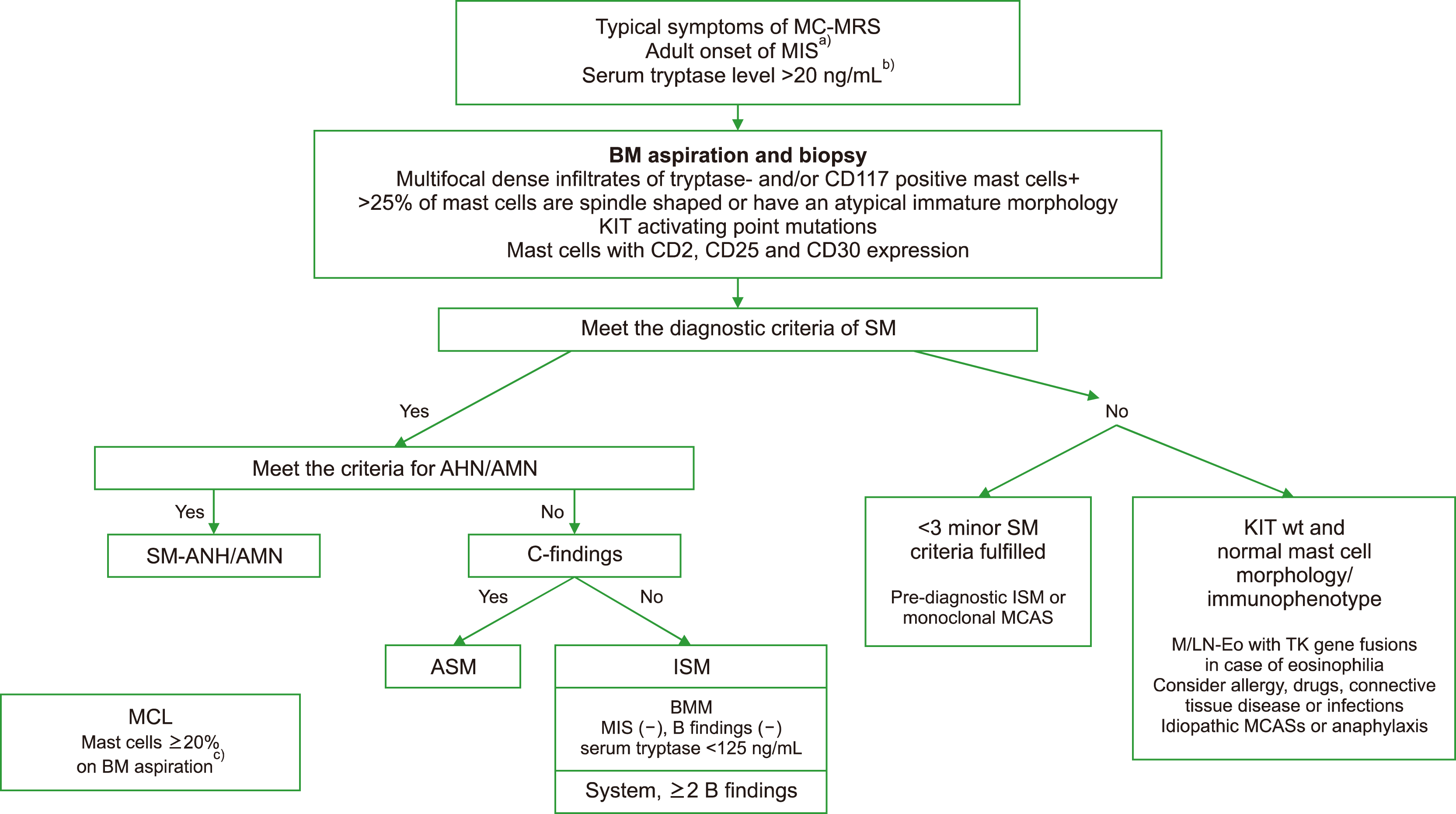
Fig. 1
Diagnostic algorithm for patients with suspected systemic mastocytosis. In all adult patients with documented mastocytosis in the skin (MIS), a complete staging, including a bone marrow (BM) examination, is required. a)Independent of the serum tryptase level; b)Basal tryptase levels at least 48 hours after resolution of all MC-MRSs; c)in ICC, a core biopsy specimen may be used to diagnose MCL if the aspirate is a dry tap.
Abbreviations: MC-MRS, mast cell-mediator related symptoms; MCAS, mast cell activation syndrome; MIS, mastocytosis in the skin; M/LN-Eo, myeloid/lymphoid neoplasm with eosinophilia.
br-58-s1-s96-f1.tif
Table 1
2022 WHO/ICC classification of mastocytosis.
WHO ICC
Cutaneous mastocytosis Cutaneous mastocytosis
Urticaria pigmentosa/maculopapular cutaneous mastocytosis Urticaria pigmentosa/maculopapular cutaneous mastocytosis
Monomorphic
Polymorphic
Diffuse cutaneous mastocytosis Diffuse cutaneous mastocytosis
Cutaneous mastocytoma Mastocytoma of skin
Isolated mastocytoma
Multilocalized mastocytoma
Systemic mastocytosis Systemic mastocytosis
Bone marrow mastocytosis (BMM)
Indolent systemic mastocytosis (ISM) Indolent systemic mastocytosis (ISM) include bone marrow mastocytosis (BMM)
Smoldering systemic mastocytosis (SSM) Smoldering systemic mastocytosis (SSM)
Aggressive systemic mastocytosis (ASM) Aggressive systemic mastocytosis (ASM)
Systemic mastocytosis with an associated hematologic neoplasm (SM-AHN) Systemic mastocytosis with an associated myeloid neoplasm (SM-AMN)
Mast cell leukemia Mast cell leukemia
Mast cell sarcoma Mast cell sarcoma

Bone marrow mastocytosis (BMM) as a clinicopathologic variant in ICC classification became a new SM subtype in WHO classification.

Table 2
Molecular abnormalities in patients with CM and SM.
Molecular abnormality Reported in patients with Estimated frequency in patients with SM
KIT D816V All SM variants >90%
Also in CM 15–20%
KIT D816Y CM, ISM, SM-AHNMD <5%
KIT D816F CM <5%
KIT D816H MCL, ASM, SM-AHNMD <5%
KIT D820G ASM <5%
KIT V560G ISM <5%
KIT F522C ISM <5%
KIT E839K CM <5%
KIT V530I SM-AHNMD <5%
KIT K509I CM, SM (including familial variant) <5%
Other KIT mutations CM and/or SM variants <5%
FIP1L1/PDGFRA SM-CEL <5%
AML1/ETO SM-AML with t(8;21) <5%
JAK2 V617F SM-PMF <5%
TET2 mutations SM-AHNMD, ISM, ASM <5%∼
SRSF2 mutationsa) ASM, SM-AHNMD <5%∼
DNMT3A mutations ISM, SM-AHNMD <5%∼
ASXL1 mutationsa) ASM, SM-AHNMD <5%∼
RUNX1 mutationa) ASM, SM-AHNMD <5%∼
CBL mutations SM-AHNMD <5%∼
U2AF1 mutations SM-AHNMD <5%∼
EZH2 mutations SM-AHNMD <5%∼
RAS mutations ASM, SM-AHNMD <5%∼

Modified from Valent [26].

a)Indicates the high molecular risk gene mutations frequently used in multiparameter prognostic scoring systems for advanced SM.

Abbreviations: AHNMD, associated hematologic non-mast cell-lineage disease; AML, acute myeloid leukemia; CEL, chronic eosinophilic leukemia; PMF, primary myelofibrosis.

Table 3
Clinical manifestations of systemic mastocytosis.
Symptoms Flushing, prutirus, blistering
Anaphylaxis
Hypotension, tachycardia
Fever, night sweats
Fatigue
Abdominal clamping
Nausea, vomiting
Diarrhea
Peptic ulcer disease/GERD
Weight lossa), malabsorption
Headache, cognitive impairment, depression
Organ involvement/damage Splenomegalya)
Hepatomegaly
Portal hypertension, ascitesa)
Lymphadenopathy
Osteoporosis/osteosclerosis, pathologic fracturea)
Laboratory findings Anemia, thrombocytopeniaa)
Monocytosis
Eosinophilia
Circulating mast cells
Elevated serum tryptase
Elevated alkaline phosphatasea)
Hypoalbuminemiaa)

a)Indicates C-findings.

Table 4
Refined diagnostic criteria for systemic mastocytosis.
WHO ICC
Major criterion Multifocal dense infiltrates of mast cells (≥15 mast cells in aggregates) in bone marrow biopsies and/or in sections of other extracutaneous organ(s) Multifocal dense infiltrates of tryptase- and/or CD117 positive mast cells (≥15 mast cells in aggregates) detected in sections of bone marrow and/or other extracutaneous organ(s)d)
Minor criteria ≥25% of all mast cells are atypical cells (type I or type II) on bone marrow smears or are spindle-shaped in mast cell infiltrates detected in sections of bone marrow or other extracutaneous organsa) In bone marrow biopsy or in section of other extracutaneous organs >25% of mast cells are spindle shaped or have an atypical immature morphologye)
KIT-activating KIT point mutation(s) at codon 816 or in other critical regions of KITb) in bone marrow or another extracutaneous organ KIT D816V mutation or other activating KIT mutation detected in bone marrow, peripheral blood, or other extracutaneous organsd,f)
Mast cells in bone marrow, blood, or another extracutaneous organ express one or more of: CD2 and/or CD25 and/or CD30c) Mast cells in bone marrow, peripheral blood or other extracutaneous organs express CD25, CD2, and/or CD30, in addition to mast cell markers
Baseline serum tryptase concentration >20 ng/mL (in the case of an unrelated myeloid neoplasm, an elevated tryptase does not count as an SM criterion. In the case of a known HαT, the tryptase level should be adjusted) Elevated serum tryptase level, persistently >20 ng/mL. In cases of SM-AMN an elevated tryptase does not count as a SM minor criterion.
At least 1 major and 1 minor The presence of the major criterion
In the absence of the major criterion, 3 minor criteria In the absence of the major criterion, at least 3 of the following 4 minor criteria must be present

a)In tissue sections, an abnormal mast cell morphology counts in both a compact infiltrate and a diffuse (or mixed diffuse+compact) mast cell infiltrate. However, the spindle-shaped form does not count as an SM criterion when mast cells are lining vascular cells, fat cells, nerve cells, or the endosteal-lining cell layer. In the bone marrow smear, an atypical morphology of mast cells does not count as SM criterion when mast cells are located in or adjacent to bone marrow particles. Morphologic criteria of atypical mast cells have been described previously.

b)Any type of KIT mutation counts as minor SM criterion when published solid evidence for its transforming behavior is available.

c)All 3 markers fulfill this minor SM criterion when expression in mast cells can be confirmed by either flow cytometry or by immunohistochemistry or by both techniques.

d)In the absence of a KIT mutation particularly in cases with eosinophilia, the presence of tyrosine kinase gene fusions associated with myeloid/lymphoid neoplasm with eosinophilia (M/LN-Eo) must be excluded.

e)Round-cell well-differentiated morphology can occur in a small subset of cases. In these cases, the mast cells are often negative for CD25 and CD2 but positive for CD30.

f)To avoid “false-negative” results, use of a high sensitivity PCR assay for detection of KIT D816V mutation is recommended. If negative, exclusion of KIT mutation variants is strongly recommended in suspected SM.

Table 5
“B” findings and “C” findings in WHO and ICC diagnostic criteria.
WHO ICC
B findings Infiltration grade (MC) in BM ≥30% in histology (IHC) and/or serum tryptase ≥200 ng/mLa) and/or KIT D816V VAF ≥10% in BM or PB leukocytes High mast cell burden, >30% of BM cellularity by mast cell aggregates (assessed on BM biopsy) and serum tryptase >200 ng/mL
Signs of myeloproliferation and/or myelodysplasia without a frank AHN; normal or mildly abnormal CBCs Cytopenia (not meeting criteria for C findings) or -cytosis. Reactive causes are excluded, and criteria for other myeloid neoplasms are not met
Organomegaly without dysfunction; hepatomegaly, splenomegaly or lymphadenopathy (>2 cm) Hepatomegaly without impairment of liver function, or splenomegaly without features of hypersplenism including thrombocytopenia, and/or lymphadenopathy (>1 cm size) on palpation or imaging
C findings BM dysfunction: HB <10 g/dL, and/or PLT <100 G/L, and/or neutrophils <1 G/L
Hepatomegaly with liver dysfunction
Splenomegaly with hypersplenism
Large osterolysis (≥2 cm) with pathologic fracture±bone pain
Malabsorption with weight loss due to GI MC infiltrates

a)In the case of a known hereditary α tryptasemia (HαT), the basal serum tryptase level should be adjusted.

The diagnosis of variants of systemic mastocytosis require correlation with B and C findings. “B” findings represent burden of disease. “C”-findings represent SM induced organ damage.

Table 6
Refined diagnostic criteria for systemic mastocytosis with associated hematologic/myeloid neoplasms.
SM-AHN SM-AMNa)
Meets the diagnostic criteria for systemic mastocytosis Meets the diagnostic criteria for systemic mastocytosis
Meets the WHO criteria for myeloid AHN type, lymphoid AHN type Meets the criteria for an associated myeloid neoplasm, e.g., CMML or other MDS/MPN, MDS, MPN, AML, or other myeloid neoplasm
The associated myeloid neoplasm should be fully classified according to established criteria

a)SM-AHN is modified to SM-AMN in new ICC criteria because SM-AHN is limited to the presence of an associated myeloid neoplasm, with which it often also shares a KIT mutations and/or clonal genetic abnormalities.

Table 7
Summary of prognostic factors in multiparameter prognostic scoring systems applicable to SM.
Non-advanced SM Advanced SM
REMA [38] IPSM [41] IPSM MARS [42] GPS [43] MAPS [44]
Age >60 year
Hb, g/dL
>10
>11
Platelet×109/L
<100 a)
<150
WBC >16×109/L
Increased serum level
Baseline tryptase a)
β2-microglobulin a) a)
Alkaline phosphatase
Mutational profile
BM KIT D816V VAF>1% a)
Additional somatic mutations ASXL1a),b)
RUNX1a)
DNMT3Aa)
SRSF2
ASXL1
RUNX1
SRSF2
ASXL1
RUNX1
DNMT3A
ASXL1
RUNX1
NRAS

a)Prognostic factor only for PFS. b)A/R/D gene pathogenic VAF ≥30%, independent predictors for OS in REMA.

Table 8
Therapeutic considerations for mast cell mediator related symptoms.
Pruritis, flushing
H1 and H2 antagonist
Leukotriene antagonist
Topical glucocorticoids
Nonsteroidal anti-inflammatory drug (aspirin)
Psoralen plus UVA (PUVA) for refractory symptoms
Omalizumab
Hypotension/anaphylaxis
Intramuscular epinephrine
For attempted prophylaxis in patients with frequent life-threatening episodes consider scheduled H1 and H2 antagonists +/- glucocorticoids
Cytoreductive therapy (IFN-α or cladribine)
Headache, cognitive impairment, depression
H1 and H2 antagonist
Sodium cromolyn
Abdominal pain, cramping
H2 antagonists +/- proton pump inhibitor
Leukotriene antagonists
Sodium cromolyn
Peptic ulcer disease/GERD, nausea, vomiting
H2 antagonists +/- proton pump inhibitor
Glucocorticoids
Diarrhea
Proton pump inhibitor +/- leukotriene antagonist +/- anticholinergics
Glucocorticoids
Ascites
Glucocorticoids
Portocaval shunt or cytoreductive therapy (INFα or cladribine)
Osteopenia/osteoporosis
Calcium supplementation +/- vitamin D
Bisphosphonates
Cytoreductive therapy (IFN-α or cladribine) in severe osteoporosis at risk for pathologic fracture or severe localized bone pain

Abbreviations: H1, histamine receptor 1; H2, histamine receptor 2; IFN-α, interferon-α.

TOOLS
Similar articles