Abstract
Benign mediastinal cysts are challenging to diagnose. Although Endoscopic Ultrasound (EUS) and EUS-guided fine needle aspiration (FNA) can accurately diagnose mediastinal foregut cysts, little is known about their complications. This paper reports a rare case in which EUS-FNA performed on mediastinal hemangioma resulted in an aortic hematoma. A 29-year-old female patient was commissioned for EUS of an asymptomatic accidental mediastinal lesion. Chest CT revealed a 4.9×2.9×10.1 cm thin-walled cystic mass in the posterior mediastinum. EUS revealed a large, anechoic cystic lesion with a regular thin wall with negative Doppler. EUS-guided FNA was performed using a single-use 19-gauge aspiration needle (EZ Shot 3; Olympus, Tokyo, Japan), and approximately 70 cc of serous pinkish fluid was aspirated. The patient was in a stable condition with no signs of acute complication. One day after EUS-FNA, thoracoscopic resection for mediastinal mass was conducted. The purple and multi-loculated large cyst was removed. Upon removal, however, an aortic hematoma caused by a focal descending aortic wall injury was observed. After a few days of close observation, the patient was discharged upon stable 3D aorta angio CT findings. This paper reports a rare and severe complication of EUS-FNA, in which an aspiration needle caused a direct injury to the aorta. The injection must be performed carefully to avoid damaging the adjacent organs or digestive tract walls.
Go to : 
Mediastinal masses are relatively uncommon findings found most frequently in the anterior compartment. They include various entities demonstrating a spectrum of clinical and pathologic features.1 Although a comprehensive evaluation that includes history, examination, and laboratory studies is essential, imaging studies, such as CT, are critical to establishing a presumptive diagnosis. On the other hand, the imaging modality alone may provide an inconclusive diagnosis depending on its location and inner contents.2 This is particularly true for mediastinal cysts comprising 12 to 30% of mediastinal masses.3 Well-marginated and round lesions containing solid or liquid components are sometimes indistinguishable from degenerated tumors.4 In such cases, additional measures, such as biopsies, may be required for accurate diagnosis and optimal treatment.
The clinical use of EUS-guided fine needle aspiration (FNA) was first reported in 1992, and its clinical boundary has been expanding.5 From intraluminal gastrointestinal lesions, such as submucosal and gastrointestinal tumors,6 EUS-FNA can also diagnose extraluminal lesions, such as pancreas, spleen, adrenal glands, peritoneal cavity, nearby lymph nodes, and even mediastinum, accurately through a combined power of ultrasound and cytologic analysis from aspirates.7-11 Perhaps the most significant advantage of EUS-FNA is that its diagnostic performance does not come at the cost of a decreased safety profile. Studies have reported its diagnostic power and safety in various lesions,12-14 leading many medical institutions worldwide to adopt EUS-FNA in diagnosis and treatment.
Although the rate of complications arising from EUS-FNA is low, its number continues to rise along with the increasing number of procedures performed. With occasional infections, however, most physicians are yet to experience severe complications arising from EUS-FNA. This paper reports a rare case in which EUS-FNA, performed on mediastinal hemangioma, which was preliminarily misdiagnosed as a bronchogenic cyst that resulted in an aortic hematoma caused by aortic wall abrasion from an FNA needle injury. This case report was approved by institutional review board of The Catholic University of Korea, Seoul St. Mary's hospital (K23Z ASE0150).
Go to : 
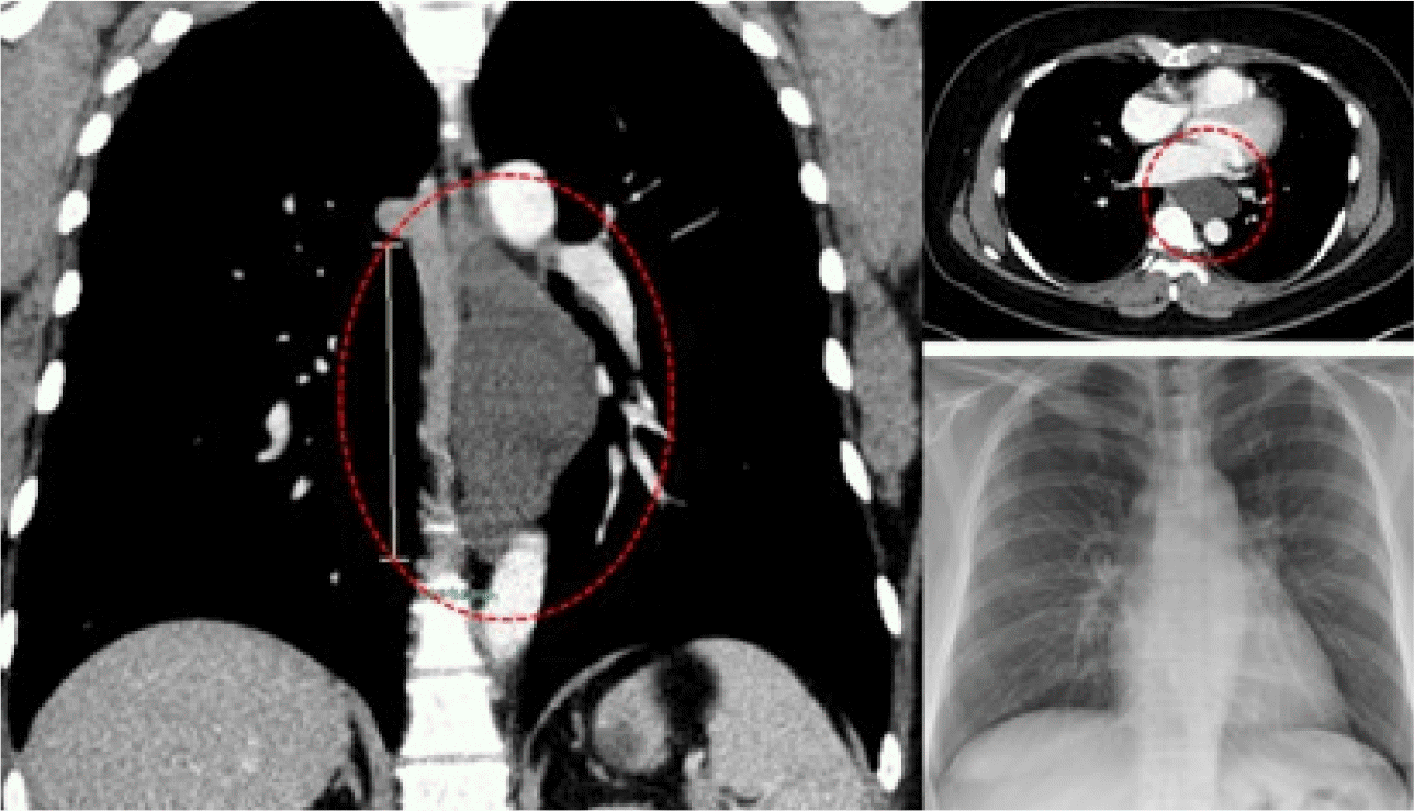
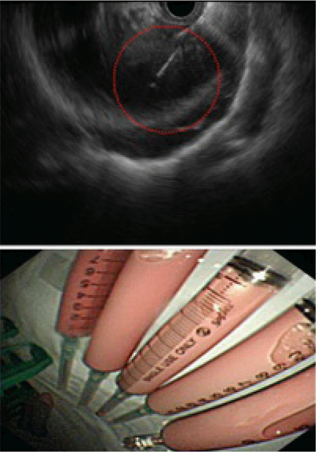
A 29-year-old female patient was commissioned by the cardiothoracic surgery department for EUS of an asymptomatic accidental mediastinal lesion. Chest CT revealed a 4.9×2.9×10.1 cm thin-walled cystic mass in the posterior mediastinum, abutting and seemingly arising from the esophagus (Fig. 1). The patient had no symptoms related to the mass and was not on any regular medication, including anticoagulants or antiplatelet agents. EUS revealed a large, anechoic cystic lesion with a regular thin wall with negative Doppler studies in the posterior mediastinum. The preliminary EUS diagnosis was an esophageal duplication cyst or a bronchogenic cyst. The thoracic surgeon requested a reduction of the cystic volume using EUS-FNA to facilitate a surgical resection. After prophylactic antibiotics treatment, a single- use 19-gauge aspiration needle (EZ Shot 3; Olympus, Tokyo, Japan) was used to aspirate approximately 70 cc of serous pinkish fluid (Fig. 2). The patient was in a stable condition with no signs of complication after the procedure.
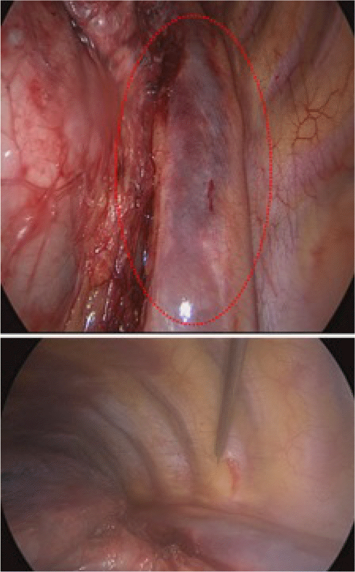
One day after EUS-FNA, the patient received a thoracoscopic resection for the mediastinal lesion. Upon entering the mediastinum, large, purple, and multiloculated cyst, well-capsulated without any connection to the esophagus, was observed in the posterior mediastinum and was then removed.
After removal, however, an aortic hematoma, which was thought to have been caused by a focal descending aortic wall injury, was observed incidentally in the surgical field (Fig. 3). Fortunately, the patient was in a stable condition with no symptoms related to the hematoma. After three days of close observation, the patient underwent 3D aorta angio CT, which revealed a small amount of fluid collection along the descending thoracic aorta as an immediate post-operational change but no significant change in the aorta wall itself. The patient was discharged and asked to make frequent visits to the outpatient clinic at short intervals until the hematoma was thought to be completely resolved.
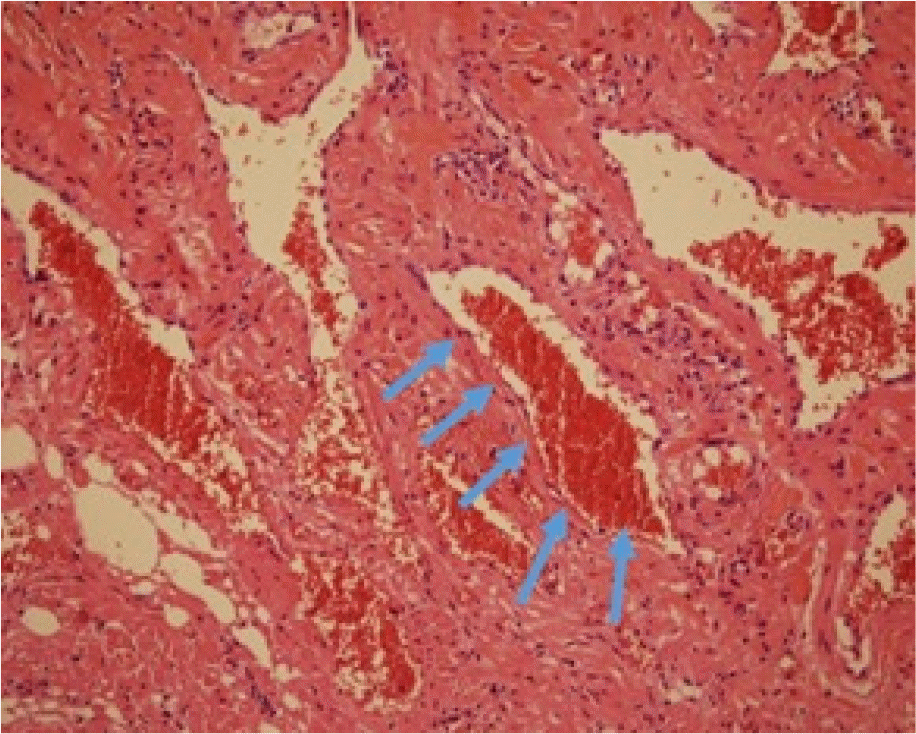
The pathology results of the surgical specimen showed that the mediastinal cyst was not just a simple cyst. It was cavernous hemangioma with thrombosis, a sporadic vascular tumor for which the treatment of choice is a complete surgical resection.15 The patient’s postoperative course was uneventful, and no recurrence has yet been noted (Fig. 4).
Go to : 
EUS provides a minimally invasive approach to diagnosing benign mediastinal cysts and may be more accurate than CT or other imaging modalities. On the other hand, a large variation in EUS appearances can sometimes make it difficult to make an accurate EUS-based diagnosis, in which FNA is required for further evaluation. Making an accurate diagnosis only with imaging diagnosis in a mediastinal cystic lesion is difficult. FNA is commonly performed to obtain tissue from masses or associated lymph nodes and to aspirate the contents of cystic structures for analysis.
While specific numbers vary according to the target sites and studies, EUS-FNA is generally considered a safe procedure. A systematic review of complications and deaths associated with EUS-FNA (51 reports and 10,941 patients) revealed a complication rate of 0.98% and a mortality rate of 0.02%. Two deaths included in the review were due to severe acute pancreatitis and cholangitis.16 EUS-FNA of the mediastinal lesion is generally considered a very safe technique. The possible complications of EUS-FNA of the mediastinal lesion are esophageal perforation, infection, or bleeding. The safety record for analyzing mediastinal lymph nodes with EUS-FNA is impressive, with no major complications reported in several hundreds of patients.17-20 A study including 168 patients with mediastinal masses/lymphadenopathy revealed no serious complications. Minor complications (e.g., transient pain or fever) occurred only in 0.006%.21 A systematic review and meta-analysis on EUS–FNA for mediastinal lymph node lesions in patients with non-small cell lung cancer (18 reports, 1,201 patients) only identified minor complications, such as sore throat and fever in 10 patients (0.8%).22 Some cases of post-procedural mediastinitis have been observed when the puncture targets are cystic lesions or necrotic lymph nodes.20,23,24 According to various studies, less than 2% of patients who undergo EUS-FNA suffer some form of hemorrhage. Most involve minor bleeding from a gastrointestinal puncture that resolves spontaneously, and transfusion and endoscopic hemostasis are rarely required (0–0.44% of all cases)18-20 to various studies, less than 2% of patients who undergo EUS-FNA undergo some form of hemorrhage. Most involve minor bleeding from a gastrointestinal puncture that resolves spontaneously, and rarely transfusion and endoscopic hemostasis are required (0–0.44% of all cases).23 Mediastinal cysts are at risk of infection during EUS-FNA, even in prophylactic antibiotic therapy, and, if infected, can lead to mediastinitis with or without sepsis.24
Hence, the case of post-procedural aortic hematoma reveals a rare yet possibly lethal complication that can result from EUS-FNA. In this patient, the needle punctured through the digestive tract wall and the thin-walled cyst at once under EUS guidance. With the needle placed successfully within the cyst, the inner cystic contents were aspirated. As the cyst shrank in size, the needle was thought to have pierced through the cystic wall and damaged the nearby arterial wall. The position of the injection needle was monitored closely via EUS during the procedure so that the needle tip remained within the cyst. The cyst wall was too thin, and the needle was too sharp. The injury was not detected during the procedure owing to the focal size of the arterial damage, and the patient did not experience any symptoms. It was only through thoracoscopy a day later that arterial hematoma was found incidentally. In general, aspiration is not recommended because of the risk of infection in the case of media static. In this patient, cyst aspiration was performed to facilitate removal by thoracoscopy by reducing the cyst size.
In conclusion, this paper reported a rare but potentially severe complication in the form of aortic hematoma after performing EUS-FNA on a benign mediastinal cyst. While diagnostically accurate and generally safe, endoscopists must still perform the procedure carefully to prevent damage to nearby organs or digestive tract walls that can lead to severe complications.
Go to : 
REFERENCES
1. Carter BW, Marom EM, Detterbeck FC. 2014; Approaching the patient with an anterior mediastinal mass: a guide for clinicians. J Thorac Oncol. 9(9 Suppl 2):S102–S109. DOI: 10.1097/JTO.0000000000000294. PMID: 25396306.
2. Carter BW, Okumura M, Detterbeck FC, Marom EM. 2014; Approaching the patient with an anterior mediastinal mass: a guide for radiologists. J Thorac Oncol. 9(9 Suppl 2):S110–S118. DOI: 10.1097/JTO.0000000000000295. PMID: 25396307.
3. Takeda S, Miyoshi S, Minami M, Ohta M, Masaoka A, Matsuda H. 2003; Clinical spectrum of mediastinal cysts. Chest. 124:125–132. DOI: 10.1378/chest.124.1.125. PMID: 12853514.
4. Jeung MY, Gasser B, Gangi A, et al. 2002; Imaging of cystic masses of the mediastinum. Radiographics. 22 Suppl 1:S79–S93. DOI: 10.1148/radiographics.22.suppl_1.g02oc09s79. PMID: 12376602.
5. Vilmann P, Jacobsen GK, Henriksen FW, Hancke S. 1992; Endoscopic ultrasonography with guided fine needle aspiration biopsy in pancreatic disease. Gastrointest Endosc. 38:172–173. DOI: 10.1016/S0016-5107(92)70385-X. PMID: 1568614.
6. Mekky MA, Yamao K, Sawaki A, et al. 2010; Diagnostic utility of EUS-guided FNA in patients with gastric submucosal tumors. Gastrointest Endosc. 71:913–919. DOI: 10.1016/j.gie.2009.11.044. PMID: 20226456.
7. Zhang L, Sanagapalli S, Stoita A. 2018; Challenges in diagnosis of pancreatic cancer. World J Gastroenterol. 24:2047–2060. DOI: 10.3748/wjg.v24.i19.2047. PMID: 29785074. PMCID: PMC5960811.
8. Iwashita T, Yasuda I, Tsurumi H, et al. 2009; Endoscopic ultrasound-guided fine needle aspiration biopsy for splenic tumor: a case series. Endoscopy. 41:179–182. DOI: 10.1055/s-0028-1119474. PMID: 19214901.
9. Martin-Cardona A, Fernandez-Esparrach G, Subtil JC, et al. On belhaf of Spanish Group for EUS-Guided TA in the adrenal gland. EUS-guided tissue acquisition in the study of the adrenal glands: Results of a nationwide multicenter study. PLoS One. 2019; 14:e0216658. DOI: 10.1371/journal.pone.0216658. PMID: 31170163. PMCID: PMC6553722.
10. Yasuda I, Goto N, Tsurumi H, et al. 2012; Endoscopic ultrasound-guided fine needle aspiration biopsy for diagnosis of lymphoproliferative disorders: feasibility of immunohistological, flow cytometric, and cytogenetic assessments. Am J Gastroenterol. 107:397–404. DOI: 10.1038/ajg.2011.350. PMID: 21989147.
11. Catalano MF, Rosenblatt ML, Chak A, Sivak MV Jr, Scheiman J, Gress F. 2002; Endoscopic ultrasound-guided fine needle aspiration in the diagnosis of mediastinal masses of unknown origin. Am J Gastroenterol. 97:2559–2565. DOI: 10.1111/j.1572-0241.2002.06023.x. PMID: 12385439.
12. Krishna NB, LaBundy JL, Saripalli S, Safdar R, Agarwal B. 2009; Diagnostic value of EUS-FNA in patients suspected of having pancreatic cancer with a focal lesion on CT scan/MRI but without obstructive jaundice. Pancreas. 38:625–630. DOI: 10.1097/MPA.0b013e3181ac35d2. PMID: 19506529.
13. Akahoshi K, Oya M, Koga T, Shiratsuchi Y. 2018; Current clinical management of gastrointestinal stromal tumor. World J Gastroenterol. 24:2806–2817. DOI: 10.3748/wjg.v24.i26.2806. PMID: 30018476. PMCID: PMC6048423.
14. Michael H, Ho S, Pollack B, Gupta M, Gress F. 2008; Diagnosis of intra-abdominal and mediastinal sarcoidosis with EUS-guided FNA. Gastrointest Endosc. 67:28–34. DOI: 10.1016/j.gie.2007.07.049. PMID: 18155422.
15. Hashimoto H, Oshika Y, Obara K, Takeshima S, Sato K, Tanaka Y. 2009; Intercostal venous hemangioma presenting as a chest wall tumor. Gen Thorac Cardiovasc Surg. 57:228–230. DOI: 10.1007/s11748-008-0344-6. PMID: 19367460.
16. Wang KX, Ben QW, Jin ZD, et al. 2011; Assessment of morbidity and mortality associated with EUS-guided FNA: a systematic review. Gastrointest Endosc. 73:283–290. DOI: 10.1016/j.gie.2010.10.045. PMID: 21295642.
17. Wiersema MJ, Vilmann P, Giovannini M, Chang KJ, Wiersema LM. 1997; Endosonography-guided fine-needle aspiration biopsy: diagnostic accuracy and complication assessment. Gastroenterology. 112:1087–1095. DOI: 10.1016/S0016-5085(97)70164-1. PMID: 9097990.
18. Fritscher-Ravens A, Sriram PV, Bobrowski C, et al. 2000; Mediastinal lymphadenopathy in patients with or without previous malignancy: EUS-FNA-based differential cytodiagnosis in 153 patients. Am J Gastroenterol. 95:2278–2284. DOI: 10.1111/j.1572-0241.2000.02243.x. PMID: 11007229.
19. Larsen SS, Krasnik M, Vilmann P, et al. 2002; Endoscopic ultrasound guided biopsy of mediastinal lesions has a major impact on patient management. Thorax. 57:98–103. DOI: 10.1136/thorax.57.2.98. PMID: 11828036. PMCID: PMC1746251.
20. Wildi SM, Hoda RS, Fickling W, et al. 2003; Diagnosis of benign cysts of the mediastinum: the role and risks of EUS and FNA. Gastrointest Endosc. 58:362–368. DOI: 10.1016/S0016-5107(03)00009-9. PMID: 14528209.
21. Al-Haddad M, Wallace MB, Woodward TA, et al. 2008; The safety of fine-needle aspiration guided by endoscopic ultrasound: a prospective study. Endoscopy. 40:204–208. DOI: 10.1055/s-2007-995336. PMID: 18058615.
22. Micames CG, McCrory DC, Pavey DA, Jowell PS, Gress FG. 2007; Endoscopic ultrasound-guided fine-needle aspiration for non-small cell lung cancer staging: A systematic review and metaanalysis. Chest. 131:539–548. DOI: 10.1378/chest.06-1437. PMID: 17296659.
23. ASGE Standards of Practice Committee. Early DS, Acosta RD, Chandrasekhara V, et al. 2013; Adverse events associated with EUS and EUS with FNA. Gastrointest Endosc. 77:839–843. DOI: 10.1016/j.gie.2013.02.018. PMID: 23684089.
24. Aerts JG, Kloover J, Los J, van der Heijden O, Janssens A, Tournoy KG. 2008; EUS-FNA of enlarged necrotic lymph nodes may cause infectious mediastinitis. J Thorac Oncol. 3:1191–1193. DOI: 10.1097/JTO.0b013e3181872752. PMID: 18827619.
Go to : 


 PDF
PDF Citation
 Print
Print


 XML Download