Journal List > Korean J Gastroenterol > v.81(4) > 1516082559

뇌-장-미생물 축

Abstract

Patients frequently report that stress causes or exacerbates gastrointestinal (GI) symptoms, indicating a functional relationship between the brain and the GI tract. The brain and GI tract are closely related embryologically and functionally, interacting in various ways. The concept of the brain–gut axis was originally established in the 19th and early 20th centuries based on physiological observations and experiments conducted in animals and humans. In recent years, with the growing recognition that gut microbiota plays a vital role in human health and disease, this concept has been expanded to the brain–gut–microbiota axis. The brain influences the motility, secretion, and immunity of the GI tract, with consequent effects on the composition and function of the gut microbiota. On the other hand, gut microbiota plays an essential role in the development and function of the brain and enteric nervous system. Although knowledge of the mechanisms through which the gut microbiota influences distant brain function is incomplete, studies have demonstrated communication between these organs through the neuronal, immune, and endocrine systems. The brain–gut–microbiota axis is an essential aspect of the pathophysiology of functional GI disorders such as irritable bowel syndrome, and is also involved in other GI diseases, including inflammatory bowel disease. This review summarizes the evolving concept of the brain–gut–microbiota axis and its implications for GI diseases, providing clinicians with new knowledge to apply in clinical practice.

서 론

임상 진료 과정에서 환자들의 위장관 증상이 스트레스에의해 발생하거나 악화되는 것을 흔히 경험하게 된다. 이것은뇌와 위장관이 기능적으로 연관되어 있다는 것을 시사하며 발생학적으로도 중추신경계와 장신경계는 모두 신경능선(neural crest) 기원으로 서로 밀접한 관계를 가지고 있다.1 이런 뇌와 장 사이의 관계에 대한 인식은 고대로부터 존재해 왔다. 당시 의학자들은 정신과 육체를 분리하지 않고 하나로 보는 전체론(holism) 개념으로 질병을 이해했기 때문에 정신이 신체에 영향을 줄 수 있다고 믿었다. 그러나 17세기 데카르트의 이원론(dualism) 이 등장하고 19세기 미생물이 질병의 원인이라는 병균이론(germ theory)이 주창되면서 질병에대한 인식이 바뀌게 되었다. 즉 환자의 정신과 신체는 서로독립적이며, 환자가 특정 증상을 호소할 때 그 기전을 설명할수 있는 기질적인 원인이 있어야 질병으로 진단하는 근대 서양의학의 사조가 정립된 것이다.2 이런 관점에서는 만성 소화불량이나 복통을 호소하더라도 혈액검사, 병리검사, 영상검사및 내시경 검사 등에서 이상소견이 발견되지 않으면 신체적질병으로 인정되지 않았고 정신 질환으로 취급되었다.
그러나 19세기와 20세기의 다양한 실험을 통해 뇌와 위장관사이의 상호작용에 대한 객관적 증거가 보고되면서 이 두 기관이양방향으로 영향을 주고 받는다는 뇌-장 축(brain-gut axis) 개념이 수립되었고, 현재는 위장관 질환의 주요 병리기전으로자리잡았다. 이 개념은 최근 인간의 건강과 질병에서 장내 미생물 무리의 역할이 알려지고 이에 대한 연구가 폭발적으로 증가하면서 뇌-장-미생물 축(brain-gut-microbiota axis)이라는 개념으로 확장되고 있다(Fig. 1).3 본 글에서는 뇌-장-미생물 축의개념과 소화기질환에서의 연구된 내용을 정리해보고자 한다.
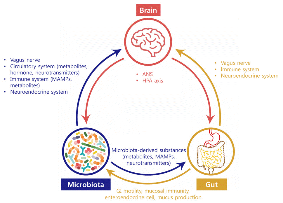
본 론

1. 뇌-장-미생물 축 개념의 발달

뇌가 위장관 기능에 영향을 줄 수 있다는 초기 연구들은위루(gastric fistula) 가 생긴 환자나 인공으로 위루를 만든 동물 모델을 대상으로 위의 움직임이나 분비를 직접 관찰함으로써 이루어졌다. 1833 년 외과 의사 William Beaumont 은 복부총상으로 발생한 영구적 위루를 통해 위를 관찰함으로써 분노나 공포 같은 정신적 변화가 위점막의 형태와 기능에 영향을준다는 것을 최초로 보고하였다.4 이후 20세기로 넘어와Pavlov 의 개실험 등을 통해 미주신경의 역할이 증명되었고,5 위루 환자들을 관찰한 연구들에서 분노, 강렬한 쾌감 또는 공포나 우울을 경험할 때는 각각 위점막이 발적 또는 창백해지거나 운동 및 분비가 증가 또는 감소되는 것이 보고되었다.5 또 1950 년대 직장경으로 대장을 관찰한 실험에서 불안감이나슬픔, 적개심, 자책감과 같은 감정적 변화가 대장 운동과 점막충혈에 영향을 주는 것이 증명되었다.6
이런 기념비적 관찰 실험을 통해서 뇌-장 축 개념이 시작되었지만, 현재의 관점에서는 당시 연구들은 실험적 설계가 엄밀하지 않았고 방법론의 타당성에 의문이 제기되고 있다. 그러나 1960-70 년대에 발달한 여러 위장관 생리 검사법과 실험기법을 통해 스트레스는 일반적으로 위운동성을 감소시키고대장운동성을 증가시킨다는 것이 정립되었다.7
뇌-장 축에서 중추신경계 내부의 기전이 관심 받게 된 것은 20세기 초 뇌하수체의 내분비 기능이 알려지고 스트레스라는개념이 확립된 이후이다.8 1950년대부터 스트레스 반응에서시상하부-뇌하수체-부신 축(hypothalamic-pituitary-adrenal axis, HPA) 의 역할에 대한 연구가 시작되었지만 1981년에서야corticotropin-releasing factor (CRF) 가 분리되었다.9 시상하부에서 분비되는 CRF 의 내분비적 기능은 ACTH 분비를 자극하는 것인데, 스트레스 반응에서도 주 매개물질로 작용해 위장관에 영향을 미친다는 것이 밝혀졌다. 스트레스에 의해 분비된CRF는 CRF1 수용체와 천골 부교감신경을 경유해 대장운동을증가시키고, CRF2 수용체와 미주신경을 경유해 위운동을 감소시킨다.10 또 대장 점막에서 비만세포 등의 염증세포 증가와염증성 사이토카인 증가에도 관여한다.11 중추신경계외에 말초의 감각 및 교감신경, 면역세포, 위장관 내분비세포 등에서도 CRF 와 연관 펩티드가 분비되어 위장관 감각과 염증에 영향을준다.12
1990 년대 후반부터는 기능적 자기공명영상(functional magnetic resonance imaging, fMRI) 을 이용해서 위장기능과 뇌의 각 영역 사이의 상관관계가 연구되기 시작했다. 기능성 위장관 질환 환자들은 정상인에 비해 위장관 자극에 대한 감각처리 영역의 과장된 반응을 보이며, 감정처리 영역의 이상과내장감각을 조절하는 영역의 활성의 감소 등이 보고되었다.13 자율신경계, HPA 축, 그리고 CRF 에 관한 연구가 뇌에서 장쪽 방향의 기전을 밝혔다면, fMRI 연구는 위장관 자극에 대한뇌의 기능적 영역의 변화를 보여줌으로써 뇌와 장의 양방향상호작용에 대한 근거를 제공하고 있다.
21세기에 들어와 장내 미생물 무리가 인간의 정상 생리와건강을 유지하는데 중요한 역할을 한다는 것이 알려졌고 많은 질환에서 장내 미생물 무리 불균형(dysbiosis) 이 동반된다는 것이 보고되었다. 이에 따라 장-뇌 축 개념도 새로운 전환기를 맞고 있는데, 스트레스와 같은 뇌의 변화가 위장관을 넘어 장내 미생물 무리까지도 영향을 줄 수 있다는 증거들이늘어나고 있다.14,15 예를 들면, 성질이 사나운 쥐와 얌전한 쥐를 같이 한 케이지에서 키우는 사회적 스트레스 실험에서 스트레스를 받은 얌전한 쥐는 장내 미생물 중에서 Bacteroides 감소와 Clostridium 증가가 관찰되었다.16 또 호주의 대학생들을 대상으로 한 연구에서는 스트레스가 낮은 학기초의 분변과 학기중간 시험기간의 분변을 채취해 장내 미생물 무리를비교했을 때 스트레스가 높은 시험기간동안 분변내 유산균의 비율이 감소되는 것을 확인하였다.17 최근에는 장내 미생물 무리가 장의 염증에 영향을 주며, 심지어 뇌의 발달과 기능에도영향을 미친다는 연구들도 보고되어 주목을 받고 있다. 이와같은 연구 결과를 근거로 기존의 뇌와 장 사이의 관계로 국한되었던 개념이 지금은 뇌-장-미생물 축으로 확장되었는데 이역시 양방향으로 상호작용하는 개념이다.

2. 뇌→장→미생물 방향의 조절기전

장내 미생물 무리는 분만 방식, 수유 방식, 식이, 운동, 약제등 다양한 요인에 의해 영향을 받는데, 중추신경계와 장신경계에 의해 조절되는 위장관의 생리적 변화도 장내 미생물 무리에 영향을 주는 중요한 요인이다.
1) 위장관 생리기능 변화를 통한 조절기전
우선 자율신경계는 위장관 생리의 변화, 즉 국소 운동 변화와 위산, 점액, 중탄산염 및 수분의 분비 등을 조절함으로써장내 미생물 무리의 조성에 영향을 미친다.14 특히 위장관 운동성의 변화에 따라 장내용물의 이동 속도가 달라지는 것은수분량, 영양소 이용, 세균 배출정도에 영향을 주기 때문에장내 미생물 무리 조성에 큰 변화를 줄 수 있다. 소장 운동성의 변화나 설사 혹은 변비와 같은 대장운동의 변화는 dysbiosis의 원인이 될 수 있다. 이런 측면에서, 장이동속도를 잘 반영하는 브리스톨 대변 형태(Bristol stool form) 는 장내 미생물 무리의 풍부도 및 조성과 아주 강력한 연관관계를 보인다.18
2) 위장관 분비물질 변화를 통한 조절기전
스트레스나 중추신경계의 영향으로 다양한 장내 분비 물질들의 분비가 조절되어 장내 미생물 무리에 영향을 줄 수 있다. 예를 들면, 스트레스는 파네스세포(Paneth cell)로부터 감염방어에 중요한 역할을 하는 α-defensin 분비를 증가시켜 미생물에 영향을 준다. 중추신경계는 장내분비세포, 면역세포, 및 신경에서 세로토닌, 사이토카인, 카테콜아민, dynorphin 의 분비에 영향을 줘 미생물을 조절할 수 있고, 항상성이 깨진 상태에서는 장점막 교감신경에서 에피네프린과 노에피네프린이 장관내로 분비되어 퀘럼 센싱(quorum-sensing) 기전을 통해 병원균의 성장과 독성을 변화시킨다.19 흥미롭게도 인간의 장내 미생물 무리는 멜라토닌에 반응해 일주기 리듬(circadian rhythm) 을 보이기도 한다.20
그러므로 스트레스나 중추신경계의 변화는 자율신경계를통한 위장관 운동변화, 점액 생산, 다양한 장분비 기능, 점막면역 반응, 직접적인 장점막 투과도 변화 등을 조절함으로써장관내에 존재하는 미생물 무리에 영향을 준다고 할 수 있다.21

3. 미생물→장→뇌 방향의 조절기전

뇌-장-미생물 방향의 신호전달과 달리 미생물-장-뇌 방향의작용은 최근에 연구되기 시작해 아직 신호전달 기전이 명확하게 밝혀지지 않았다. 현재까지의 연구로 보면 신경계, 면역계, 내분비계를 통해 신호를 전달하는 것으로 생각된다.
1) 신경계를 통한 조절기전
미주신경(vagus nerve) 은 구심성(afferent) 섬유가 80%로 구성되어 장관과 뇌 사이의 신호를 전달하는 중요한 통로이다.21 장내 미생물 무리는 단쇄 지방산(short chain fatty acid) 같은 대사체를 생산하거나 세포벽의 peptidoglycan 혹은lipopolysaccharide (LPS) 등이 미생물 관련 분자 패턴(Microbe-associated molecular patterns, MAMPs) 으로 작용해 점막하 구심성 신경 수용체로 직접 신호를 전달할 수있다. 또 장내 미생물 무리가 장내분비세포를 자극해 세로토닌을 분비시키고 분비된 세로토닌은 확산(diffusion) 기전으로신경에 신호를 전달하는 간접적인 기전이 있다.14,23 장내분비세포 중에서도 미주신경 말단과 시냅스를 형성하고 있는 neuropod 세포를 장내 미생물 무리가 자극하는 경우 확산이 아닌신경전달과 유사한 직접적인 방식으로 신호를 전달하게 된다.24
2) 면역계를 통한 조절기전
말초 면역계의 변화가 신경학적 기능과 행동에 영향을 미칠 수 있다는 증거들이 늘어나고 있는데, 면역 자극원으로 작용할 수 있는 장내 미생물 무리도 면역계를 통해서 뇌에 영향을 미친다.23 장내 미생물의 MAMPs 는 다양한 면역세포, 특히 대식세포, 호중구, 수지상 세포와 같은 선천 면역 세포를활성화시킨다. 이렇게 활성화된 면역세포들이 생산한 IL-1α, IL-1β, TNF-α, IL-6 와 같은 염증성 사이토카인은 뇌혈관장벽을 통과해 뉴런과 신경교세포, 특히 미세아교세포에 발현되는수용체에 작용해 뇌기능에 영향을 주게 된다.23 LPS 는 직접뇌혈관장벽을 통과해 영향을 미칠 수도 있다.25
3) 내분비계를 통한 조절기전
위장관내 환경변화에 따라 장내분비세포에서 방출된 신호전달 분자들은 혈중 이동으로 중추신경계에서 식이와 연관된 행동들을 조절하거나 미주신경 말단에 작용하여 뇌기능에 영향을 줄 수 있는데 이 기전은 미생물-장-뇌 축의 주요 통로로작용한다.14 예를 들면, 미생물이 생산한 단쇄지방산은 L 세포의 free fatty acid receptor 를 통해 glucagon like peptide-1 (GLP-1) 의 발현 및 분비를 조절한다.26 또 미생물이 대사시킨 2차 담즙산은 L 세포의 G protein-coupled bile acid receptor (TGR5)를 활성화해 Peptide YY 와 GLP-1을 분비시킨다.27
위장관 기능에 중요한 세로토닌의 분비 역시 장내 미생물무리의 영향을 받을 수 있다. 단쇄지방산 같은 다양한 미생물생산 대사체가 장크롬화세포에서 tryptophan hydroxylase-1 (Tph1) 발현을 증가시켜 세로토닌 생산을 증가시키거나 미생물이 tryptophan 을 대사해 생산한 indole 이 장크롬화세포 수용체를 자극해 세로토닌 분비를 촉진시킬 수 있다.28,29
흥미롭게도 장내 미생물 무리는 세로토닌, 도파민, 에피네프린과 노에피네프린, γ-아미노뷰티르산, 아세틸콜린과 같은신경전달물질을 직접 생산할 수 있다. 그러나 신경전달물질은분자량이 커서 뇌혈관장벽을 통과하지 못하기 때문에 말초에서 생산된 신경전달물질이 중추신경계에 직접 작용하기는 어렵고, 뇌혈관장벽을 통과할 수 있는 전구물질의 대사나 생산을 장내 미생물 무리가 조절해 영향을 미칠 가능성이 있다.23

4. 장내 미생물 무리와 뇌 및 장신경계 발달 사이의 관계

출생 후 장내 미생물 무리가 발달하는 시기는 뇌와 위장관의 기능이 발달되는 시기와 부분적으로 일치해 뇌, 위장관, 그리고 장내 미생물 무리의 발달이 병렬적으로 진행된다. 최근 장내 미생물 무리가 다양한 기전으로 뇌에 영향을 준다는사실이 밝혀지면서 뇌의 발달과 퇴행성 뇌질환에서 역할이 주목 받고 있다.
먼저 장내 미생물 무리가 뇌발달에 미치는 영향을 살펴보면, 출생 직후 장내 미생물 무리는 직접 혹은 간접적인 방식으로 뇌신경 발생, 수초화 및 미세아교세포(microglia) 활성화, 뇌혈관장벽 형성 등에 필수적인 역할을 한다.30 무균쥐에서는 정상쥐와는 달리 불안 반응, 비정상적인 움직임, 스트레스 반응 강화, 기억력 장애를 보이고, HPA 축의 이상을 나타낸다.30,31 이런 이상 소견은 정상 장내 미생물 무리나 유익균을조기에 이식하면 회복된다. 그러나 병원균을 이식하거나 이유기 이후에 이식하면 회복되지 않아 뇌 발달에 장내 미생물의 종류와 작용 시점이 결정적이라는 것을 시사한다.32 장내 미생물 무리의 중추신경계 발달에 대한 영향을 인간 대상으로 연구하기는 어려운데, 일부 코호트 연구에서 유아기의 항생제노출은 인지 발달에 부정적인 영향을 미친다고 보고되었다.33
장내 미생물 무리는 장신경계 발달에도 필수적이라는 증거들이 보고되고 있다. 무균쥐는 장신경의 수와 기능적 아형이적고 내인성 일차 구심 신경(intrinsic primary afferent neurons) 의 흥분성이 억제되어 위장관 운동능이 감소된 특징을보인다.31,34 무균쥐에 정상 장내 미생물 무리를 이식하면 신경의 흥분도 회복, 장신경의 화학적 특성(coding) 변경, 그리고 아교세포 밀도 정상화 등이 나타나며, 결과적으로 장운동 이상이 회복되고 위장관 생리가 정상화된다.35 이러한 결과는 장내 미생물 무리가 장신경계의 가소성(plasticity) 에 중요한 역할을 한다는 것을 시사한다.34
장내 미생물 무리가 뇌기능에 영향을 준다는 연구 결과들은 자폐 스펙트럼 장애 같은 발달장애나 치매 같은 퇴행성뇌질환에서 장내 미생물 기반 치료법 개발의 근거가 되고 있다. 또한, 심한 dysbiosis 는 장기능 이상을 초래하여 증상을유발하는 원인이 될 수 있기 때문에 여러 소화기 질환의 병리기전으로 인정받고 있다. 그러나 장내 미생물무리가 뇌와 장의 발달 및 기능에 미치는 영향은 주로 무균쥐에서 관찰된것으로 인간에게 직접 적용하기 어렵다는 문제점이 있기 때문에 그 해석에 주의가 필요하다. 예를 들면 쥐실험에서는 항생제 투여로 행동의 변화가 유발되지만 임상 연구에서는 항생제사용과 행동변화 사이의 연관성이나 인과관계가 분명하지 않다.36 또 항생제가 투여되는 경우는 감염을 치료하기 위한 것이므로 감정변화나 행동장애가 관찰된다고 하더라도 감염으로 인한 염증성 사이토카인의 영향일 가능성이 있다.37,38

5. 소화기질환에서 뇌-장-미생물 축의 역할

1) 과민성장증후군
뇌-장-미생물 축에 대한 많은 연구들은 우울과 불안이 높은 빈도로 동반되고 스트레스에 의해 흔히 증상이 악화되는 과민성장증후군을 대상으로 수행되었다. 과민성장증후군의 병리기전은 다양한 요소들이 복잡하게 작용하는 생물정신사회적모델(biopsychosocial model) 로 설명되는데 여기에 정신적스트레스와 장내 미생물 무리가 주요 기전으로 자리잡고 있다.39,40 과민성장증후군 환자의 경우 스트레스는 정상인과 다르거나 과장된 위장관 운동반응을 유발하고(Table 1)41-44 내장 감각에도 영향을 미칠 수 있어 내장과민성의 원인이 될수 있다.45
과민성장증후군의 병리기전 중 장 운동과 감각의 변화에이어 최근 밝혀진 장투과성의 증가, 장의 신경-면역학적 활성화, 장내 미생물 무리 불균형 등은 뇌-장-미생물 축의 중요성을 시사한다. 기존에 밝혀진 병리기전도 장내 미생물 무리의이상으로 연결되는데, 그 예로 과민성장증후군의 위험요인으로 잘 알려진 어린 시절의 학대나 외상을 들 수 있다. 동물실험을 통해 출생 직후 어미 분리(maternal sepration) 스트레스에 노출된 쥐는 정상쥐보다 배변 횟수가 증가, 내장과민성발생, HPA 축의 이상, 염증성 사이토카인의 증가가 관찰된다.46 이렇게 어린시절 스트레스를 받은 동물은 장내 미생물무리의 조성도 정상쥐와 다르다는 것이 확인되어 어린시절의 스트레와 dysbiosis 및 위장관 증상 사이의 관계를 보여주었다.47
그러나 임상연구마다 변화된 미생물의 종류나 증감이 달라서 일관된 변화 패턴을 관찰하기 어렵고 또 환자들 사이에서도 차이가 심해 미생물 조성변화 자체를 바이오마커로 삼기는어렵다. 최근에는 장내 미생물 무리 조성의 변화와 함께 대사체 생산이나 담즙산 대사가 새로운 병인으로 제시되고 있기때문에48-50 향후 단순한 미생물 조성의 변화가 아닌 기능적변화와 대사체를 조합한 진정한 의미의 장내 미생물 무리 기반 바이오마커를 찾는 노력이 있어야 할 것이다.
2) 염증성장질환
염증성장질환은 만성적인 장관 염증이 특징인 질환으로서 고유한 dysbiosis와 함께 치료 예후와 연관된 특정 미생물이보고되었고, 분변이식술도 치료방법으로 시도되고 있다.51,52 이렇게 장내 미생물 무리와 장염 사이의 관계가 잘 알려진염증성장질환에 뇌-장 축 개념이 더해져 뇌-장-미생물 축이작용한다는 연구들이 보고되고 있다. 역학연구에서 과민성장증후군 처럼 염증성장질환 환자도 정상인에 비해 불안이나 우울증이 많이 동반된다고 알려졌다.53 반대로 불안이나 우울증을 가진 사람에서 염증성장질환이 잘 발생하는데, 우울증 환자에서 크론병은 2.1배, 궤양성대장염은 2.2배 더 많이 발생하는 것이 보고되었다.54
장염증과 정신의학적 증상의 양방향 상관성은 전임상 및임상 연구에서도 확인할 수 있다. 예를 들면, 장염 유발 쥐를 회복시킨 후 우울증을 유도하면 다시 염증이 활성화되고55 궤양성대장염 환자에서 정신적 스트레스가 혈중 및 대장점막의 염증성 사이토카인을 증가시킨다는 보고는 뇌-장 축 방향의영향을 시사한다.56 이는 불안이나 우울로 발생하는 중추 신경계의 신호가 원심성으로 장의 생리 및 면역 기능에 영향을미쳐 dysbiosis와 만성 염증을 유발하는 기전으로 설명된다.57 반면, 쥐에서 위장관 염증을 유발하면 불안증상이 같이나타나는 실험결과나58 항 TNF-α 제제로 염증성장질환 환자의 장염을 치료하면 우울증도 함께 호전된다는 보고는 장-뇌축 방향의 영항을 시사한다.59 이는 장의 만성 염증으로 구심성 신경이 자극되면 중추 신경계의 변연계에 작용해 정신의학적 증상을 유발했을 것으로 추정된다.
이러한 연구들은 장-미생물 사이의 상호작용이 중요한 염증성장질환이 뇌까지도 상호작용하는 뇌-장-미생물 축 질환임을 시사한다.

6. 뇌-장-미생물 축의 치료적 적용

뇌-장-미생물 축의 대표적 질환인 과민성장증후군은 과거부터 점진적 근육 이완 기법, 최면치료, 인지행동치료, 대인관계정신치료 등 다양한 정신의학적 치료가 시도되어 왔다. 이런 정신의학적 치료를 내과적 치료와 같이 시행한 경우 내과적 치료만 받은 경우에 비해 유의하게 높은 치료 효과가 보고되었다. 흥미롭게도 과민성장증후군 환자에서 정신치료에 의해 직장 과민성이 호전되는 것은 복통 감소와는 연관이 없었고 우울 감소와 연관되었다.60 과민성장증후군에 대한 정신의학적 치료는 메타분석에서 효과적인 것으로 나타났으나 연구의 질이 낮고 치료 효과가 과장되었다는 문제점이 제기되었다.61
기능성소화기질환 치료에는 정신의학적 치료보다 더 흔하게 신경조절제가 사용되며, 이는 우울 및 불안에 대한 효과외에 그 자체가 진통 효과를 가진다는 사실에 기반한 것이다. 주로 삼환계항우울제와 세로토닌재흡수차단제가 이런 목적으로 사용되며, 세로토닌-노르에피네프린 재흡수 차단제,62 시냅스 전 α2 아드레날린 (α2-adrenergic) 수용체 길항제,63 항불안제 등이 대장 감각 역치 조절, 오심, 위적응 개선 등의 기전으로 사용되고 있다. 흥미롭게도 항우울제로 우울증을 치료한경우가 치료하지 않았던 경우보다 염증성장질환의 발생 빈도가 낮았고,52 염증성장질환 환자에서 항우울제를 사용했던 환자는 사용하지 않은 환자보다 내과적 치료 단계를 올려야 했던 비율이 낮은 경향성을 보였다.64 이는 뇌가 장염증에 미치는 영향을 시사하는 소견으로 신경조절제를 통증 조절뿐만 아니라 염증 조절의 측면에서도 사용이 가능함을 시사한다.
반대로 장내 미생물 무리를 조절함으로써 정신증상이나 뇌질환을 치료할 가능성도 제시되었다. 가장 먼저 시도된 것은 프로바이오틱스로서 정신증상을 개선시키는 프로바이오틱스를 사이코바이오틱스(psychobiotics) 로 부른다. 대표적인 사이코바이오틱스는 Lactobacillus helveticus R0052과 Bifidobacterium longum R0175 복합제로 경도의 우울 혹은 불안증이 있는자원자,65 주요우울증 환자66 스트레스성 위장관 질환자에서67 우울 및 불안, 신체화장애, 분노, 적대감의 호전과 24시간 요중코티솔 감소, 수면의 질 개선68 등이 보고되었다. 이 외에도 Lactobacillus casei Shirota 나69 Bifidobacterium animalis 가 포함된 발효유70도 불안이나 뇌 인지기능에 영향을 주는것이 보고되었다. 그러나 최근 메타분석에서 주요 우울증을제외한 다른 정신 질환에는 효과가 없었고, 기존 프로바이오틱스 임상연구의 방법론적 문제가 제기되어왔기 때문에 모든 정신과적 증상에 적용 가능한지는 더 검증이 필요할 것으로 보인다.71-73
분변이식술은 난치성 Clostridioides difficile 감염의 효과적인 치료법으로서 최근 국내 임상 가이드라인까지 제정되었고, 장내 미생물 무리가 뇌 기능에 미치는 역할이 알려지면서 난치성 뇌질환의 치료 목적으로도 시도되고 있다.74 중추신경계질환에 대한 분변이식술 적용은 다양한 소화기증상과 dysbiosis 가 동반되는 자폐 스펙트럼 장애에서 먼저 시도되었다. 7-16세의자폐 스펙트럼 환자 18명을 대상으로 한 소규모 공개 연구(open label study) 에서 분변이식술 후 자폐 증상 지표와 위장관 증상이 호전되었고 이 효과는 2년 후까지 지속되었다.75,76 이 외에도파킨슨병, 치매, 우울증, 뇌전증, 근위축성 측색 경화증, 만성피로 증후군, 다발성 경화증 등에 대해서 분변이식술 임상이진행중이다.77 우울과 불안을 가진 과민성장증후군 환자를 대상으로 한 연구에서는 분변이식술 시행 후 장증상 개선과 함께우울 증상의 호전과 피로 감소 및 삶의 질 개선이 보고되었지만그 결과가 일관되지 않았다.78-80
그러나 분변이식술을 비롯한 장내 미생물 기반 치료는 불명확한 기전, 용량 조절이 용이하지 않은 점, 알 수 없는 부작용 발생 가능성 등의 여러 제한점들과 잘 설계된 대규모 임상연구 부족으로 아직까지 C. difficile 장염을 제외한 질환에서는 분명하게 인정받지 못하고 있다. 향후 미생물-장-뇌 축에대한 기전 연구가 더 많아지고 다양한 임상 연구 결과가 쌓이면 효과적인 장내 미생물 무리 기반 치료제가 개발되어 장질환뿐만 아니라 뇌질환 치료에도 사용될 것으로 기대된다.

결 론

고대의 의사들이 환자의 정신과 육체를 분리하지 않고 하나로 치료하던 전체론적 관점은 현대에 이르러 다시 뇌-장-미생물 축의 개념으로 부활하고 있다. 이 새로운 개념을 기반으로 한 연구들을 통해 여러가지 난치성 질환의 병인을 규명하는데 진보를 이룬 것처럼 향후 다양한 소화기질환에서도 적용될 것으로 기대된다. 그러나 최신 메타지노믹스 기법으로 얻어진 방대한 데이터가 쏟아지고 있음에도 불구하고 연구마다 결과가 일관되지 않으며, 각 질환에서 어떤 미생물이 얼마나영향력을 가지고 있는지 아직 명확하지 않다. 게다가 대부분의 연구는 세균에 국한된 것으로 바이러스나 곰팡이, 박테리오파지, 혹은 아직 모르는 미생물에 대한 연구는 매우 부족한실정이다.
이러한 제한점에도 불구하고 뇌-장-미생물 축이 다양한 질병 발생의 과정에 중요한 역할을 하는 것은 분명하다. 또한, 치료적인 측면에서 기존의 정신의학적 치료나 신경조절제와전혀 다른 장내 미생물 기반 치료 방법이 등장하면서 새로운 전환기를 맞고 있다. 그러므로 임상의사들은 과민성장증후군이나 염증성장질환을 포함한 다양한 소화기 질환을 치료할 때최선의 치료결과를 얻기 위해 뇌-장-미생물 축에 대한 최신지식을 습득하고 임상에서 적용하도록 노력해야 할 것이다.

Notes

Financial support

None.

Conflict of interest

None.

REFERENCES

1. Goldstein AM, Hofstra RM, Burns AJ. 2013; Building a brain in the gut: development of the enteric nervous system. Clin Genet. 83:307–316. DOI: 10.1111/cge.12054. PMID: 23167617. PMCID: PMC3721665.
2. Lucas G. 2018; Gut thinking: the gut microbiome and mental health beyond the head. Microb Ecol Health Dis. 29:1548250. DOI: 10.1080/16512235.2018.1548250. PMID: 30532687. PMCID: PMC6282467.
3. Infographic for clinicians: Brain-gut-microbiota axis. [Internet]. Available from: http://www.ksgm.org/rang_board/list.html?num=474&code=m_reference. Seoul: The Korean Society of Neurogastroenterology and Motility/Brain Gut Axis Research Group;cited 2023 Mar 1.
4. Beaumont W. 1977; Experiments and observations on the gastric juice and the physiology of digestion. Nutr Rev. 35:144–145. DOI: 10.1111/j.1753-4887.1977.tb06570.x. PMID: 327355.
5. Drossman DA. 2016; Functional gastrointestinal disorders: History, pathophysiology, clinical features and Rome IV. Gastroenterology. 150:1262–1279.e2. DOI: 10.1053/j.gastro.2016.02.032. PMID: 27144617.
6. Almy TP. 1951; Experimental studies on the irritable colon. Am J Med. 10:60–67. DOI: 10.1016/0002-9343(51)90219-7. PMID: 14799500.
7. Mönnikes H, Tebbe JJ, Hildebrandt M, et al. 2001; Role of stress in functional gastrointestinal disorders. Evidence for stress-induced alterations in gastrointestinal motility and sensitivity. Dig Dis. 19:201–211. DOI: 10.1159/000050681. PMID: 11752838.
8. Selye H. 1946; The general adaptation syndrome and the diseases of adaptation. J Clin Endocrinol Metab. 6:117–230. DOI: 10.1210/jcem-6-2-117. PMID: 21025115.
9. Guillemin R. 2011; Neuroendocrinology: a short historical review. Ann N Y Acad Sci. 1220:1–5. DOI: 10.1111/j.1749-6632.2010.05936.x. PMID: 21388398.
10. Taché Y, Bonaz B. 2007; Corticotropin-releasing factor receptors and stress-related alterations of gut motor function. J Clin Invest. 117:33–40. DOI: 10.1172/JCI30085. PMID: 17200704. PMCID: PMC1716215.
11. Kim YS, Lee MY, Ryu HS, et al. 2014; Regional differences in chronic stress-induced alterations in mast cell and protease-activated receptor-2-positive cell numbers in the colon of Ws/Ws rats. J Neurogastroenterol Motil. 20:54–63. DOI: 10.5056/jnm.2014.20.1.54. PMID: 24466445. PMCID: PMC3895609.
12. Buckinx R, Adriaensen D, Nassauw LV, Timmermans JP. 2011; Corticotrophin-releasing factor, related peptides, and receptors in the normal and inflamed gastrointestinal tract. Front Neurosci. 5:54. DOI: 10.3389/fnins.2011.00054. PMID: 21541251. PMCID: PMC3082851.
13. Kano M, Dupont P, Aziz Q, Fukudo S. 2018; Understanding neurogastroenterology from neuroimaging perspective: A comprehensive review of functional and structural brain imaging in functional gastrointestinal disorders. J Neurogastroenterol Motil. 24:512–527. DOI: 10.5056/jnm18072. PMID: 30041284. PMCID: PMC6175554.
14. Martin CR, Osadchiy V, Kalani A, Mayer EA. 2018; The brain-gut-microbiome axis. Cell Mol Gastroenterol Hepatol. 6:133–148. DOI: 10.1016/j.jcmgh.2018.04.003. PMID: 30023410. PMCID: PMC6047317.
15. Carabotti M, Scirocco A, Maselli MA, Severi C. 2015; The gut-brain axis: interactions between enteric microbiota, central and enteric nervous systems. Ann Gastroenterol. 28:203–209.
16. Bailey MT, Dowd SE, Galley JD, Hufnagle AR, Allen RG, Lyte M. 2011; Exposure to a social stressor alters the structure of the intestinal microbiota: implications for stressor-induced immunomodulation. Brain Behav Immun. 25:397–407. DOI: 10.1016/j.bbi.2010.10.023. PMID: 21040780. PMCID: PMC3039072.
17. Knowles SR, Nelson EA, Palombo EA. 2008; Investigating the role of perceived stress on bacterial flora activity and salivary cortisol secretion: a possible mechanism underlying susceptibility to illness. Biol Psychol. 77:132–137. DOI: 10.1016/j.biopsycho.2007.09.010. PMID: 18023961.
18. Vandeputte D, Falony G, Vieira-Silva S, Tito RY, Joossens M, Raes J. 2016; Stool consistency is strongly associated with gut microbiota richness and composition, enterotypes and bacterial growth rates. Gut. 65:57–62. DOI: 10.1136/gutjnl-2015-309618. PMID: 26069274. PMCID: PMC4717365.
19. Rhee SH, Pothoulakis C, Mayer EA. 2009; Principles and clinical implications of the brain-gut-enteric microbiota axis. Nat Rev Gastroenterol Hepatol. 6:306–314. DOI: 10.1038/nrgastro.2009.35. PMID: 19404271. PMCID: PMC3817714.
20. Paulose JK, Wright JM, Patel AG, Cassone VM. 2016; Human gut bacteria are sensitive to melatonin and express endogenous circadian rhythmicity. PLoS One. 11:e0146643. DOI: 10.1371/journal.pone.0146643. PMID: 26751389. PMCID: PMC4709092.
21. Quigley EMM. 2017; Gut microbiome as a clinical tool in gastrointestinal disease management: are we there yet? Nat Rev Gastroenterol Hepatol. 14:315–320. DOI: 10.1038/nrgastro.2017.29. PMID: 28356581.
22. Bonaz B, Bazin T, Pellissier S. 2018; The vagus nerve at the interface of the microbiota-gut-brain axis. Front Neurosci. 12:49. DOI: 10.3389/fnins.2018.00049. PMID: 29467611. PMCID: PMC5808284.
23. Sampson TR, Mazmanian SK. 2015; Control of brain development, function, and behavior by the microbiome. Cell Host Microbe. 17:565–576. DOI: 10.1016/j.chom.2015.04.011. PMID: 25974299. PMCID: PMC4442490.
24. Kaelberer MM, Buchanan KL, Klein ME, et al. 2018; A gut-brain neural circuit for nutrient sensory transduction. Science. 361:eaat5236. DOI: 10.1126/science.aat5236. PMID: 30237325. PMCID: PMC6417812.
25. Haba R, Shintani N, Onaka Y, et al. 2012; Lipopolysaccharide affects exploratory behaviors toward novel objects by impairing cognition and/or motivation in mice: Possible role of activation of the central amygdala. Behav Brain Res. 228:423–431. DOI: 10.1016/j.bbr.2011.12.027. PMID: 22209851.
26. Tolhurst G, Heffron H, Lam YS, et al. 2012; Short-chain fatty acids stimulate glucagon-like peptide-1 secretion via the G-protein-coupled receptor FFAR2. Diabetes. 61:364–371. DOI: 10.2337/db11-1019. PMID: 22190648. PMCID: PMC3266401.
27. Ridlon JM, Kang DJ, Hylemon PB. 2006; Bile salt biotransformations by human intestinal bacteria. J Lipid Res. 47:241–259. DOI: 10.1194/jlr.R500013-JLR200. PMID: 16299351.
28. Yano JM, Yu K, Donaldson GP, et al. 2015; Indigenous bacteria from the gut microbiota regulate host serotonin biosynthesis. Cell. 161:264–276. DOI: 10.1016/j.cell.2015.02.047. PMID: 25860609. PMCID: PMC4393509.
29. Ye L, Bae M, Cassilly CD, et al. 2021; Enteroendocrine cells sense bacterial tryptophan catabolites to activate enteric and vagal neuronal pathways. Cell Host Microbe. 29:179–196.e9. DOI: 10.1016/j.chom.2020.11.011. PMID: 33352109. PMCID: PMC7997396.
30. Sharon G, Sampson TR, Geschwind DH, Mazmanian SK. 2016; The central nervous system and the gut microbiome. Cell. 167:915–932. DOI: 10.1016/j.cell.2016.10.027. PMID: 27814521. PMCID: PMC5127403.
31. Spichak S, Guzzetta KE, O'Leary OF, Clarke G, Dinan TG, Cryan JF. 2018; Without a bug's life: Germ-free rodents to interrogate microbiota-gut-neuroimmune interactions. Drug Discov Today Dis Models. 28:79–93. DOI: 10.1016/j.ddmod.2019.08.002.
32. Sudo N, Chida Y, Aiba Y, et al. 2004; Postnatal microbial colonization programs the hypothalamic-pituitary-adrenal system for stress response in mice. J Physiol. 558:263–275. DOI: 10.1113/jphysiol.2004.063388. PMID: 15133062. PMCID: PMC1664925.
33. Slykerman RF, Hood F, Wickens K, et al. Probiotic in Pregnancy Study Group. Effect of Lactobacillus rhamnosus HN001 in pregnancy on postpartum symptoms of depression and anxiety: A randomised double-blind placebo-controlled trial. EBioMedicine. 2017; 24:159–165. Probiotic in Pregnancy Study Group. DOI: 10.1016/j.ebiom.2017.09.013. PMID: 28943228. PMCID: PMC5652021.
34. Margolis KG, Cryan JF, Mayer EA. 2021; The microbiota-gut-brain axis: From motility to mood. Gastroenterology. 160:1486–1501. DOI: 10.1053/j.gastro.2020.10.066. PMID: 33493503. PMCID: PMC8634751.
35. De Vadder F, Grasset E, Mannerås Holm L, et al. 2018; Gut microbiota regulates maturation of the adult enteric nervous system via enteric serotonin networks. Proc Natl Acad Sci U S A. 115:6458–6463. DOI: 10.1073/pnas.1720017115. PMID: 29866843. PMCID: PMC6016808.
36. Ceylani T, Jakubowska-Doğru E, Gurbanov R, Teker HT, Gozen AG. 2018; The effects of repeated antibiotic administration to juvenile BALB/c mice on the microbiota status and animal behavior at the adult age. Heliyon. 4:e00644. DOI: 10.1016/j.heliyon.2018.e00644. PMID: 29872772. PMCID: PMC5986162.
37. Dinan K, Dinan T. 2022; Antibiotics and mental health: The good, the bad and the ugly. J Intern Med. 292:858–869. DOI: 10.1111/joim.13543. PMID: 35819136. PMCID: PMC9796968.
38. Ishii W, Komine-Aizawa S, Hayakawa S. 2016; Antibiotics or infection itself? The possible importance of inflammatory cytokines on mental states. J Clin Psychiatry. 77:e1653. DOI: 10.4088/JCP.16lr11074. PMID: 28086015.
39. Van Oudenhove L, Crowell MD, Drossman DA, et al. Biopsychosocial aspects of functional gastrointestinal disorders. Gastroenterology. 2016; Feb. 18. doi: 10.1053/j.gastro.2016.02.027. DOI: 10.1053/j.gastro.2016.02.027. PMID: 27144624. PMCID: PMC8809487.
40. Creed F. 2022; Risk factors for self-reported irritable bowel syndrome with prior psychiatric disorder: The lifelines cohort study. J Neurogastroenterol Motil. 28:442–453. DOI: 10.5056/jnm21041. PMID: 35799238. PMCID: PMC9274465.
41. Welgan P, Meshkinpour H, Hoehler F. 1985; The effect of stress on colon motor and electrical activity in irritable bowel syndrome. Psychosom Med. 47:139–149. DOI: 10.1097/00006842-198503000-00005. PMID: 4048360.
42. Narducci F, Snape WJ Jr, Battle WM, London RL, Cohen S. 1985; Increased colonic motility during exposure to a stressful situation. Dig Dis Sci. 30:40–44. DOI: 10.1007/BF01318369. PMID: 3965273.
43. Welgan P, Meshkinpour H, Beeler M. 1988; Effect of anger on colon motor and myoelectric activity in irritable bowel syndrome. Gastroenterology. 94:1150–1156. DOI: 10.1016/0016-5085(88)90006-6. PMID: 3350284.
44. Fukudo S, Nomura T, Muranaka M, Taguchi F. 1993; Brain-gut response to stress and cholinergic stimulation in irritable bowel syndrome. A preliminary study. J Clin Gastroenterol. 17:133–141. DOI: 10.1097/00004836-199309000-00009. PMID: 8031340.
45. Murray CD, Flynn J, Ratcliffe L, Jacyna MR, Kamm MA, Emmanuel AV. 2004; Effect of acute physical and psychological stress on gut autonomic innervation in irritable bowel syndrome. Gastroenterology. 127:1695–1703. DOI: 10.1053/j.gastro.2004.08.057. PMID: 15578507.
46. O'Mahony SM, Marchesi JR, Scully P, et al. 2009; Early life stress alters behavior, immunity, and microbiota in rats: implications for irritable bowel syndrome and psychiatric illnesses. Biol Psychiatry. 65:263–267. DOI: 10.1016/j.biopsych.2008.06.026. PMID: 18723164.
47. Enqi W, Jingzhu S, Lingpeng P, et al. 2020; Comparison of the gut microbiota disturbance in rat models of irritable bowel syndrome induced by maternal separation and multiple early-life adversity. Front Cell Infect Microbiol. 10:581974. DOI: 10.3389/fcimb.2020.581974. PMID: 33520732. PMCID: PMC7840688.
48. Enck P, Aziz Q, Barbara G, et al. 2016; Irritable bowel syndrome. Nat Rev Dis Primers. 2:16014. DOI: 10.1038/nrdp.2016.14. PMID: 27159638. PMCID: PMC5001845.
49. Jiang W, Wu J, Zhu S, Xin L, Yu C, Shen Z. 2022; The role of short chain fatty acids in irritable bowel syndrome. J Neurogastroenterol Motil. 28:540–548. DOI: 10.5056/jnm22093. PMID: 36250361. PMCID: PMC9577580.
50. Min YW, Rezaie A, Pimentel M. 2022; Bile acid and gut microbiota in irritable bowel syndrome. J Neurogastroenterol Motil. 28:549–561. DOI: 10.5056/jnm22129. PMID: 36250362. PMCID: PMC9577585.
51. Pittayanon R, Lau JT, Leontiadis GI, et al. 2020; Differences in gut microbiota in patients with vs without inflammatory bowel diseases: A systematic review. Gastroenterology. 158:930–946.e1. DOI: 10.1053/j.gastro.2019.11.294. PMID: 31812509.
52. Jo HG, Seo GS. 2021; [Efficacy and safety of fecal microbiota transplantation and prospect of microbe-based therapies for inflammatory bowel disease]. Korean J Gastroenterol. 78:31–36. Korean. DOI: 10.4166/kjg.2021.089. PMID: 34312355.
53. Mikocka-Walus A, Knowles SR, Keefer L, Graff L. 2016; Controversies revisited: A systematic review of the comorbidity of depression and anxiety with inflammatory bowel diseases. Inflamm Bowel Dis. 22:752–762. DOI: 10.1097/MIB.0000000000000620. PMID: 26841224.
54. Frolkis AD, Vallerand IA, Shaheen AA, et al. 2019; Depression increases the risk of inflammatory bowel disease, which may be mitigated by the use of antidepressants in the treatment of depression. Gut. 68:1606–1612. DOI: 10.1136/gutjnl-2018-317182. PMID: 30337374.
55. Ghia JE, Blennerhassett P, Deng Y, Verdu EF, Khan WI, Collins SM. 2009; Reactivation of inflammatory bowel disease in a mouse model of depression. Gastroenterology. 136:2280–2288.e1-4. DOI: 10.1053/j.gastro.2009.02.069. PMID: 19272381.
56. Mawdsley JE, Macey MG, Feakins RM, Langmead L, Rampton DS. 2006; The effect of acute psychologic stress on systemic and rectal mucosal measures of inflammation in ulcerative colitis. Gastroenterology. 131:410–419. DOI: 10.1053/j.gastro.2006.05.017. PMID: 16890594.
57. Gracie DJ, Hamlin PJ, Ford AC. 2019; The influence of the brain-gut axis in inflammatory bowel disease and possible implications for treatment. Lancet Gastroenterol Hepatol. 4:632–642. DOI: 10.1016/S2468-1253(19)30089-5. PMID: 31122802.
58. Bercik P, Verdu EF, Foster JA, et al. 2010; Chronic gastrointestinal inflammation induces anxiety-like behavior and alters central nervous system biochemistry in mice. Gastroenterology. 139:2102–2112.e1. DOI: 10.1053/j.gastro.2010.06.063. PMID: 20600016.
59. Horst S, Chao A, Rosen M, et al. 2015; Treatment with immunosuppressive therapy may improve depressive symptoms in patients with inflammatory bowel disease. Dig Dis Sci. 60:465–470. DOI: 10.1007/s10620-014-3375-0. PMID: 25274158.
60. Guthrie E, Barlow J, Fernandes L, et al. North of England IBS Research Group. Changes in tolerance to rectal distension correlate with changes in psychological state in patients with severe irritable bowel syndrome. Psychosom Med. 2004; 66:578–582. North of England IBS Research Group. DOI: 10.1097/01.psy.0000128899.22514.c0. PMID: 15272106.
61. Ford AC, Lacy BE, Harris LA, Quigley EMM, Moayyedi P. 2019; Effect of antidepressants and psychological therapies in irritable bowel syndrome: An updated systematic review and meta-analysis. Am J Gastroenterol. 114:21–39. DOI: 10.1038/s41395-018-0222-5. PMID: 30177784.
62. Ghia JE, Blennerhassett P, Collins SM. 2008; Impaired parasympathetic function increases susceptibility to inflammatory bowel disease in a mouse model of depression. J Clin Invest. 118:2209–2218. DOI: 10.1172/JCI32849. PMID: 18451995. PMCID: PMC2350428.
63. Iglesias-Escabi IM, Kleesattel D, McDaniel LS, et al. 2022; Effect of mirtazapine on nausea in children with functional nausea and functional dyspepsia postprandial distress syndrome. Paediatr Drugs. 24:155–161. DOI: 10.1007/s40272-022-00494-2. PMID: 35188625.
64. Hall BJ, Hamlin PJ, Gracie DJ, Ford AC. 2018; The effect of antidepressants on the course of inflammatory bowel disease. Can J Gastroenterol Hepatol. 2018:2047242. DOI: 10.1155/2018/2047242. PMID: 30271765. PMCID: PMC6151237.
65. Messaoudi M, Lalonde R, Violle N, et al. 2011; Assessment of psycho-tropic-like properties of a probiotic formulation (Lactobacillus helveticus R0052 and Bifidobacterium longum R0175) in rats and human subjects. Br J Nutr. 105:755–764. DOI: 10.1017/S0007114510004319. PMID: 20974015.
66. Kazemi A, Noorbala AA, Azam K, Eskandari MH, Djafarian K. 2019; Effect of probiotic and prebiotic vs placebo on psychological outcomes in patients with major depressive disorder: A randomized clinical trial. Clin Nutr. 38:522–528. DOI: 10.1016/j.clnu.2018.04.010. PMID: 29731182.
67. Diop L, Guillou S, Durand H. 2008; Probiotic food supplement reduces stress-induced gastrointestinal symptoms in volunteers: a double-blind, placebo-controlled, randomized trial. Nutr Res. 28:1–5. DOI: 10.1016/j.nutres.2007.10.001. PMID: 19083380.
68. Wallace CJK, Milev RV. 2021; The efficacy, safety, and tolerability of probiotics on depression: Clinical results from an open-label pilot study. Front Psychiatry. 12:618279. DOI: 10.3389/fpsyt.2021.618279. PMID: 33658952. PMCID: PMC7917127.
69. Benton D, Williams C, Brown A. 2007; Impact of consuming a milk drink containing a probiotic on mood and cognition. Eur J Clin Nutr. 61:355–361. DOI: 10.1038/sj.ejcn.1602546. PMID: 17151594.
70. Tillisch K, Labus J, Kilpatrick L, et al. 2013; Consumption of fermented milk product with probiotic modulates brain activity. Gastroenterology. 144:1394–1401.e1-4. DOI: 10.1053/j.gastro.2013.02.043. PMID: 23474283. PMCID: PMC3839572.
71. Goh KK, Liu YW, Kuo PH, Chung YE, Lu ML, Chen CH. 2019; Effect of probiotics on depressive symptoms: A meta-analysis of human studies. Psychiatry Res. 282:112568. DOI: 10.1016/j.psychres.2019.112568. PMID: 31563280.
72. Dehghani F, Abdollahi S, Shidfar F, Clark CCT, Soltani S. Probiotics supplementation and brain-derived neurotrophic factor (BDNF): a systematic review and meta-analysis of randomized controlled trials. Nutr Neurosci. 2022; Aug. 22. doi: 10.1080/1028415X.2022.2110664. DOI: 10.1080/1028415X.2022.2110664. PMID: 35996352.
73. Quigley EMM. 2022; Clinical trials of probiotics in patients with irritable bowel syndrome: Some points to consider. J Neurogastroenterol Motil. 28:204–211. DOI: 10.5056/jnm22012. PMID: 35189598. PMCID: PMC8978119.
74. Gweon TG, Lee YJ, Kim KO, et al. Gut Microbiota and Therapy Research Group Under the Korean Society of Neurogastroenterology and Motility. Clinical practice guidelines for fecal microbiota transplantation in Korea. J Neurogastroenterol Motil. 2022; 28:28–42. DOI: 10.5056/jnm21221. PMID: 34980687. PMCID: PMC8748844.
75. Kang DW, Adams JB, Gregory AC, et al. 2017; Microbiota Transfer Therapy alters gut ecosystem and improves gastrointestinal and autism symptoms: an open-label study. Microbiome. 5:10. DOI: 10.1186/s40168-016-0225-7. PMID: 28122648. PMCID: PMC5264285.
76. Kang DW, Adams JB, Coleman DM, et al. 2019; Long-term benefit of Microbiota Transfer Therapy on autism symptoms and gut microbiota. Sci Rep. 9:5821. DOI: 10.1038/s41598-019-42183-0. PMID: 30967657. PMCID: PMC6456593.
77. Wortelboer K, Nieuwdorp M, Herrema H. 2019; Fecal microbiota transplantation beyond Clostridioides difficile infections. EBioMedicine. 44:716–729. DOI: 10.1016/j.ebiom.2019.05.066. PMID: 31201141. PMCID: PMC6606746.
78. Mizuno S, Masaoka T, Naganuma M, et al. 2017; Bifidobacterium-rich fecal donor may be a positive predictor for successful fecal microbiota transplantation in patients with irritable bowel syndrome. Digestion. 96:29–38. DOI: 10.1159/000471919. PMID: 28628918. PMCID: PMC5637308.
79. Lahtinen P, Jalanka J, Hartikainen A, et al. 2020; Randomised clinical trial: faecal microbiota transplantation versus autologous placebo administered via colonoscopy in irritable bowel syndrome. Aliment Pharmacol Ther. 51:1321–1331. DOI: 10.1111/apt.15740. PMID: 32343000.
80. Johnsen PH, Hilpüsch F, Valle PC, Goll R. 2020; The effect of fecal microbiota transplantation on IBS related quality of life and fatigue in moderate to severe non-constipated irritable bowel: Secondary endpoints of a double blind, randomized, placebo-controlled trial. EBioMedicine. 51:102562. DOI: 10.1016/j.ebiom.2019.11.023. PMID: 31877418. PMCID: PMC6931102.

Fig. 1
Brain–gut–microbiota axis. ANS, autonomic nervous system; HPA axis, hypothalamus-pituitary-adrenal axis; MAMPs, microbe-associated molecular patterns. Data from the Infographic for clinicians: Brain-gut-microbiota axis.3
kjg-81-4-145-f1.tif
Table 1
Effects of Psychological Stress on Colonic Motility in Healthy Controls and IBS Patients with Irritable Bowel Syndrome
Stressor Parameter Effect

Normal IBS
Mental arithmetic test, cold pressor, fear41 Motor activity
2–4 cpm slow-wave activity


Ice-water immersion, stroop test, ball sorting42 Motility index
Spike potentials

↑↑
Anger stressor43 Motor activity
Spike activity

↑↑
↑↑
Mental arithmetic test, metronome sound44 Motility index
TOOLS
Similar articles