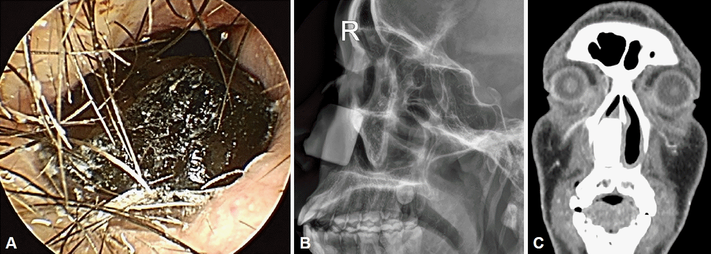
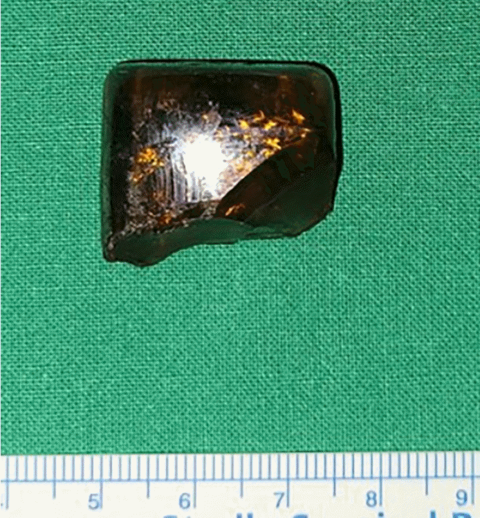
We identified several lessons from this case. First, an impacted foreign body in the anterior nasal cavity could induce tooth pain. Tooth pain of nonodontogenic causes could originate from various reasons, including referred pain from the sinonasal cavity. The anterior two-thirds of the nose are innervated by branches of the maxillary division V2 of the trigeminal nerve [
5,
6]. Pain perceived by the patient could actually be of sinonasal cavity origin [
5]. Our patient perceived continuous throbbing tooth pain in his right anterior tooth region, and the pain was relieved after foreign body removal. Therefore, we hypothesized that the hard, large foreign body, which was impacted in the anterior nasal cavity and pushed on the nasal floor and nasal vestibule area, might have induced referred pain through stimulation of the trigeminal nerve. Second, a glass foreign body demonstrates high intensity on imaging (slightly higher intensity than bone). In our patient, the glass foreign body showed higher density than the surrounding bony structures. For foreign bodies in the nasal cavity, conventional plain radiography is usually the initial imaging modality. However, CT is the most accurate tool, particularly when a nonopaque object is anticipated or the foreign body is located inside pneumatized areas [
7]. A previous study reported that cone-beam CT (CBCT) was equally accurate in visualizing metal, stone, and tooth particles. Furthermore, CBCT was better than CT in detecting glass particles in the periosteum. Magnetic resonance imaging is more sophisticated, but it cannot be applied initially in cases with a suspected foreign body of unknown composition [
7]. Therefore, in cases of foreign bodies in the sinonasal cavity whose composition is not accurately known, we suggest that at least CBCT should be considered as an initial imaging tool. Finally, if it is not possible to determine the type of material based on imaging, the clinician should prepare for the possibility that the material could be broken during surgical removal, especially if the foreign body is large and impacted in the nasal cavity. In our case, as the glass foreign body was tightly impacted in the anterior nasal cavity, we grasped it strongly and applied a substantial amount of strength to take it out. Fortunately, the surface of the glass material was very smooth and it was quite thick, as we identified on preoperative CT images; therefore, we were able to remove it in a single block without breaking it. If the foreign body is broken into multiple pieces, some of the pieces may not be completely removed. Glass may be present for weeks or years, causing chronic sinus pain or pain of unknown origin. Therefore, CT images should be carefully reviewed regarding the size, thickness, and condition of the surface area, and the possibility of the material breaking should be considered before surgery.


 PDF
PDF Citation
 Print
Print


 XML Download