

 PDF
PDF Citation
 Print
Print


The ABO blood group shows phenotypic variants, largely owing to its molecular genetic basis [1]. As an independent tool in the clinical laboratory, serology can reveal ABO discrepancies and ABO genotyping is essential for correct determination of the blood group. The cis-AB blood group largely explains the ABO discrepancies in the East-Asian population [2]. The cis-AB allele results in a mutated enzyme with both A and B glycosyltransferase activities and leads to decreased levels of both antigens, which is presented as various phenotypes [3-5]. The first phenotypes identified were A2B3, A2B, and A1B3, which are associated with the cis-AB01/O, cis-AB01/B, and cis-AB01/A genotypes, respectively [6]. Further studies revealed more phenotypes, including A1Bel, A1Bx, A1Bm, AintB3, AintB, and A1 [7].
In this study, we performed a retrospective investigation of the ABO grouping test results of two medical institutions in Korea. When weak or unusual expression of antigens was observed, the ABO genotyping results and medical records were evaluated. In selected samples for which the cause of ABO discrepancy remained unresolved, we looked for genetic variants in the full ABO coding region and promoter region. We reviewed the serology and genotyping results of 39 samples from Seoul St. Mary’s Hospital and 8 samples from Incheon St. Mary’s Hospital from January 2016 to June 2021. Additionally, we included pedigree analyses of two families. This study was approved by the Institutional Review Board (IRB) of the Catholic Medical Center (Seoul, Korea) and the need for informed consent was waived (XC22RADI0073).
Serological tests were routinely performed using a column agglutination technique using ORTHO BioVue System (Ortho Clinical Diagnostics, Pencoed, UK) on an automatic analyzer, ORTHO VISION (Ortho Clinical Diagnostics, Raritan, NJ, USA). In the presence of weak or unusual expression of the A or B antigens, manual tube methods using murine monoclonal anti-A, anti-B (Shinyang Diagnostics, Seoul, Korea) were employed, and serum typing was done with A1 and B cells (MIRR. SciTech Corp., Seoul, Korea) using a plate method. To further identify ABO subgroups, anti-A1 lectin obtained from Dolichos biflorus seeds and anti-H from Ulex europaeus seeds (Lorne Laboratories Ltd, Berkshire, UK) were used.
For ABO genotyping, DNA was extracted from EDTA blood samples using a QIAsymphony DSP DNA Kit (Qiagen, Hilden, Germany). PCR was run using a C1000 Touch Thermal Cycler (Bio-Rad, Hercules, CA, USA) with a mixture of primer pairs (Table 1) for amplification. The products were purified using the ExoSAP-IT PCR Product Cleanup Reagent (Thermo Fisher Scientific, Waltham, MA, USA). A Big Dye Terminator Cycle Sequencing Kit (Applied Biosystems, Foster City, CA, USA) and an ABI 3500XL DX Genetic Analyzer (Applied Biosystems) were used for Sanger sequencing according to the manufacturer’s instructions. Sequencing data were analyzed using the ABO reference sequence (NM_020469.2) with Sequencher v.5 (Gene Codes Corp, Ann Arbor, MI, USA). The ABO alleles were classified according to the nomenclature used by the International Society of Blood Transfusion.
A total of 47 ABO serology and genotyping results were reviewed. These were referred for ABO genotyping test as they all showed a ABO discrepancy—defined as a mismatch between the cell and serum typing or presence of a weaker strength of agglutination (≤3+ in cell typing; ≤2+ in serum typing), as described in a previous study [8]. Results were further classified as ‘phenotype-genotype matches’ or ‘phenotype-genotype mismatches’ (Table 2). The frequency of phenotypes consistent with their genotypes was 72.3% (34/47 cases). Among them, 23 cases demonstrated a cis-AB allele by genotyping, with cis-AB/O being the most common genotype. All samples with A2B3 phenotype had cis-AB/O alleles but not necessarily vice versa. Cis-AB.01/O.01.01 and cis-AB.01/O.01.02 genotypes were also associated with A1B3 or AB phenotypes. All cis-AB.01/B.01 patients expressed A1B3 phenotypes. Consistent with previous studies, two patients with cis-AB.01/A1.02 genotype demonstrated A phenotype. One patient with cis-AB.01/A1.02 genotype did not show agglutination with anti-B in cell typing but showed weak agglutination with B cell in serum typing, and was thus categorized as A1Bel. Other weak ABO subgroups included Ax/Aweak, an uncommon phenotype with a 0.01% frequency in a large-scale study [9].
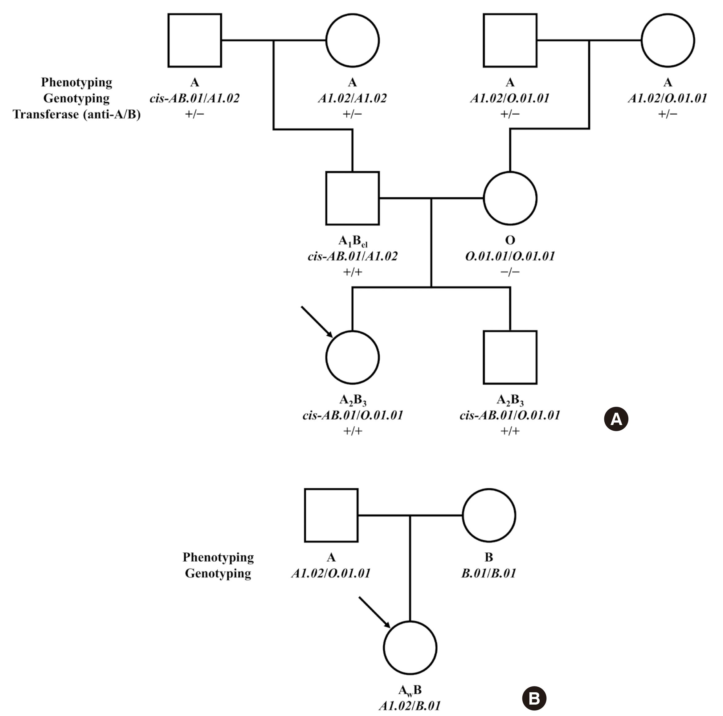
The frequency of phenotypes inconsistent with their genotypes was 27.7% (13/47 cases). Serology showed five distinct A subgroups (A2, A3, Aw, Ael, Aint), but genotypes of all samples included the common A1.02 allele. Direct sequencing of exons 3 to 5 was performed for a patient who showed Ael phenotype but A1.02/O.01.01 genotype, and no other variants were detected. Whole exon sequencing including the promoter region was recommended to the clinical department. The serology result of another patient showed AwB, but her genotype was A1.02/B.01. This discrepancy was confirmed by pedigree analysis of her father’s and mother’s genotyping results (Fig. 1B). The patient continued to show weakened A antigen expression in follow-up testing. Furthermore, while two different B subgroups (B3, Bw) were expressed, 6 out of 7 patients had the consensus B.01 allele. One patient with Bw phenotype initially showed 6 possible genotypes in sequencing. Further analysis of exons 3 to 5 showed additional variants, and the patient’s genotype was narrowed down to either B.01/O.01.02 or B3.02/O.01.35. Further studies such as allele specific sequencing and family studies could have located the allele that contained the variants.
Medical records were evaluated for any medical factors that may have affected the test results. Weak but variable A or B antigen expression was noted in patients with hematological diseases (N=5), including juvenile myelomonocytic leukemia, Burkitt lymphoma, high-grade B-cell lymphoma, acute myeloid leukemia, and myelodysplastic syndrome. All patients showed weak antigen expression at initial diagnosis, and none of them received out-of-group transfusion prior to serological typing. Interestingly, one patient with myelodysplastic syndrome evolved into leukemic transformation 9 months after initial diagnosis. One patient who showed weak B antigen expression was diagnosed with renal cell carcinoma.
Pregnancy was suspected to cause weak antigen expression in 3 patients. Notably, one multipara, who showed normal AB blood grouping results, exhibited weak A antigen expression (3+ in cell typing, +/- in serum typing) during her third pregnancy. When ABO genotyping was performed, she was diagnosed as cis-AB.01/B.01. Repeating sample testing 4 years after her third delivery expressed a normal AB. This patient showed a simultaneous presence of medical factor and weak ABO subgroup allele. Other women were unattainable for sample testing to be repeated after delivery.
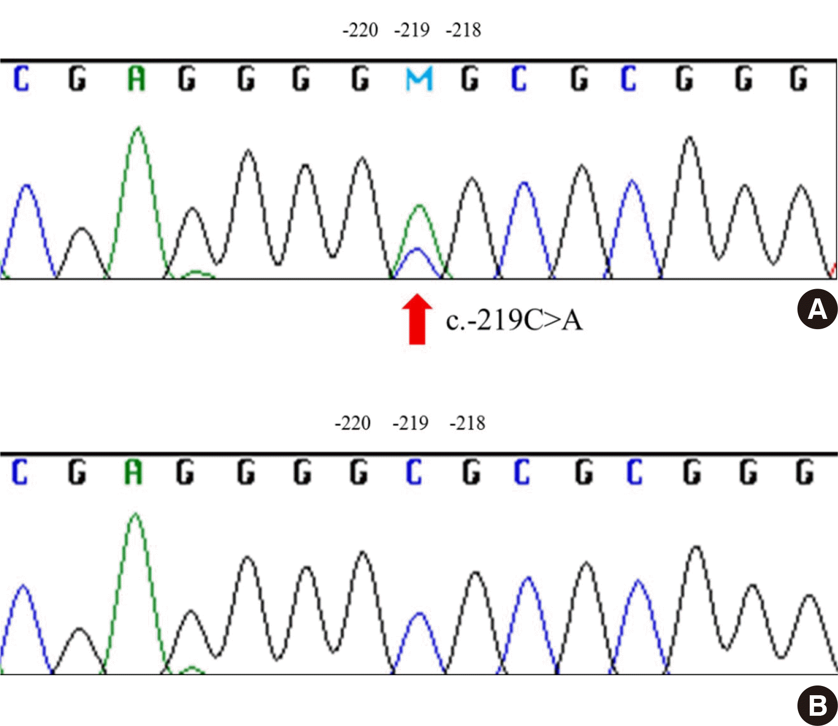
Pedigree analysis was performed for one family that showed cis-AB inheritance across three generations. Interestingly, both the father and paternal grandfather had cis-AB.01/A1.02 genotype with different phenotypes: A1Bel, A (Fig. 1A), and the results were the same after retesting. Therefore, whole exon sequencing including promoter region was performed, and a single-point mutation in the promoter region (-219 C>A) was found in one of the two patients (Fig. 2). In transferase analysis, the patient who had a mutation in the promoter region had A transferase but not B transferase, which led to A phenotype. The other patient who did not possess the mutation had both A and B transferases and presented as A1Bel. We suggest that the expression of the ABO gene was suppressed by the mutation of the promoter region, resulting in A phenotype even though the patient’s genotype was cis-AB.01/A1.02. This in turn implies that the single point mutation was on the cis-AB-allele in the individual.
The ABO gene, located on chromosome 9q34, consists of seven coding exons, with the largest open reading frame located in exons 6 and 7 [10]. As analysis of only exons 6 and 7 is sometimes insufficient, further sequencing of other exons, introns, and the promoter region is required [11, 12]. In 1997, Kominato et al. first verified the essential role of the promoter sequence in the regulation of ABO gene transcription using KATO III, the human gastric cancer cell line [13, 14]. Recent studies revealed cases where mutations of the promoter sequence downregulate the promoter activity, thereby decreasing A or B antigen expression. Isa et al. [15] discovered two single-point mutations (-76G>C and -86G>T) in the promoter on the A-allele in three A3 patients with A1.01/O.01.01 and A1.01/O.01.02 as well as on the B-allele in a B3 patient with B.01/O.01.02. These findings were verified using a luciferase assay that compared the promoter activity of reporter plasmids carrying the promoters with mutations in the -76 and -68 regions with that of a wild type promoter [15]. Additionally, Hellberg et al. [16] reported a mutation in the -72 region in a patient who showed phenotype B3 but B.01/O.01.01 genotype.
In conclusion, the prevalence of the ABO subgroups demonstrated in this study was consistent with that of similar previously published studies. Medical factors, including hematologic disorders, malignancy, and pregnancy also affected the ABO discrepancies, as noted previously [17]. This study further aimed to focus on the effect of a mutation in the promoter region on ABO gene expression. Further evaluation of the promoter region could help differentiate ABO discrepancies that remain unresolved.
 XML Download