Abstract
Background
The clinical phenotype of hemophilia A (HA) does not always correlate with severity. Similarly, the presence of inhibitors does not necessarily increase the risk of bleeding. This paradox between clinical and laboratory findings may be partially attributed to non-modifiable factors, such as blood group, which is known to influence FVIII levels in healthy individuals. Our aim was to assess the effect of ABO blood group antigens on FVIII levels across the severity spectrum of HA and risk of inhibitor development.
Methods
Data of consecutive patients with HA who visited the coagulation unit of a northern Indian tertiary care hospital between 2010‒2021 were reviewed. Patients with missing blood group data, transfusion histories, or baseline FVIII levels were excluded.
Results
Mild, moderate, and severe HA was present in 41 (6.9%), 72 (12.2%), and 479 (80.9%) patients, respectively. There were no differences in the FVIII levels among the various blood groups across the HA severity spectrum. Inhibitors were administered to 35 patients (5.9%). In the multivariate analysis, blood group A was an independent risk factor for the development of inhibitors (adjusted odds ratio 2.70, P=0.04) after adjusting for age at onset of bleeding, FVIII transfusion, age at first FVIII transfusion, and severity of HA.
The central role of ABO blood group antigens in transfusion and transplantation has been well-established for decades [1]. However, in addition to expression in red blood cells, ABO blood group antigens are highly expressed in various cells and tissues, including epithelial cells, platelets, and vascular endothelium [1, 2]. ABO antigens have been recognized as risk factors for various diseases such as peptic ulcers, cardiovascular disease, and malignancies [1-3]. In addition to their roles in maintaining cell membrane integrity, mediating cell adhesion, and acting as receptors for extracellular ligands, ABO antigens help in hemostasis. It has been observed that von Willebrand factor (vWF) levels are 25% higher in individuals with non-O blood groups [4]. In healthy individuals, the ABO blood group antigen-mediated variation in the glycosylation of vWF affects its clearance and concentration, thereby leading to a change in the FVIII concentration [5]. It is reasonable to hypothesize that ABO blood group antigens influence residual FVIII levels in patients with hemophilia A (HA). However, this remains underexplored, and previously reported literature is largely confined to patients with non-severe HA [6, 7].
The development of neutralizing alloantibodies is a major hurdle in HA management [8]. Various nongenetic and environmental risk factors govern the development of HA inhibitors. However, the role of ABO blood groups in determining FVIII immunogenicity and the risk of developing HA inhibitors in HA has not been well studied [9-11]. With this in mind, we retrospectively reviewed the role of the ABO blood group in determining residual FVIII activity and the risk of inhibitor development in patients with HA.
The data of all consecutive patients with HA who visited the coagulation unit of the Department of Hematology in a North Indian tertiary care hospital between 2010–2021 were reviewed. Patients with missing blood group data, transfusion histories, or baseline FVIII levels were excluded.
The clinical details of the patients, including demographic factors, site, and precipitant(s) of bleeding were retrieved from the records. The ABO blood group type was also noted in the review of the available records. Details regarding family history were obtained by analyzing the pedigree chart. The history of the type of transfusion product received was also noted in cases where details were available.
Investigations performed included prothrombin time (PT), activated partial thromboplastin time (aPTT), fibrinogen level, mixing studies, factor VIII (FVIII) assay, inhibitor screening test, inhibitor assay, vWF antigen (vWF:Ag) and vWF glycoprotein 1b receptor (vWF:GPIbR) assay. These tests were run on the fully automated coagulation analyzers [STA Compact, STA-R Evolution (both, Diagnostica Stago, Asnieres, France) and/or ACL TOP 500 CTS (Instrumentation Laboratory, Bedford, MA, USA)] as per instructions from the manufacturer in the kit and depending upon instrument availability. The inhibitor assay was performed using the classical Bethesda assay (CBA). The Nijmegen-modified Bethesda assay (NBA) was performed as indicated. High-titer inhibitors were defined as inhibitor titers >5 Bethesda units. The correlation between the residual factor VIII levels and inhibitor development was also studied.
Quantitative data are presented as mean±standard deviation or median with interquartile range, as applicable. Qualitative data are expressed as proportions and percentages. Quantitative data between the two groups were compared using the t-test for parametric data and the Mann–Whitney U test for non-parametric data. Quantitative data between three or more groups were compared using the Kruskal–Wallis test with Dunn’s test for multiple comparisons. Statistical comparisons of qualitative data between two groups were assessed using Fisher’s exact test, whereas qualitative data between three or more groups were analyzed using the chi-squared test. Correlations were assessed using Pearson correlation coefficients. Multivariate logistic regression analysis for risk factors for developing inhibitors was performed by incorporating the variables of age at onset of bleeding, FVIII transfusion, age at first FVIII transfusion, blood group, and severity of HA using a stepwise model where variables were entered into the model if P<0.05 and removed if P>0.1. Multicollinearity was ruled out by assessing the variance inflation factors. All statistical tests were two-sided, and a P-value <0.05 was considered statistically significant.
Of the 680 patients whose data were reviewed, 88 were excluded because of non-availability of baseline FVIII levels (N=8) or blood groups (N=80). Finally, 592 HA patients were included in this study, among whom 41 (6.9%), 72 (12.2%), and 479 (80.9%) had mild, moderate, and severe disease, respectively. Median age at the onset of bleeding was 3 (2–8) years. A history of FVIII transfusion was observed in 479 patients (80.9%). These patients received FVIII on demand, and none of the patients received prophylactic therapy. Overall, intra-articular bleeding was the most common site of bleeding (89%). No difference in the site of bleeding was noted among the three severity subgroups. The most common blood group in our cohort was O (N=203, 34.3%), followed by B (N=179, 30.2%), AB (N=106, 17.9%), and A (N=104, 17.6%). The demographic profiles and clinical parameters of the patients are shown in Table 1. Spontaneous bleeding was significantly more common in patients in blood group A (53.8%) than in those in the non-A blood group (42.2%, P=0.04), as shown in Supplementary Table 1. However, there was no difference in the sites of bleeding among patients with HA in different blood groups (Supplementary Table 2).
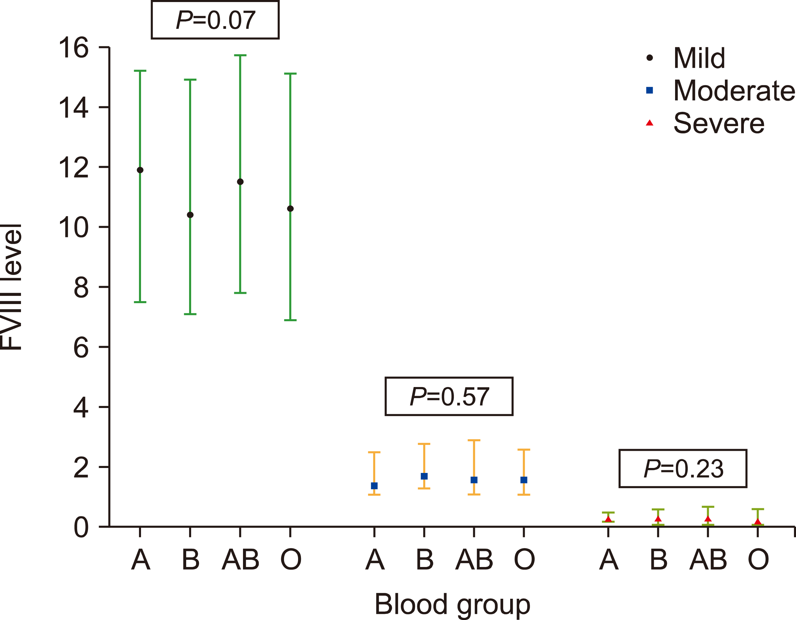
The median FVIII levels in mild, moderate, and severe HA were 10.7% (7.1–16.58), 1.6% (1.2–2.5), and 0.25% (0.1–0.6), respectively. In mild HA, there was a trend towards higher FVIII levels in blood groups A (11.9%) and AB (11.5%) than in blood groups O (10.6%) and B (10.4%), although the difference was not statistically significant (P=0.07). No significant differences in FVIII levels were observed between different blood groups with moderate or severe HA (Fig. 1).
In the whole cohort, there was no correlation between vWF and FVIII levels [r, 0.12; 95% confidence interval (CI), -0.16 to 0.41; P=0.79]. Furthermore, there was no difference in vWF levels among the mild, moderate, and severe HA groups (Table 1). vWF levels were significantly lower in patients with blood group O [134.1 (81.7–176.4)] compared to those with non-O blood groups [154.9 (91.6–195.6), P=0.03]. There were no significant differences in vWF levels between the other blood groups (Supplementary Table 3). There was no difference in vWF levels between HA patients with or without inhibitors [142.7 (98.8–192.3) vs. 148 (95.8–190.1), P=0.66].
FVIII inhibitors were administered to all 35 patients. The overall prevalence of the inhibitors was 5.9%. The prevalence of different blood groups among the inhibitor-positive patients was as follows: A (N=13, 37.1%), B (N=8, 22.9%), AB (N=5, 14.3%), and O (N=9, 25.7%). Blood-group A patients had higher odds of developing inhibitors (OR, 3.03; 95% CI, 1.48–6.23; P=0.003) compared to non-A blood groups (Table 2). None of the other blood groups differed significantly between the patients with and without inhibitors. In the multivariate analysis, blood group A remained an independent predictor of the development of inhibitors (adjusted OR, 2.70; P=0.04) after adjusting for age at onset of bleeding, FVIII transfusion, age at first FVIII transfusion, and severity of HA (Table 3).
Among patients with high-titer inhibitors (N=28), most patients had blood group A (N=12, 42.8%), followed by blood group B (N=6, 21.4%), O (N=5, 17.9%), and AB (N=5, 17.9%). There was no difference in the proportion of patients with high- or low-titer inhibitors among the various blood groups (Supplementary Table 4).
The role of blood group antigens in governing FVIII levels in healthy individuals is well established. Indeed, healthy individuals in the non-O blood group had 25–35% higher levels of FVIII than those in the O blood group [4, 5]. This may explain the higher risk of thrombotic events, including stroke, in the non-O blood group [1-3]. However, literature regarding the effect of blood group antigens on residual FVIII levels in HA is sparse and largely limited to patients with non-severe HA. Rejtő et al. [6] and Loomans et al. [7] reported no differences in FVIII antigen and activity levels between the non-O and O blood groups in patients with non-severe HA. However, we observed a higher trend in residual FVIII levels in patients with blood groups A and AB and mild HA only. We did not observe any statistically significant differences in FVIII levels among blood groups with mild, moderate, or severe HA. These observations suggest that blood group antigens do not play a substantial role in determining residual FVIII levels in patients with HA (irrespective of severity), unlike what is observed in the general population.
We observed lower vWF levels in patients with blood group O, as previously reported [4, 12]. Recent data suggest that patients with blood group A may have an increased severity of bleeding when bleeding is of an unknown cause [13]. In our cohort of patients with HA, spontaneous bleeding was more common in patients with blood group A, and there was no difference in the site of bleeding among the various blood groups. This suggests that patients with HA and blood group O may not be predisposed to more severe bleeding. This is possibly because in HA, the major factors governing the risk and severity of bleeding are FVIII levels and the presence of inhibitors instead of vWF levels. The risk of developing inhibitors in various blood groups is controversial, and contradictory results have been reported in different studies. The data from these studies are summarized in Table 4 [9-11].
Two previous studies from Italy reported that, in comparison to non-O blood groups, blood group O is protective against the development of inhibitors and is also associated with low-titer inhibitors [9, 10]. Although blood group O did not show a statistically significant association with inhibitor development in our study, it had the lowest odds of inhibitor development. Furthermore, the most common blood group in patients without inhibitors was blood group O, suggesting that blood group O may play a putative protective role against the development of FVIII inhibitors, which needs to be explored in further studies. The roles of genetic mutations, ethnicity, and geographic variation in modulating the association between blood group antigens and inhibitors need to be examined in detail. In our study, blood group A was independently associated with an increased risk of developing inhibitors, with an adjusted odds ratio of 2.71 after adjusting for age at onset of bleeding, FVIII transfusion, age at first FVIII transfusion, and severity of HA. The only other study from India that examined the association between blood group and the development of inhibitors in 300 patients with HA corroborates our findings that patients with blood group A have a higher risk of inhibitor development (Table 4) [11].
The reasons for the observed association between blood group antigens and the risk of inhibitor development have not been sufficiently investigated. The binding of vWF to the circulating FVIII stabilizes it and prevents its decay [14]. Clearance of the FVIII-vWF complex by the low-density lipoprotein receptor-related protein-1 receptor on macrophages was slower in those with blood group A than in those with blood group O [14, 15]. This difference may result in a substantially longer half-life of infused FVIII in HA patients with blood group A than in those with blood group O, which may translate into a higher risk of inhibitor development.
The present study is among the largest studies on patients with HA treated with inhibitors in the Indian subcontinent. However, we acknowledge the limitations of our study, including its retrospective design. We did not have data concerning the amount and type (plasma-derived or recombinant) of FVIII transfused. Genetic analyses were not performed in the present real-world observational study. We acknowledge the caveats in interpreting our data on the relationship between bleeding phenotype and blood groups. All of our HA patients had a history of bleeding at presentation, and we cannot comment on the influence of blood groups on the prospective risk of bleeding in these patients. We did not have adequate data for the formal calculation of bleeding assessment scores, such as the Vincenza or ISTH scores.
REFERENCES
1. Ewald DR, Sumner SC. 2016; Blood type biochemistry and human disease. Wiley Interdiscip Rev Syst Biol Med. 8:517–35. DOI: 10.1002/wsbm.1355. PMID: 27599872. PMCID: PMC5061611.

2. Liumbruno GM, Franchini M. 2013; Beyond immunohaematology: the role of the ABO blood group in human diseases. Blood Transfus. 11:491–9. DOI: 10.2450/2013.0152-13. PMID: 24120598. PMCID: PMC3827391.
3. Wu O, Bayoumi N, Vickers MA, Clark P. 2008; ABO(H) blood groups and vascular disease: a systematic review and meta-analysis. J Thromb Haemost. 6:62–9. DOI: 10.1111/j.1538-7836.2007.02818.x. PMID: 17973651.
4. Jenkins PV, O'Donnell JS. 2006; ABO blood group determines plasma von Willebrand factor levels: a biologic function after all? Transfusion. 46:1836–44. DOI: 10.1111/j.1537-2995.2006.00975.x. PMID: 17002642.
5. Lenting PJ, Christophe OD, Denis CV. 2015; von Willebrand factor biosynthesis, secretion, and clearance: connecting the far ends. Blood. 125:2019–28. DOI: 10.1182/blood-2014-06-528406. PMID: 25712991.

6. Rejtő J, Königsbrügge O, Grilz E, et al. 2020; Influence of blood group, von Willebrand factor levels, and age on factor VIII levels in non-severe haemophilia A. J Thromb Haemost. 18:1081–6. DOI: 10.1111/jth.14770. PMID: 32073230. PMCID: PMC7318586.
7. Loomans JI, van Velzen AS, Eckhardt CL, et al. 2017; Variation in baseline factor VIII concentration in a retrospective cohort of mild/moderate hemophilia A patients carrying identical F8 mutations. J Thromb Haemost. 15:246–54. DOI: 10.1111/jth.13581. PMID: 27943580.
8. Srivastava A, Santagostino E, Dougall A, et al. 2020; WFH guidelines for the management of hemophilia, 3rd edition. Haemophilia . 26(Suppl 6):1–158. DOI: 10.1111/hae.14046. PMID: 32744769.

9. Franchini M, Coppola A, Santoro C, et al. 2021; ABO blood group and inhibitor risk in severe hemophilia A patients: a study from the Italian Association of Hemophilia Centers. Semin Thromb Hemost. 47:84–9. DOI: 10.1055/s-0040-1718870. PMID: 33525041.

10. Franchini M, Coppola A, Mengoli C, et al. 2017; Blood group O protects against inhibitor development in severe hemophilia A patients. Semin Thromb Hemost. 43:69–74. DOI: 10.1055/s-0036-1592166. PMID: 27825181.
11. Arshad S, Singh A, Awasthi NP, Kumari S, Husain N. 2018; Clinicopathological parameters influencing inhibitor development in patients with hemophilia A receiving on-demand therapy. Ther Adv Hematol. 9:213–26. DOI: 10.1177/2040620718785363. PMID: 30181842. PMCID: PMC6116755.
12. Desch KC. 2018; Regulation of plasma von Willebrand factor. F1000Res. 7:96. DOI: 10.12688/f1000research.13056.1. PMID: 29416854. PMCID: PMC5782404. PMID: 0234ff01d6e94056bd14507d01010b9d.
13. Mehic D, Hofer S, Jungbauer C, et al. 2020; Association of ABO blood group with bleeding severity in patients with bleeding of unknown cause. Blood Adv. 4:5157–64. DOI: 10.1182/bloodadvances.2020002452. PMID: 33095871. PMCID: PMC7594405.
14. Franchini M, Crestani S, Frattini F, Sissa C, Bonfanti C. 2014; ABO blood group and von Willebrand factor: biological implications. Clin Chem Lab Med. 52:1273–6. DOI: 10.1515/cclm-2014-0564. PMID: 24945431.
15. Kepa S, Horvath B, Reitter-Pfoertner S, et al. 2015; Parameters influencing FVIII pharmacokinetics in patients with severe and moderate haemophilia A. Haemophilia. 21:343–50. DOI: 10.1111/hae.12592. PMID: 25582282.

Fig. 1
FVIII levels in different blood groups across the severity spectrum of HA (P-value calculated using Kruskal–Wallis test with Dunn’s correction).

Table 1
Clinicopathological characteristics of study population.
Table 2
ABO blood groups and risk of inhibitor development.
Table 3
Risk factors for development of inhibitors (N=592).
Table 4
Association of various blood groups and inhibitor development among HA patients in different studies.
| Parameters | Arshad et al. 2018 [11] | Fanchino et al. 2016 [10] | Franchino et al. 2021 [9] | Present study | ||||
|---|---|---|---|---|---|---|---|---|
| Total HA patients | 300 | 209 | 287 | 592 | ||||
| HA patients with inhibitors (N, %) | 29 (9.6%) | 56 (26.8%) | 85 (29.6%) | 35 (5.9%) | ||||
| Low titer inhibitor | 17 (58.6%) | 12 (5.7%) | 21 (24.7%) | 7 (20%) | ||||
| High titer inhibitor | 12 (41.4%) | 44 (21.1%) | 64 (75.3%) | 28 (80%) | ||||
| ABO blood group, N (%) | With inhibitor | Without inhibitor | With inhibitor | Without inhibitor | With inhibitor | Without inhibitor | With inhibitor | Without inhibitor |
| A | 19 (65.5%) | 54 (20%) | 30 (31.9%) | 64 (68.1%) | 39 (45.9%) | 62 (30.7%) | 13 (37.5%) | 196 (95.6%) |
| B | 2 (6.8%) | 98 (36.1%) | 9 (39.1%) | 14 (60.9%) | 13 (15.3%) | 23 (11.4%) | 8 (22.9%) | 173 (30.6%) |
| AB | 2 (6.8%) | 43 (16%) | 1 (25.0%) | 3 (75.0%) | 7 (8.2%) | 6 (3.0%) | 5 (14.3%) | 104 (18.4%) |
| O | 6 (20.6%) | 76 (28%) | 16 (18.2%) | 72 (81.8%) | 26 (30.6%) | 111 (55.0%) | 9 (25.7%) | 196 (95.6%) |
| P |
O vs. A: 0.02a) O vs. B: 0.12 O vs. AB: 0.52 A vs. B: 0.005a) A vs. AB: 0.49 B vs. AB: 0.05 |
O vs. A: 0.033 O vs. B: 0.032 O vs. non-O: 0.017 |
<0.001 |
O vs. non-O: 0.28 A vs. non-A: 0.003a) B vs. non-B: 0.33 AB vs. non-AB: 0.57 |
||||


 PDF
PDF Citation
 Print
Print


 XML Download